This website uses cookies to ensure you get the best experience on our website.


- Table of Contents
Plan chromogenic ADH5 IHC in paraffin sections around the generally cytoplasmic tissue pattern (HPA tissue IHC). The catalog antibody's IHC range is 0.5–1 μg/ml (datasheet RP1111), and liver hepatocytes and kidney tubule cells provide high-staining comparison tissues (HPA tissue IHC).

Expected localisation, validated protocols, controls and antibodies — the at-a-glance facts below, then the full design guide.
| Expected localisation | Cytoplasm expected; tissue staining generally cytoplasmic (UniProt; HPA tissue IHC) | |
| Staining pattern | General cytoplasmic staining across tissue cell types (HPA tissue IHC) | |
| Antigen retrieval | Citrate pH 6 HIER, heat-mediated (datasheet RP1111) | |
| Positive control | Epididymis+4 more · see all | |
| Negative control | Bone marrow+4 more · see all |
| Fixation | Keep fixation consistent across sections (standard IHC practice; not target-specific) | |
| Caveat | In liver, endogenous peroxidase may mimic DAB staining (HPA tissue IHC; standard IHC practice) | |
| Regulation | Staining regulation is not established (UniProt) | |
| Isoform / epitope | No annotated isoforms; cytoplasmic, with no TM segment (UniProt) |
The catalog antibody uses citrate pH 6 heat retrieval (datasheet RP1111). The published ADH5 IHC protocols below cover iBAT (PMC8640996) and breast tissue (PMC12000288).
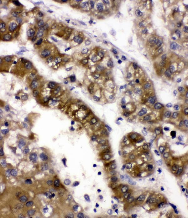
| Sample | Paraffin-embedded human liver cancer tissues; fixative not specified (datasheet RP1111) |
| Fixation | Image fixative and duration unreported (datasheet RP1111); verify before use. |
| Sectioning | 4–5 µm sections on charged slides (standard) |
| Deparaffinisation | Xylene, graded ethanol series to water (standard) |
| Antigen retrieval | Heat retrieval: Citrate pH 6, 20 min (datasheet RP1111) |
| Peroxidase block | 3% H2O2, 10 min, room temperature (standard) |
| Blocking | 10% goat serum (datasheet RP1111) |
| Primary antibody | Rabbit anti-ADH5, 0.5-1μg/ml (datasheet RP1111) |
| Primary incubation | Overnight at 4 °C (datasheet RP1111) |
| Detection | Streptavidin-biotin complex (SABC), DAB chromogen (datasheet RP1111) |
| Counterstain | Hematoxylin, blue, dehydrate and mount (standard) |
| Expected result | ADH5-positive staining in glandular cells of epididymis (HPA tissue IHC: High). HPA tissue profile: General cytoplasmic expression. No signal in the no-primary control. |
ADH5 is a cytoplasmic protein with no annotated transmembrane segment (UniProt P11766: subcellular location; topology). In paraffin-section IHC, expect cytoplasmic staining in hepatocytes, kidney tubular cells, epididymal glandular cells, smooth muscle cells, and cells in seminiferous ducts; HPA reports these as high (HPA: tissue IHC). HPA rates its tissue pattern Approved, with medium agreement between staining and RNA data and external verification pending (HPA: reliability).
| Cytoplasmic signal in hepatocytes or kidney tubular cells (HPA: High in both cell types). | This matches the reported compartment and provides a useful positive reference (UniProt P11766: Cytoplasm; HPA: tissue IHC). Judge signal in the named cells, because a tissue-level positive result does not make every cell in the section positive. |
| Predominantly nuclear, membranous, or extracellular signal, with little cytoplasmic signal (UniProt P11766: Cytoplasm). | This does not match the annotated location; treat it as a possible staining artefact or antibody specificity issue, not evidence of a new ADH5 location (UniProt P11766: Cytoplasm; general IHC interpretation). Review controls before assigning that pattern to ADH5. |
| Strong signal in skeletal-muscle myocytes or lymph-node germinal-center cells (HPA: Not detected in those cells). | The reported HPA pattern does not support those cells as strong positives. Consider cross-reactivity or chromogenic detection background, then compare the same run with a reported positive cell population (HPA: tissue IHC; general IHC practice). |
| Diffuse color across cells and surrounding tissue, without clear cell boundaries (general IHC observation). | A uniform haze is difficult to score as ADH5 because the expected pattern is cellular and cytoplasmic (UniProt P11766: Cytoplasm). Check a no-primary control for background from the detection system and review blocking and wash conditions (general IHC practice). |
| No convincing cytoplasmic signal in hepatocytes or kidney tubular cells (HPA: High in both). | This conflicts with the reported high IHC levels but does not alone prove ADH5 is absent (HPA: tissue IHC; general IHC interpretation). Check section quality, the positive control, antibody dilution, retrieval conditions, and detection reagents before scoring a negative result (general IHC practice). |
| Compartment and topology (UniProt P11766). | ADH5 is annotated in the cytoplasm and has no transmembrane segment; use cytoplasmic staining as the localisation check (UniProt P11766). The record provides no basis for expecting a membrane pattern. |
| Cell-specific tissue pattern (HPA: tissue IHC). | HPA reports high staining in selected glandular, tubular, hepatocyte, smooth-muscle, and seminiferous-duct cell populations; adipocytes are medium and skeletal-muscle myocytes are not detected (HPA: tissue IHC). Score the specified cells rather than the whole tissue uniformly. |
| Strength of IHC evidence (HPA: reliability; antibodies). | HPA rates the tissue profile Approved, with medium staining–RNA consistency and external verification pending; HPA044578 and HPA061919 are each IHC Approved (HPA: tissue IHC; antibodies). These ratings support use as references without guaranteeing a result in every specimen. |
| Can IF/ICC confirm the same pattern? (HPA: subcellular data). | HPA lists no main ICC-IF location and no cell lines with ICC-IF images for ADH5; its tissue IHC pattern cannot establish an IF/ICC result (HPA: subcellular; tissue IHC). Use this page's cytoplasmic expectation as a hypothesis from UniProt, not an IF validation claim (UniProt P11766: Cytoplasm). |
| Situation | Likely cause | Next action |
|---|---|---|
| A reported high-staining cell population is blank (HPA: tissue IHC). | The run may have insufficient detectable signal; a blank section alone cannot distinguish assay failure from specimen variation (general IHC interpretation). | Check a same-run positive tissue with the named cell population, then review catalog-antibody dilution, retrieval, and detection controls (HPA: tissue IHC; general IHC practice). |
| Nuclei dominate the signal (UniProt P11766: Cytoplasm). | The compartment conflicts with the annotated cytoplasmic location; nonspecific staining is possible (UniProt P11766; general IHC interpretation). | Compare with a no-primary control and an HPA-reported high cytoplasmic cell population before scoring the nuclear signal as ADH5 (HPA: tissue IHC; general IHC practice). |
| Color appears in cells HPA reports as not detected (HPA: tissue IHC). | Possible antibody cross-reactivity or detection-system background; HPA's cell-specific finding is a reference, not proof that every specimen is negative (HPA: tissue IHC; general IHC interpretation). | Check the no-primary control, assess cellular localisation, and compare with a reported high-staining population in the same run (UniProt P11766: Cytoplasm; HPA: tissue IHC; general IHC practice). |
| Diffuse staining obscures cell boundaries (general IHC observation). | Background may arise from detection activity or insufficient blocking or washing (general IHC practice). | Inspect the no-primary control; if it also shows color, review endogenous-enzyme blocking, wash steps, and chromogen development (general chromogenic IHC practice). |
| Signal is weak in adipocytes or other medium-staining cells (HPA: tissue IHC). | The reference level is medium, so weak signal there is less decisive than loss of signal in an HPA-reported high population (HPA: tissue IHC; general IHC interpretation). | Assess a high-staining reference cell population and keep exposure or chromogen-development conditions consistent across comparisons (HPA: tissue IHC; general IHC practice). |
| Different sections give conflicting staining intensity (general IHC observation). | Section or run variation may affect comparison; HPA also reports medium staining–RNA consistency and pending external verification (general IHC practice; HPA: reliability). | Compare cell-specific patterns with matched positive and no-primary controls, and document retrieval and detection conditions before assigning biological meaning (HPA: tissue IHC; general IHC practice). |


Comprehensive Human Protein Atlas IHC scoring per tissue (reliability: Approved — Medium consistency between antibody staining and RNA expression data. Pending external verification.). Rows are taken directly from the HPA tissue chart — click any row's HPA link to view the source.
| Tissue | Cell type | Level | Evidence | Source |
|---|---|---|---|---|
| Epididymis | Glandular cells | High | Protein (IHC) | HPA → |
| Kidney | Cells in tubules | High | Protein (IHC) | HPA → |
| Liver | Hepatocytes | High | Protein (IHC) | HPA → |
| Smooth muscle | Smooth muscle cells | High | Protein (IHC) | HPA → |
| Testis | Cells in seminiferous ducts | High | Protein (IHC) | HPA → |
| Tissue | Cell type | Level | Evidence | Source |
|---|---|---|---|---|
| Bone marrow | Hematopoietic cells | Not detected | Protein (IHC) | HPA → |
| Caudate | Glial cells | Not detected | Protein (IHC) | HPA → |
| Cerebellum | Cells in granular layer | Not detected | Protein (IHC) | HPA → |
| Lymph node | Germinal center cells | Not detected | Protein (IHC) | HPA → |
| Skeletal muscle | Myocytes | Not detected | Protein (IHC) | HPA → |
Use cytoplasmic staining, matched controls and consistent processing to assess ADH5 in paraffin sections (UniProt P11766; HPA tissue IHC).
RP1111 has real ADH5 IHC data from paraffin-embedded human liver cancer tissue and IF/ICC data from A431 cells (RP1111 image captions).
RP1111 will render with its human liver cancer paraffin-section IHC figure (RP1111 IHC image caption). Additional captions document human mammary cancer paraffin sections and A431 IF/ICC; the catalog lists Human, Mouse, and Rat reactivity (RP1111 IHC and IF image captions; RP1111 catalog reactivity).
Which to pick: Choose RP1111 for tissue IHC: its own figure documents staining in human liver cancer paraffin sections after citrate retrieval at pH 6 for 20 minutes (RP1111 IHC image caption). For IF/ICC, RP1111 has an A431 cell image using 2 μg/mL antibody (RP1111 IF image caption). RP1111 lists Human, Mouse, and Rat reactivity, but the supplied tissue images show human samples; the IHC caption does not report the fixative (RP1111 catalog reactivity; RP1111 IHC image captions).