This website uses cookies to ensure you get the best experience on our website.


- Table of Contents
Plan AFF4 paraffin IHC around an expected nuclear tissue pattern (HPA tissue IHC). The IHC-validated antibody has a recommended dilution of 2–5 μg/mL (datasheet A03824); interpret staining with the reported low consistency between antibody staining and RNA expression in mind (HPA tissue IHC).

Expected localisation, validated protocols, controls and antibodies — the at-a-glance facts below, then the full design guide.
| Expected localisation | Nuclear staining in tissue (HPA tissue IHC) | |
| Staining pattern | Nuclear staining across many tissue cell types (HPA tissue IHC) | |
| Antigen retrieval | EDTA pH 8.0 HIER, heat-mediated (datasheet A03824) | |
| Positive control | Caudate+4 more · see all | |
| Negative control | Ovary |
| Fixation | Keep fixation consistent across sections (standard IHC practice; not target-specific) | |
| Caveat | Staining and RNA expression show low consistency (HPA tissue IHC) | |
| Regulation | High in glia and early spermatids (HPA tissue IHC) | |
| Isoform / epitope | Three isoforms; epitope coverage is unreported (UniProt) |
The catalog antibody’s IHC-P protocol (datasheet: A03824) is accompanied by a published protocol for a human HNSCC tissue array (PMC6031063).
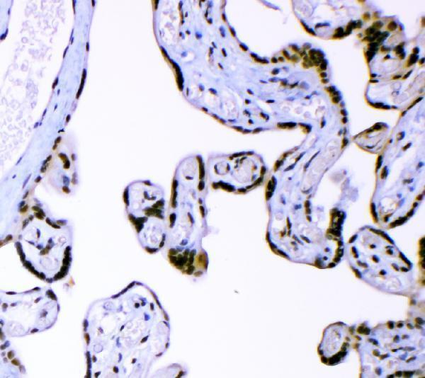
| Sample | Paraffin-embedded human placenta tissue; fixative not specified (datasheet A03824) |
| Fixation | Image fixative and duration unreported (datasheet A03824); verify before use. |
| Sectioning | 4–5 µm sections on charged slides (standard) |
| Deparaffinisation | Xylene, graded ethanol series to water (standard) |
| Antigen retrieval | Heat retrieval: EDTA pH 8.0 (datasheet A03824); 20 min, 95–100 °C (standard) |
| Peroxidase block | 3% H2O2, 10 min, room temperature (standard) |
| Blocking | 10% goat serum (datasheet A03824) |
| Primary antibody | Rabbit anti-AFF4, 2-5μg/ml (datasheet A03824) |
| Primary incubation | Overnight at 4 °C (datasheet A03824) |
| Detection | HRP-conjugated secondary, DAB chromogen (datasheet A03824) |
| Counterstain | Hematoxylin, blue, dehydrate and mount (standard) |
| Expected result | AFF4-positive staining in glial cells of caudate (HPA tissue IHC: High). HPA tissue profile: Ubiquitous nuclear expression. No signal in the no-primary control. |
AFF4 is a nuclear protein associated with transcriptionally active chromatin and has no transmembrane segment (UniProt Q9UHB7). Expect nuclear staining across many cell types, with high staining reported in glial cells in several brain regions and round or early spermatids in testis (HPA: tissue IHC). HPA describes ubiquitous nuclear expression, but its tissue IHC reliability is Approved with low consistency between antibody staining and RNA expression (HPA: tissue IHC).
| Distinct nuclear signal in the expected cells, with less staining outside nuclei. | This fits AFF4 localisation to the nucleus and chromosome (UniProt Q9UHB7) and HPA's ubiquitous nuclear IHC profile (HPA: tissue IHC). High signal in glial cells of the caudate, cerebral cortex or hippocampus, or in round or early spermatids, is consistent with the reported cell-level observations (HPA: High in those cells). Compare cells within the same section; intensity alone cannot establish antibody specificity. |
| Strong membrane or predominantly cytoplasmic staining, with little nuclear signal. | That compartment pattern conflicts with AFF4's nuclear localisation and lack of a transmembrane segment (UniProt Q9UHB7). Treat it as suspect staining and inspect the negative control and detection chemistry. A weak cytoplasmic haze accompanying convincing nuclear signal has a different interpretation from a dominant nonnuclear pattern; assess the nuclear result before calling the section positive. |
| Signal concentrates in a cell type reported as unstained, such as ovarian stroma, while expected nuclei are weak. | HPA reports ovarian stromal cells as not detected (HPA: tissue IHC), so a strong isolated result there deserves scrutiny for cross-reactivity or endogenous detection activity. This is a comparison, not an absolute biological exclusion: HPA also reports low consistency between staining and RNA data (HPA: reliability description). Check morphology and controls before assigning the signal to AFF4. |
| Diffuse colour covers tissue, empty spaces or many cells without clear nuclear boundaries. | The pattern gives poor evidence of AFF4 because its expected localisation is nuclear (UniProt Q9UHB7; HPA: tissue IHC). General IHC practice: diffuse background can arise from nonspecific binding or detection chemistry. Compare a matched negative control, inspect wash and blocking steps, and score only signal that can be assigned to intact cells and their nuclei. |
| No nuclear signal appears in a section chosen for its reported high staining. | A caudate section with glial cells or testis with round or early spermatids offers an HPA-reported high-staining comparison (HPA: tissue IHC). Absence there raises a workflow or specimen question, but does not by itself prove AFF4 is absent: HPA rates tissue IHC Approved while reporting low staining–RNA consistency (HPA: reliability description). Confirm the relevant cell population is present. |
| Tissue and cell context | HPA reports high staining in selected brain glial cells and round or early spermatids, medium staining in adrenal glandular cells and bone marrow hematopoietic cells, and no detection in ovarian stroma (HPA: tissue IHC). Choose comparison areas by cell type, not tissue name alone; HPA's low staining–RNA consistency limits how strongly any single intensity should be interpreted (HPA: reliability description). |
| Antibody validation | The tissue IHC profile is Approved, with an explicit low-consistency caveat (HPA: tissue IHC). HPA023690 is listed as IHC Approved, while HPA029634 and HPA076229 are listed as ICC Supported without IHC status (HPA: antibody validation). An ICC result from those entries does not independently validate a chromogenic paraffin-section result. |
| Isoforms and antigen identity | UniProt lists 3 AFF4 isoforms and a full-length chain spanning residues 1–1163, with no signal peptide or propeptide annotated (UniProt Q9UHB7). The supplied sources do not map the IHC antibody epitope to an isoform. If a section disagrees with the expected pattern, check the antibody's stated immunogen and application information before attributing it to isoform expression. |
| IF/ICC Q&A: what localisation is expected? | HPA reports mainly nucleoplasmic signal, with additional nucleolar fibrillar-center and nuclear-body localisation in ICC-IF (HPA: subcellular, supported). These finer structures can guide interpretation of an IF image, but chromogenic IHC need only show a credible nuclear pattern; the ICC-IF observations do not specify an IHC protocol or guarantee that subnuclear detail will resolve in paraffin sections. |
| Situation | Likely cause | Next action |
|---|---|---|
| Expected high-staining cells are present, but their nuclei are blank. | The result conflicts with reported high staining in those cells (HPA: tissue IHC); the primary or detection workflow may have failed. | General IHC practice: verify the primary antibody's IHC-P instructions, the stated retrieval and dilution, and the detection controls. Recheck cell identity and section quality before recording a negative AFF4 call. |
| Only cytoplasm or cell borders stain. | Predominantly extranuclear staining conflicts with AFF4's nuclear location and lack of a transmembrane segment (UniProt Q9UHB7). | Review matched negative controls, antibody application information and cell boundaries. Score nuclear and extranuclear staining separately; avoid treating a bright border as a positive AFF4 nucleus. |
| Ovarian stromal cells stain strongly. | That differs from HPA's not-detected observation for ovarian stroma (HPA: tissue IHC), although HPA notes low staining–RNA consistency (HPA: reliability description). | Confirm stromal identity and examine a negative detection control for endogenous activity. Compare with an HPA-reported high-staining cell population before interpreting the isolated result. |
| Brown colour is spread across the section without cell-specific nuclear contrast. | Diffuse background obscures the nuclear pattern expected for AFF4 (UniProt Q9UHB7; HPA: tissue IHC). | General IHC practice: inspect blocking, washing, detection timing and a no-primary control. Reduce background through the validated workflow, then reassess whether identifiable nuclei retain signal. |
| Nuclear staining is present but weaker than expected in a reported high-staining tissue. | The sampled area may lack the relevant high-staining cells; HPA levels are cell-specific and its tissue IHC has low staining–RNA consistency (HPA: tissue IHC). | Compare like cell types across sections and record the distribution as well as intensity. Confirm the selected glial or spermatid population before changing the interpretation or technical conditions. |
| IF shows nuclear dots, but chromogenic IHC looks broadly nuclear. | ICC-IF reports additional nuclear-body and nucleolar fibrillar-center localisation alongside mainly nucleoplasmic AFF4 (HPA: subcellular, supported). | Interpret each assay at its available resolution. For this IHC section, judge nuclear staining against tissue and cell context (HPA: tissue IHC); use the separate IF/ICC guide for imaging-specific decisions. |


Comprehensive Human Protein Atlas IHC scoring per tissue (reliability: Approved — Low consistency between antibody staining and RNA expression data.). Rows are taken directly from the HPA tissue chart — click any row's HPA link to view the source.
| Tissue | Cell type | Level | Evidence | Source |
|---|---|---|---|---|
| Caudate | Glial cells | High | Protein (IHC) | HPA → |
| Cerebellum | Cells in molecular layer | High | Protein (IHC) | HPA → |
| Cerebral cortex | Glial cells | High | Protein (IHC) | HPA → |
| Hippocampus | Glial cells | High | Protein (IHC) | HPA → |
| Testis | Round or early spermatids | High | Protein (IHC) | HPA → |
| Tissue | Cell type | Level | Evidence | Source |
|---|---|---|---|---|
| Ovary | Ovarian stroma cells | Not detected | Protein (IHC) | HPA → |
Troubleshoot AFF4 staining in paraffin sections by checking nuclear signal, retrieval, controls and scoring; use IF as a complementary localisation check.
A03824 has a human placenta paraffin-section IHC image (catalog image caption); its listed IHC reactivity covers human, mouse, and rat (catalog applications/reactivity). No IF image is supplied (catalog image list).
A03824 is listed for IHC in human, mouse, and rat samples (catalog applications/reactivity). Its image shows chromogenic IHC in a human placenta paraffin section (catalog image caption).
Which to pick: Choose A03824 for paraffin-section tissue IHC: its own image documents EDTA pH 8.0 retrieval and DAB detection in human placenta (catalog image caption). A03824 has no listed IF/ICC validation or IF image, so this payload supports no IF/ICC pick (catalog applications/image list). A03824 lists mouse and rat IHC reactivity, but the supplied image demonstrates human tissue only; the fixative is unreported (catalog applications/reactivity; catalog image caption).