This website uses cookies to ensure you get the best experience on our website.
- Table of Contents
1 Citations 8 Q&As
Facts about Afamin.
Binds vitamin E (PubMed:15952736, PubMed:12463752). May transfer vitamin E in your body fluids under conditions where the lipoprotein system isn't sufficient (PubMed:15952736).
Human | |
---|---|
Gene Name: | AFM |
Uniprot: | P43652 |
Entrez: | 173 |
Belongs to: |
---|
ALB/AFP/VDB family |
Afamin; AFM; ALB2; ALB2alpha-Alb; ALBA; ALBAalpha-albumin; ALF; Alpha-Alb; Alpha-albumin; MGC125338; MGC125339
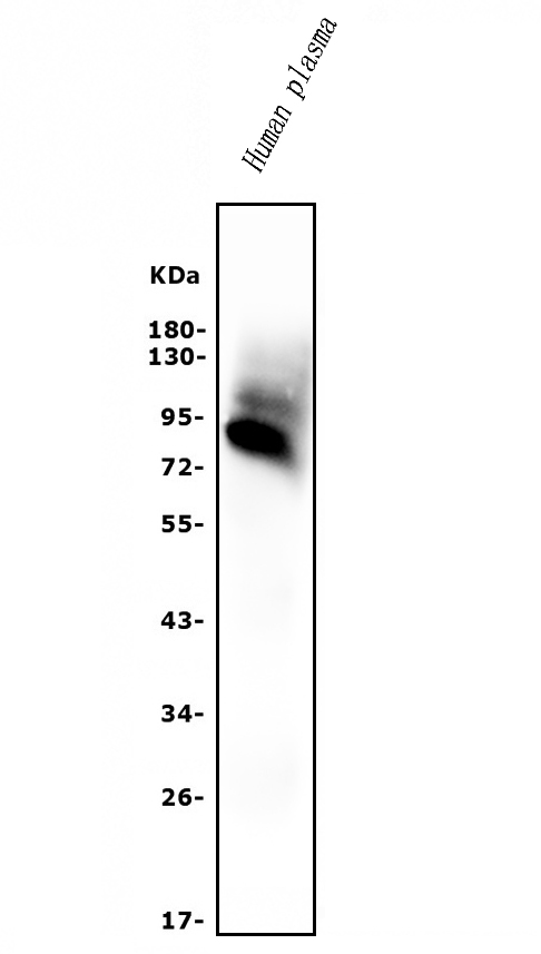
Mass (kDA):
69.069 kDA
Human | |
---|---|
Location: | 4q13.3 |
Sequence: | 4; NC_000004.12 (73481745..73504001) |
High level detected in plasma but also in extravascular fluids such as follicular and cerebrospinal fluids (at protein level).
Secreted.
Are you interested in knowing more about AFM or Nanostructures? Check out Boster's gene infographics which give you basic information about each gene in mouse and human. Then, use the search bar for genes to find a gene that is of interest to you. If you are familiar with AFM spectroscopy techniques you can skip to step 2 and explore the Boster gene infographics.
The AFM is a powerful tool to analyze materials with various physical properties. The AFM marker can detect nanoscale characteristics within materials. Its sensitivities are excellent for physical characteristics and contrasts, but lacks sensitivity for chemical information. The material phase magnitude is determined by the Molecular Force Spectroscopy Topography. Subsurface structures aren't considered to be relevant. Increasing resolution and size limits the capability of the AFM to determine the atomic structure.
The AFM marker is a single-molecule probe which allows researchers to study proteins that have non-mechanical properties. Scientists can use this technique to gain a better understanding of the functional properties and functions of proteins. Every protein has its own free energy landscape in a high-dimensional space. A mechanical force applied to a protein tilts its energy landscape, leading it to test variations along its response coordinate.
Bulk materials can also be distinguished with the AFM marker. The bulk material is colored blue or brown while the reference material, which is glass, is white. In Figure 2c, the histogram of Fattr shows distinct distributions of the samples of the material. By using this method, Khorasani and colleagues were able to detect nanoparticles in a boehmite/epoxy nanocomposite.
The AFM marker has a number of advantages. While it is extremely reproducible, it does have some limitations. The biotin-streptavidin tetramer bonds to the protein in such a way that it is difficult to distinguish between a valid and non-specific interaction. The elastic linker that connects the protein to the surface assists in removing the short-range non-specific adhesion, however, it doesn't eliminate all the background signals. Additionally the protein domains function as internal control mechanisms to block the development of unwanted signals.
AFM-SMFS can also detect proteins that are not specifically absorbable. The N-effect, which can result in underestimating unfolding forces in the early stages of events, may also alter the energy landscape parameters. This is why it is suggested that researchers take into account the N-effect when conducting the study of stretching polyproteins. This scenario demonstrates that more domains are formed at the beginning of a polyprotein stretching curve, and that the force required for them to be unfolded is lower in these domains.
The probability of adhesion is also crucial. AFM indicates that bodies made of the same material attract most. The lower attraction occurs when the materials are not similar. The higher the frequency of adhesion the more intense the attraction. Repulsion is also possible in the event that there are dissimilar materials within a specified medium. Adhesion probability can be used to enhance the efficiency of AFM.
AFM is not a good tool to extract complex structures because of its high sensitivity. The spectral data that is derived from single molecules isn't reliable. To gain reliable information about the properties of a molecule it is important to choose valid trajectories. When conducting AFM it is crucial to know the impact of the multiple-bond structure of the molecule of interest.
The AFM marker is an effective instrument to assess the surface properties of living and dead cells. It has opened a new possibility for research in many disciplines, including the study the quality of dental materials, molecular interactions preventive dentistry and implant biocompatibility. In addition to its numerous applications it also provides AFM will further shed light on the the mechanisms and mechanics of the individual organelles.
The AFM test revealed that SWNTs were well-attached and dispersed. This confirmed that EDC was successfully attached to SWNTs. The morphology of the surface of the scaffolds was predicted to be nanoscale. The surface topography of scaffolds could be altered to modify cell adhesion and proliferation. AFM was used to estimate the surface roughness of SWNTs/EDC composites.
The most beneficial uses of the AFM marker for nanostructured material is determining the precise nature of nanomaterials. They may be multi-dimensional or even discrete and have a variety of physiochemical properties. Biomarkers are a promising tool for detecting the presence of cancerous cells. As the world's most frequent cancer lung cancer is an serious health issue that requires early detection. The issue of delayed diagnosis is a major issue, but the field is entering a new phase. Nanomaterials with biosensors will allow a more precise diagnosis of the disease.
AFM AFM is a sensitive method that analyzes biomechanical properties. AFM uses the force tip to apply forces to a biological sample. The force applied is known as an "indentation" and the sample's mechanical properties are determined by analyzing the deformation. Biomechanical properties of various cells are assessed using an AFM. This technique is ideal for the investigation of cell wall properties in plant tissue.
AFM can also be used to gauge the stiffness of tissue. This is vital because stiffness and ageing are both related. AFM can assist researchers in identifying more subtle variations in stiffness with the process of aging. Because the diameter of the tip of AFM is similar to scanning electron microscopy, it is able to detect these variations. AFM can also be used to measure the stiffness in tissues. Its features allow researchers to determine the stiffness of worms' bulk.
The ability of an organism to adapt to changes in pressure is contingent on the extent to which it is able to withstand pressure from its environment. For instance the hydrostatic skeleton an organism may act as a biomarker of age which can be used to determine the overall health status of an organism. Additionally, high-throughput methods with AFM can quantify health biomarkers in unprecedented quantities. These techniques can identify genetic factors that impact health and help to improve health.
Atomic Force Microscopy is a sophisticated method for analyzing cells and tissues. The method is capable of simultaneously measuring the biomechanical properties in three dimensions. These measurements include the Young's modulus as well as the cell wall's turgor pressure. When used in conjunction with other methods, AFM is a powerful biophysical tool. It is increasingly being used in medical applications.
The Sader method is one of the most widely used calibration procedures. This method is also known as the reference method for cantilevers and was initially developed by John Sader for V-shaped cantilevers and later expanded to rectangular cantilevers. It does not require an acquisition force curve on a stiff substrate. There are two kinds: the Sader and the contact methods. The acquisition of the force curve on the solid substrate is essential for the former.
PMID: 7517938 by Lichenstein H.S., et al. Afamin is a new member of the albumin, alpha-fetoprotein, and vitamin D-binding protein gene family.
PMID: 8755513 by Nishio H., et al. Complete structure of the human alpha-albumin gene, a new member of the serum albumin multigene family.
*More publications can be found for each product on its corresponding product page