This website uses cookies to ensure you get the best experience on our website.


- Table of Contents
This AGR2 paraffin IHC guide focuses on cytoplasmic staining in glandular tissues (HPA tissue IHC). Use bronchial respiratory epithelium as a positive control and adipocytes as a negative control (HPA tissue IHC); titrate the catalog antibody at 2–5 μg/ml (datasheet A02922-2).

Expected localisation, validated protocols, controls and antibodies — the at-a-glance facts below, then the full design guide.
| Expected localisation | Cytoplasmic staining in glandular tissues (HPA tissue IHC) | |
| Staining pattern | Cytoplasmic staining in glandular and urothelial epithelia (HPA tissue IHC) | |
| Antigen retrieval | EDTA pH 8.0 HIER, heat-mediated (datasheet A02922-2) | |
| Positive control | Appendix+4 more · see all | |
| Negative control | Adipose tissue+4 more · see all |
| Fixation | Keep fixation consistent across samples (standard IHC practice; not target-specific) | |
| Caveat | Secreted AGR2 may stain beyond producer cells (UniProt) | |
| Regulation | Enhanced RNA in cervix, intestine, stomach (HPA tissue RNA) | |
| Isoform / epitope | No isoforms annotated; mature chain excludes signal peptide (UniProt) |
The catalog antibody uses heat-mediated EDTA pH 8.0 retrieval (datasheet A02922-2). Four published AGR2 IHC protocols cover human and canine tumor tissue (PMC4496185; PMC2361240; PMC8460385; PMC8532596).
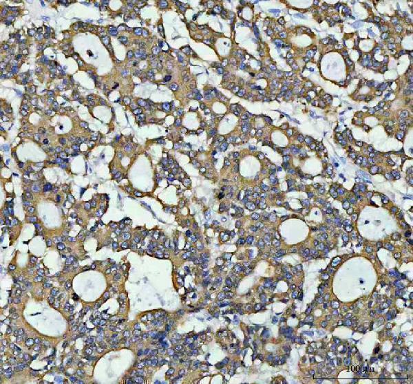
| Sample | Paraffin-embedded human colorectal adenocarcinoma tissue; fixative not specified (datasheet A02922-2) |
| Fixation | Image fixative and duration unreported (datasheet A02922-2); verify before use. |
| Sectioning | 4–5 µm sections on charged slides (standard) |
| Deparaffinisation | Xylene, graded ethanol series to water (standard) |
| Antigen retrieval | Heat retrieval: EDTA pH 8.0 (datasheet A02922-2); 20 min, 95–100 °C (standard) |
| Peroxidase block | 3% H2O2, 10 min, room temperature (standard) |
| Blocking | 10% goat serum (datasheet A02922-2) |
| Primary antibody | Rabbit anti-AGR2, 2-5μg/ml (datasheet A02922-2) |
| Primary incubation | Overnight at 4 °C (datasheet A02922-2) |
| Detection | HRP-conjugated secondary, DAB chromogen (datasheet A02922-2) |
| Counterstain | Hematoxylin, blue, dehydrate and mount (standard) |
| Expected result | AGR2-positive staining in endocrine cells of appendix (HPA tissue IHC: High). HPA tissue profile: Cytoplasmic expression in glandular tissues, including glands in GI, female and male genitalia, and urothelial epithelia. No signal in the no-primary control. |
AGR2 is secreted and also localizes to the endoplasmic reticulum; it has no transmembrane segment (UniProt O95994). In paraffin-section IHC, expect chiefly cytoplasmic staining in glandular and other listed epithelial cells (HPA tissue IHC: Enhanced reliability; medium consistency with RNA). Endoplasmic reticulum localization is supported separately by ICC-IF (HPA subcellular). Interpret the pattern by cell type and compartment, since staining strength varies across the listed tissues (HPA tissue IHC).
| Clear cytoplasmic staining in cervical or endometrial glandular cells, or bronchial respiratory epithelial cells (HPA tissue IHC: High). | This matches the reported IHC distribution (HPA tissue IHC). Score the relevant cells and their cytoplasm; staining elsewhere in the section should be assessed separately (standard IHC practice). HPA’s Enhanced tissue rating carries a medium RNA–staining consistency caveat, so the pattern supports interpretation without proving specificity in every specimen (HPA tissue IHC). |
| Predominantly nuclear staining with little cytoplasmic signal in an expected positive cell population (HPA tissue IHC; HPA subcellular). | A nuclear-dominant result conflicts with reported cytoplasmic tissue staining and supported endoplasmic reticulum localization (HPA tissue IHC; HPA subcellular). Treat it as suspect, then compare with an expected positive tissue and detection controls before assigning AGR2 expression (standard IHC practice). |
| Strong staining in esophageal squamous epithelial cells or heart cardiomyocytes (HPA tissue IHC: Not detected). | This is outside those cell types’ reported HPA pattern (HPA tissue IHC). Check whether staining persists in a no-primary control; persistence suggests endogenous detection activity or background, while primary-dependent staining warrants a specificity check (standard IHC practice). |
| Broad, low-contrast color across tissue and surrounding areas, without a clear cytoplasmic cell pattern (HPA tissue IHC). | The result cannot be scored confidently against the reported cell-restricted cytoplasmic pattern (HPA tissue IHC). Compare a no-primary control and review blocking, washing, detection and counterstain conditions for nonspecific signal (standard IHC practice). |
| No staining in bronchial respiratory epithelium or cervical glandular cells expected to stain strongly (HPA tissue IHC: High). | A negative result in an expected positive cell population raises a run-performance question before it supports biological absence (HPA tissue IHC; standard IHC practice). Verify that the positive cells are present, then review the catalog antibody’s IHC-P conditions and the detection controls (standard IHC practice). |
| Cell-specific tissue distribution (HPA tissue IHC). | High staining is reported in cervical, endometrial, epididymal and fallopian-tube glandular cells, bronchial respiratory epithelial cells, and appendix, colon and duodenum endocrine cells (HPA tissue IHC). Interpret the named cell population, not the tissue label alone (standard IHC practice). |
| Low and absent comparison sites (HPA tissue IHC). | Kidney distal tubules and breast glandular cells are listed as low, whereas adipocytes and esophageal squamous epithelial cells are Not detected (HPA tissue IHC). A weak kidney or breast result is therefore less decisive as a run control than an HPA High cell population (HPA tissue IHC; standard IHC practice). |
| Localization and topology (UniProt O95994; HPA subcellular). | UniProt lists AGR2 as secreted and endoplasmic reticulum localized, with no transmembrane segment; HPA supports endoplasmic reticulum localization by ICC-IF (UniProt O95994; HPA subcellular). These records support a cytoplasmic tissue pattern and do not establish a membrane-staining requirement (HPA tissue IHC; UniProt O95994). |
| Processing and antibody evidence (UniProt O95994; HPA antibodies). | UniProt records a signal peptide at residues 1–20 and a chain spanning 21–175; no epitope is supplied, so the effect of processing on this antibody’s IHC signal is undetermined (UniProt O95994). HPA007912 has Enhanced IHC validation, which supports its reported pattern without establishing performance in every preparation (HPA antibodies). |
| IF/ICC expectation? (HPA subcellular; HPA antibodies). | For the separate IF/ICC guide, expect endoplasmic reticulum localization: HPA calls it supported and lists A-549, RT-4 and U2OS ICC-IF images (HPA subcellular). HPA007912 is Approved for ICC; that status does not supply an IF/ICC protocol here (HPA antibodies). |
| Situation | Likely cause | Next action |
|---|---|---|
| No signal in an expected positive IHC section (HPA tissue IHC: High). | The target cell population may be absent from the viewed area, or the IHC run may have failed (standard IHC practice). | Find the named positive cells on the section; check a positive run control, reagent sequence and the catalog antibody’s IHC-P conditions before scoring the specimen negative (HPA tissue IHC; standard IHC practice). |
| Weak signal in kidney distal tubules or breast glandular cells (HPA tissue IHC: Low). | Low staining is reported for those cell populations, so weak signal alone need not indicate a failed assay (HPA tissue IHC). | Compare with a High population, such as bronchial respiratory epithelial or cervical glandular cells, under the same run conditions before changing detection settings (HPA tissue IHC; standard IHC practice). |
| Nuclear-dominant color in otherwise positive cells (HPA tissue IHC; HPA subcellular). | The compartment disagrees with HPA’s cytoplasmic tissue profile and supported endoplasmic reticulum localization; the supplied records do not identify a specific technical cause (HPA tissue IHC; HPA subcellular). | Check a no-primary control, inspect the counterstain and compare the cytoplasmic pattern in a known positive tissue before interpreting nuclear color as AGR2 (standard IHC practice). |
| Color in HPA Not detected cell types, such as adipocytes or esophageal squamous epithelium (HPA tissue IHC). | Possible explanations include background, endogenous detection activity or primary-antibody cross-reactivity; the image alone cannot distinguish them (standard IHC practice). | Run a no-primary control and compare its distribution with the test slide; if staining depends on the primary antibody, reassess specificity using the expected positive and negative cell patterns (HPA tissue IHC; standard IHC practice). |
| Diffuse color obscures cell boundaries and compartments (HPA tissue IHC). | Excess nonspecific staining or detection background can obscure the expected cytoplasmic, cell-specific pattern (HPA tissue IHC; standard IHC practice). | Review blocking, washes, primary-antibody dilution and detection conditions against the catalog IHC-P protocol; use a no-primary control to locate background from the detection steps (standard IHC practice). |
| Different apparent positivity across two glandular tissues (HPA tissue IHC). | HPA reports High, Low and Not detected staining by specified cell population, and its tissue IHC reliability notes medium consistency with RNA (HPA tissue IHC). | Identify and score the relevant cell types in each section, then compare their reported HPA levels; do not treat a whole-tissue difference alone as proof of an antibody problem (HPA tissue IHC; standard IHC practice). |
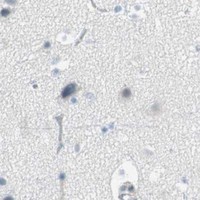


Comprehensive Human Protein Atlas IHC scoring per tissue (reliability: Enhanced — Medium consistency between antibody staining and RNA expression data.). Rows are taken directly from the HPA tissue chart — click any row's HPA link to view the source.
| Tissue | Cell type | Level | Evidence | Source |
|---|---|---|---|---|
| Appendix | Endocrine cells | High | Protein (IHC) | HPA → |
| Bronchus | Respiratory epithelial cells | High | Protein (IHC) | HPA → |
| Cervix | Glandular cells | High | Protein (IHC) | HPA → |
| Colon | Endocrine cells | High | Protein (IHC) | HPA → |
| Duodenum | Endocrine cells | High | Protein (IHC) | HPA → |
| Tissue | Cell type | Level | Evidence | Source |
|---|---|---|---|---|
| Adipose tissue | Adipocytes | Not detected | Protein (IHC) | HPA → |
| Adrenal gland | Glandular cells | Not detected | Protein (IHC) | HPA → |
| Bone marrow | Hematopoietic cells | Not detected | Protein (IHC) | HPA → |
| Caudate | Glial cells | Not detected | Protein (IHC) | HPA → |
| Cerebellum | Cells in granular layer | Not detected | Protein (IHC) | HPA → |
Troubleshoot AGR2 staining in paraffin-section chromogenic IHC by checking retrieval, staining controls, cellular localisation and scoring consistency.
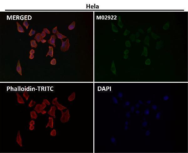
Two anti-AGR2 antibodies have paraffin-section IHC images in human tissue (catalog IHC captions); both list IF/ICC applications, and one has an IF image (catalog applications; M02922 IF caption).
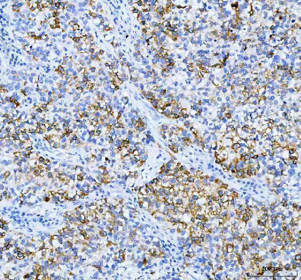
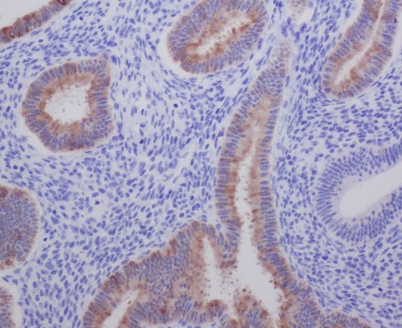
A02922-2 has IHC images from human colorectal adenocarcinoma, non-small cell lung cancer, and bladder infiltrating urothelial carcinoma with squamous differentiation (A02922-2 IHC captions). M02922 has an IHC image from paraffin-embedded human uterus and an IF image at 1:50 dilution (M02922 IHC and IF captions).
Which to pick: For tissue IHC, choose A02922-2 for its documented paraffin-section examples or M02922 for its human uterus example; neither caption reports the fixative (A02922-2 and M02922 IHC captions). For IF/ICC, M02922 is the rabbit monoclonal with an IF image, while A02922-2 also lists IF/ICC at 5 μg/ml (M02922 catalog and IF caption; A02922-2 catalog). For cross-species work, A02922-2 lists human, mouse, and rat reactivity, although its supplied IHC images show human tissue only (A02922-2 catalog and IHC captions).