Click image to see more details
-

-
-
-
-
+3
Product Info Summary
| SKU: | A02922-2 |
|---|---|
| Size: | 100 μg/vial |
| Reactive Species: | Human, Mouse, Rat |
| Host: | Rabbit |
| Application: | Flow Cytometry, IP, IF, IHC, ICC, WB |
Customers Who Bought This Also Bought

Product info
Product Name
Anti-Anterior Gradient 2/AGR2 Antibody Picoband®
SKU/Catalog Number
A02922-2
Size
100 μg/vial
Form
Lyophilized
Description
Boster Bio Anti-Anterior Gradient 2/AGR2 Antibody Picoband® catalog # A02922-2. Tested in Flow Cytometry, IP, ICC/IF, IHC, WB applications. This antibody reacts with Human, Mouse, Rat. The brand Picoband indicates this is a premium antibody that guarantees superior quality, high affinity, and strong signals with minimal background in Western blot applications. Only our best-performing antibodies are designated as Picoband, ensuring unmatched performance.
Storage & Handling
Store at -20˚C for one year from date of receipt. After reconstitution, at 4˚C for one month. It can also be aliquotted and stored frozen at -20˚C for six months. Avoid repeated freeze-thaw cycles.
Cite This Product
Anti-Anterior Gradient 2/AGR2 Antibody Picoband® (Boster Biological Technology, Pleasanton CA, USA, Catalog # A02922-2)
Host
Rabbit
Contents
Each vial contains 4 mg Trehalose, 0.9 mg NaCl and 0.2 mg Na2HPO4.
Clonality
Polyclonal
Isotype
Rabbit IgG
Immunogen
E.coli-derived human Anterior Gradient 2 recombinant protein (Position: R21-L175). Human Anterior Gradient 2 shares 93.5% amino acid (aa) sequence identity with mouse Anterior Gradient 2.
Cross-reactivity
No cross-reactivity with other proteins
Reactive Species
A02922-2 is reactive to AGR2 in Human, Mouse, Rat
Observed Molecular Weight
17 kDa
Calculated molecular weight
20.0 kDa
Background of AGR2
Anterior gradient protein 2 homolog (AGR-2), also known as secreted cement gland protein XAG-2 homolog, is a protein that in humans is encoded by the AGR2 gene. Anterior gradient homolog 2 was originally discovered in Xenopus laevis. In Xenopus AGR2 plays a role in cement gland differentiation, but in human cancer cell lines high levels of AGR2 correlate with downregulation of the p53 response, cell migration, and cell transformation. However, there have been other observations that AGR2 can repress growth and proliferation.
Antibody Validation
Boster validates all antibodies on WB, IHC, ICC, Immunofluorescence, and ELISA with known positive control and negative samples to ensure specificity and high affinity, including thorough antibody incubations.
Application & Images
Applications
A02922-2 is guaranteed for Flow Cytometry, IP, IF, IHC, ICC, WB Boster Guarantee
Recommend Dilution
| Application | Dilution | Species |
|---|---|---|
| Western blot | 0.1-0.5μg/ml | Human, Mouse, Rat |
| Immunohistochemistry (Paraffin-embedded Section) | 2-5μg/ml | Human |
| Immunocytochemistry/Immunofluorescence | 5 μg/ml | Human |
| Immunoprecipitation | 0.5-2 μg/ml | Human |
| Flow Cytometry(Fixed) | 1-3 μg/1x106 cells | Human |
Tested application
Suggested blocking solution with 5% non-fat milk or BSA; (*)Recommended protein loading: 20-40 µg per lane
Use TE buffer pH 9.0 for antigen retrieval; (*) citrate buffer pH 6.0 is an alternative.
Validation Images & Assay Conditions
Click image to see more details
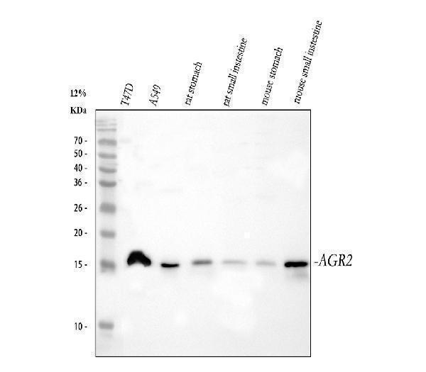
Western blot analysis of AGR2 using anti-AGR2 antibody (A02922-2).
Electrophoresis was performed on a 5-20% SDS-PAGE gel at 70V (Stacking gel) / 90V (Resolving gel) for 2-3 hours. The sample well of each lane was loaded with 30 ug of sample under reducing conditions.
Lane 1: human T47D whole cell lysates,
Lane 2: human A549 whole cell lysates,
Lane 3: rat stomach tissue lysates,
Lane 4: rat small intestine tissue lysates,
Lane 5: mouse stomach tissue lysates,
Lane 6: mouse small intestine tissue lysates.
After electrophoresis, proteins were transferred to a nitrocellulose membrane at 150 mA for 50-90 minutes. Blocked the membrane with 5% non-fat milk/TBS for 1.5 hour at RT. The membrane was incubated with rabbit anti-AGR2 antigen affinity purified polyclonal antibody (Catalog # A02922-2) at 0.5 μg/mL overnight at 4°C, then washed with TBS-0.1%Tween 3 times with 5 minutes each and probed with a goat anti-rabbit IgG-HRP secondary antibody at a dilution of 1:5000 for 1.5 hour at RT. The signal is developed using an Enhanced Chemiluminescent detection (ECL) kit (Catalog # EK1002) with Tanon 5200 system. A specific band was detected for AGR2 at approximately 17 kDa. The expected band size for AGR2 is at 20 kDa.

Click image to see more details
IHC analysis of AGR2 using anti-AGR2 antibody (A02922-2).
AGR2 was detected in a paraffin-embedded section of human colorectal adenocarcinoma tissue. Heat mediated antigen retrieval was performed in EDTA buffer (pH 8.0, epitope retrieval solution). The tissue section was blocked with 10% goat serum. The tissue section was then incubated with 2 μg/ml rabbit anti-AGR2 Antibody (A02922-2) overnight at 4°C. Peroxidase Conjugated Goat Anti-rabbit IgG was used as secondary antibody and incubated for 30 minutes at 37°C. The tissue section was developed using HRP Conjugated Rabbit IgG Super Vision Assay Kit (Catalog # SV0002) with DAB as the chromogen.

Click image to see more details
IHC analysis of AGR2 using anti-AGR2 antibody (A02922-2).
AGR2 was detected in a paraffin-embedded section of human non-small cell lung cancer tissue. Heat mediated antigen retrieval was performed in EDTA buffer (pH 8.0, epitope retrieval solution). The tissue section was blocked with 10% goat serum. The tissue section was then incubated with 2 μg/ml rabbit anti-AGR2 Antibody (A02922-2) overnight at 4°C. Peroxidase Conjugated Goat Anti-rabbit IgG was used as secondary antibody and incubated for 30 minutes at 37°C. The tissue section was developed using HRP Conjugated Rabbit IgG Super Vision Assay Kit (Catalog # SV0002) with DAB as the chromogen.

Click image to see more details
IHC analysis of AGR2 using anti-AGR2 antibody (A02922-2).
AGR2 was detected in a paraffin-embedded section of human bladder infiltrating urothelial carcinoma with squamous differentiation tissue. Heat mediated antigen retrieval was performed in EDTA buffer (pH 8.0, epitope retrieval solution). The tissue section was blocked with 10% goat serum. The tissue section was then incubated with 2 μg/ml rabbit anti-AGR2 Antibody (A02922-2) overnight at 4°C. Peroxidase Conjugated Goat Anti-rabbit IgG was used as secondary antibody and incubated for 30 minutes at 37°C. The tissue section was developed using HRP Conjugated Rabbit IgG Super Vision Assay Kit (Catalog # SV0002) with DAB as the chromogen.

Click image to see more details
IF analysis of AGR2 using anti-AGR2 antibody (A02922-2).
AGR2 was detected in an immunocytochemical section of U2OS cells. Enzyme antigen retrieval was performed using IHC enzyme antigen retrieval reagent (AR0022) for 15 mins. The cells were blocked with 10% goat serum. And then incubated with 5 μg/mL rabbit anti-AGR2 Antibody (A02922-2) overnight at 4°C. DyLight®488 Conjugated Goat Anti-Rabbit IgG (BA1127) was used as secondary antibody at 1:500 dilution and incubated for 30 minutes at 37°C. The section was counterstained with DAPI. Visualize using a fluorescence microscope and filter sets appropriate for the label used.

Click image to see more details
Immunoprecipitating (IP) AGR2 in A549 whole cell lysate.
Western blot analysis of AGR2 using anti-AGR2 antibody (A02922-2);
Lane 1: A549 whole cell lysates (30ug);
Lane 2: Rabbit control IgG instead of anti-AGR2 antibody in A549 whole cell lysate;
Lane 3: anti-AGR2 antibody (2μg) + A549 whole cell lysate (500μg).
After electrophoresis, proteins were transferred to a membrane. Then the membrane was incubated with rabbit anti-AGR2 antigen affinity purified polyclonal antibody (A02922-2) at a dilution of 0.5 μg/mL and probed with a goat anti-rabbit IgG-HRP secondary antibody (Catalog # BA1054). The signal is developed using ECL Plus Western Blotting Substrate (Catalog # AR1196-200). A specific band was detected for AGR2 at approximately 17 kDa. The expected band size for AGR2 is at 20 kDa.

Click image to see more details
Flow Cytometry analysis of CACO-2 cells using anti-AGR2 antibody (A02922-2).
Overlay histogram showing CACO-2 cells stained with A02922-2 (Blue line). The cells were fixed with 4% paraformaldehyde and blocked with 10% normal goat serum. And then incubated with rabbit anti-AGR2 Antibody (A02922-2, 1 μg/1x106 cells) for 30 min at 20°C. DyLight®488 conjugated goat anti-rabbit IgG (BA1127, 5-10 μg/1x106 cells) was used as secondary antibody for 30 minutes at 20°C. Isotype control antibody (Green line) was rabbit IgG (1 μg/1x106) used under the same conditions. Unlabelled sample (Red line) was also used as a control.
Specific Publications For Anti-Anterior Gradient 2/AGR2 Antibody Picoband® (A02922-2)
Loading publications
Recommended Resources
Here are featured tools and databases that you might find useful.
- Boster's Pathways Library
- Protein Databases
- Bioscience Research Protocol Resources
- Data Processing & Analysis Software
- Photo Editing Software
- Scientific Literature Resources
- Research Paper Management Tools
- Molecular Biology Software
- Primer Design Tools
- Bioinformatics Tools
- Phylogenetic Tree Analysis
Customer Reviews
Have you used Anti-Anterior Gradient 2/AGR2 Antibody Picoband®?
Share your experimental results or join a short interview to earn up to $1,000 in product credits or other rewards.
0 Reviews For Anti-Anterior Gradient 2/AGR2 Antibody Picoband®
Customer Q&As
Have a question?
Find answers in Q&As, reviews.
Can't find your answer?
Submit your question
4 Customer Q&As for Anti-Anterior Gradient 2/AGR2 Antibody Picoband®
Question
We are currently using anti-Anterior Gradient 2/AGR2 antibody A02922-2 for human tissue, and we are satisfied with the IHC results. The species of reactivity given in the datasheet says human, mouse, rat. Is it true that the antibody can work on zebrafish tissues as well?
Verified Customer
Verified customer
Asked: 2020-03-25
Answer
The anti-Anterior Gradient 2/AGR2 antibody (A02922-2) has not been validated for cross reactivity specifically with zebrafish tissues, but there is a good chance of cross reactivity. We have an innovator award program that if you test this antibody and show it works in zebrafish you can get your next antibody for free. Please contact me if I can help you with anything.
Boster Scientific Support
Answered: 2020-03-25
Question
We appreciate helping with my inquiry over the phone. Here are the WB image, lot number and protocol we used for mucosa of sigmoid colon using anti-Anterior Gradient 2/AGR2 antibody A02922-2. Let me know if you need anything else.
Verified Customer
Verified customer
Asked: 2018-07-27
Answer
Thanks for the data. You have provided everything we needed. Our lab team are working to resolve your inquiry as quickly as possible, and we appreciate your patience and understanding! Please let me know if there is anything you need in the meantime.
Boster Scientific Support
Answered: 2018-07-27
Question
I see that the anti-Anterior Gradient 2/AGR2 antibody A02922-2 works with IHC, what is the protocol used to produce the result images on the product page?
Verified Customer
Verified customer
Asked: 2018-07-12
Answer
You can find protocols for IHC on the "support/technical resources" section of our navigation menu. If you have any further questions, please send an email to support@bosterbio.com
Boster Scientific Support
Answered: 2018-07-12
Question
I was wanting to use your anti-Anterior Gradient 2/AGR2 antibody for IHC for rat mucosa of sigmoid colon on frozen tissues, but I want to know if it has been validated for this particular application. Has this antibody been validated and is this antibody a good choice for rat mucosa of sigmoid colon identification?
M. Rodriguez
Verified customer
Asked: 2017-01-25
Answer
It shows on the product datasheet, A02922-2 anti-Anterior Gradient 2/AGR2 antibody has been validated for IHC, WB on human, mouse, rat tissues. We have an innovator award program that if you test this antibody and show it works in rat mucosa of sigmoid colon in IHC-frozen, you can get your next antibody for free.
Boster Scientific Support
Answered: 2017-01-25
