This website uses cookies to ensure you get the best experience on our website.


- Table of Contents
Plan AGR3 chromogenic IHC on paraffin sections using bronchial ciliated cells as a positive control (HPA tissue IHC). This guide covers the catalog antibody’s 1:100–1:300 IHC dilution range (datasheet) and cytoplasmic and membranous scoring (HPA tissue IHC).

Expected localisation, validated protocols, controls and antibodies — the at-a-glance facts below, then the full design guide.
| Expected localisation | Cytoplasmic and membranous tissue staining (HPA tissue IHC) | |
| Staining pattern | Cytoplasmic and membranous staining in glandular cells (HPA tissue IHC) | |
| Antigen retrieval | Citrate pH 6.0 HIER, 95–98 °C, 20 min (rule: cytoplasmic / membrane antigen) | |
| Positive control | Appendix+4 more · see all | |
| Negative control | Adipose tissue+4 more · see all |
| Fixation | Keep fixation consistent across paraffin sections (standard IHC practice; not target-specific); AGR3-specific effects are unknown. | |
| Caveat | Lung macrophages may complicate epithelial scoring (HPA tissue IHC) | |
| Regulation | Increases with airway epithelial differentiation (UniProt) | |
| Isoform / epitope | No annotated isoforms; signal peptide 1–21 is removed (UniProt) |
The catalog antibody’s IHC-P protocol is followed by four published AGR3 IHC workflows (PMC4485854; PMC3095702; PMC6962492; PMC8232749).
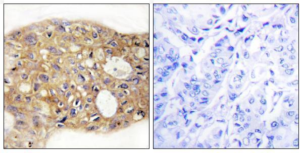
| Sample | Paraffin-embedded human breast carcinoma tissue; fixative not specified (datasheet A05950-1) |
| Fixation | Image fixative and duration unreported (datasheet A05950-1); verify before use. |
| Sectioning | 4–5 µm sections on charged slides (standard) |
| Deparaffinisation | Xylene, graded ethanol series to water (standard) |
| Antigen retrieval | Heat-induced epitope retrieval in citrate buffer, pH 6.0, 20 min at 95–98 °C (standard rule: cytoplasmic / membrane antigen) |
| Peroxidase block | 3% H2O2, 10 min, room temperature (standard) |
| Blocking | 10% normal serum of the secondary host, 30 min, room temperature (standard) |
| Primary antibody | Rabbit anti-AGR3, 1:100 - 1:300 (datasheet A05950-1) |
| Primary incubation | Overnight at 4 °C (standard) |
| Detection | HRP-polymer secondary, DAB chromogen 5–10 min (standard) |
| Counterstain | Hematoxylin, blue, dehydrate and mount (standard) |
| Expected result | AGR3-positive staining in enterocytes of appendix (HPA tissue IHC: High). HPA tissue profile: Distinct cytoplasmic and membranous expression in glandular cells in several tissues. No signal in the no-primary control. |
AGR3 is mainly an endoplasmic reticulum protein, also found in cytoplasm, and has no transmembrane segment (UniProt Q8TD06). In paraffin IHC, expect cytoplasmic and membranous staining in selected epithelial cells, especially airway and fallopian tube ciliated cells (HPA tissue IHC: High; HPA profile). HPA rates the tissue pattern Enhanced for consistency with RNA data, pending external verification (HPA tissue IHC: reliability).
| Distinct staining in bronchial or fallopian tube ciliated cell bodies. | This matches high staining in those cells (HPA tissue IHC: High). A cytoplasmic signal fits the endoplasmic reticulum location (UniProt Q8TD06; HPA subcellular). Assess cell identity and compartment together; color alone does not establish specificity. |
| Cytoplasmic and apparent membrane staining in glandular epithelium. | This is within the reported tissue IHC profile (HPA tissue IHC: cytoplasmic and membranous). AGR3 has no annotated transmembrane segment (UniProt Q8TD06 topology), so apparent membrane staining should be interpreted as an observed IHC pattern, not proof that AGR3 spans the membrane. |
| Predominantly nuclear staining with little epithelial cytoplasmic signal. | A nuclear dominant pattern does not match the reported endoplasmic reticulum location or tissue IHC profile (UniProt Q8TD06; HPA subcellular; HPA tissue IHC). Treat it as suspect and check the detection and counterstain controls before assigning it to AGR3. |
| Staining appears in an unexpected cell population. | Check the exact tissue and cell type against HPA before calling it cross-reactivity: lung macrophages and colon goblet cells are reported High (HPA tissue IHC), while UniProt reports airway ciliated cells and no detection in mucous cells (UniProt Q8TD06). Persistent unsupported staining warrants specificity controls. |
| No signal in bronchial or fallopian tube ciliated cells. | Those cell bodies are reported High (HPA tissue IHC), so absence in an intact known-positive section raises a technical or specimen question. Confirm that the expected cells are present, then review staining-run controls; a negative result alone cannot identify the failed step. |
| Cell type and tissue | Expression is uneven: bronchial and fallopian tube ciliated cells, appendix and duodenal enterocytes, and cervical glandular cells are High, whereas salivary gland glandular cells and efferent duct ciliated cells are Low (HPA tissue IHC). Specify the cell population when scoring. |
| Compartment and topology | Endoplasmic reticulum localization supports cytoplasmic staining (UniProt Q8TD06; HPA subcellular). HPA also describes a membranous tissue IHC pattern (HPA tissue IHC); with no transmembrane segment annotated, that pattern alone cannot establish surface localization (UniProt Q8TD06 topology). |
| Cell-type disagreement across sources | Colon goblet cells are High in HPA tissue IHC, while UniProt reports no detection in airway mucous cells (HPA tissue IHC; UniProt Q8TD06). These observations concern different tissues; record tissue and cell identity instead of extending either statement to all mucous cells. |
| Antibody and evidence scope | HPA053942 has Enhanced IHC validation, and the HPA tissue profile is Enhanced for staining and RNA consistency, pending external verification (HPA antibodies; HPA tissue IHC). These ratings support pattern interpretation but do not validate every new specimen or staining run. |
| Protein processing | UniProt annotates a signal peptide at residues 1–21 and a mature chain at 22–166, with no annotated glycosylation sites or isoforms (UniProt Q8TD06). The payload does not identify the antibody epitope, so processing cannot be used here to predict retrieval needs or staining strength. |
| Situation | Likely cause | Next action |
|---|---|---|
| Known-positive ciliated epithelium is blank. | The target cells may be absent from the section, or the staining run may have failed; HPA reports High signal in bronchial and fallopian tube ciliated cell bodies (HPA tissue IHC). | Verify the cells on the counterstained section and inspect the run's positive control; then review the routine IHC sequence, including retrieval, primary-antibody application and chromogenic detection (general IHC practice). |
| Most structures show diffuse color. | Background can obscure the selective epithelial pattern reported for AGR3 (HPA tissue IHC: profile); the appearance alone cannot distinguish nonspecific binding from detection background. | Compare with an appropriate no-primary control, then review blocking, washes and chromogen development for the run (general IHC practice). Judge AGR3 only after the expected cell populations remain discernible. |
| Signal is predominantly nuclear. | Nuclear dominance conflicts with the reported endoplasmic reticulum localization and cytoplasmic tissue pattern (UniProt Q8TD06; HPA subcellular; HPA tissue IHC). | Check nuclear counterstain and detection controls, then reassess whether cytoplasmic signal is present in HPA High ciliated cells (HPA tissue IHC; general IHC practice). Do not score nuclear color alone as AGR3. |
| Unexpected cells stain strongly. | The result may reflect a documented tissue-specific cell pattern or nonspecific signal: lung macrophages and colon goblet cells are High in HPA tissue IHC, despite UniProt's airway ciliated-cell emphasis (HPA tissue IHC; UniProt Q8TD06). | Identify the stained tissue and cells first. If the population is unsupported, compare no-primary and known-positive controls and review antibody specificity before interpreting the signal (general IHC practice). |
| Only faint signal appears in a proposed positive control. | The chosen cell type may have a low reported level: salivary gland glandular cells, efferent duct ciliated cells and placental trophoblastic cells are Low (HPA tissue IHC). | Use a section containing an HPA High cell population to assess the run, and compare like cell types when scoring specimens (HPA tissue IHC; general IHC practice). |
| Can an IF/ICC image settle an ambiguous IHC result? | HPA ICC-IF supports mainly endoplasmic reticulum localization with additional vesicles, while its tissue IHC profile describes cytoplasmic and membranous staining (HPA subcellular; HPA tissue IHC). The assays show different aspects of localization. | Use ICC-IF as compartment context only; resolve the paraffin-section result with its cell pattern and IHC controls (HPA subcellular; HPA tissue IHC; general IHC practice). IF/ICC procedures belong in the separate guide. |


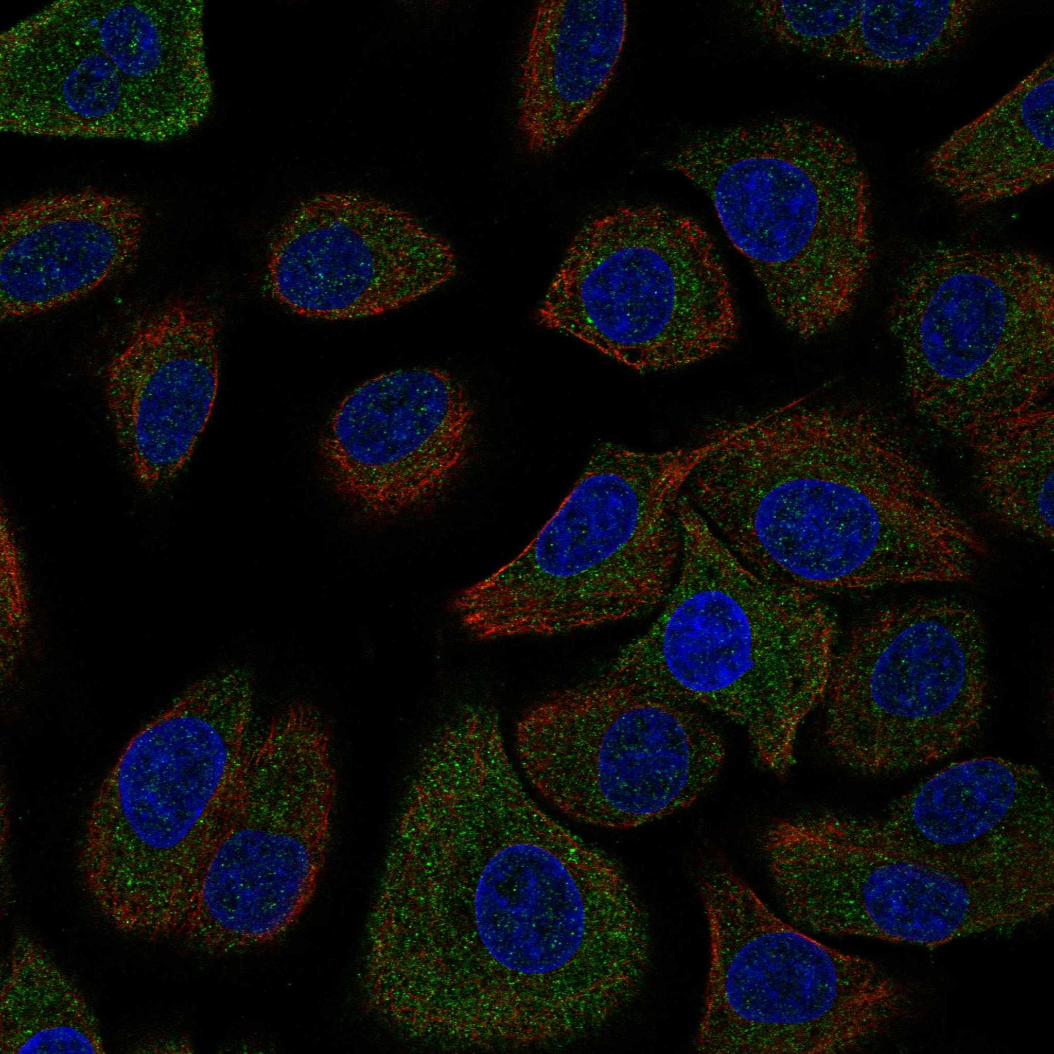
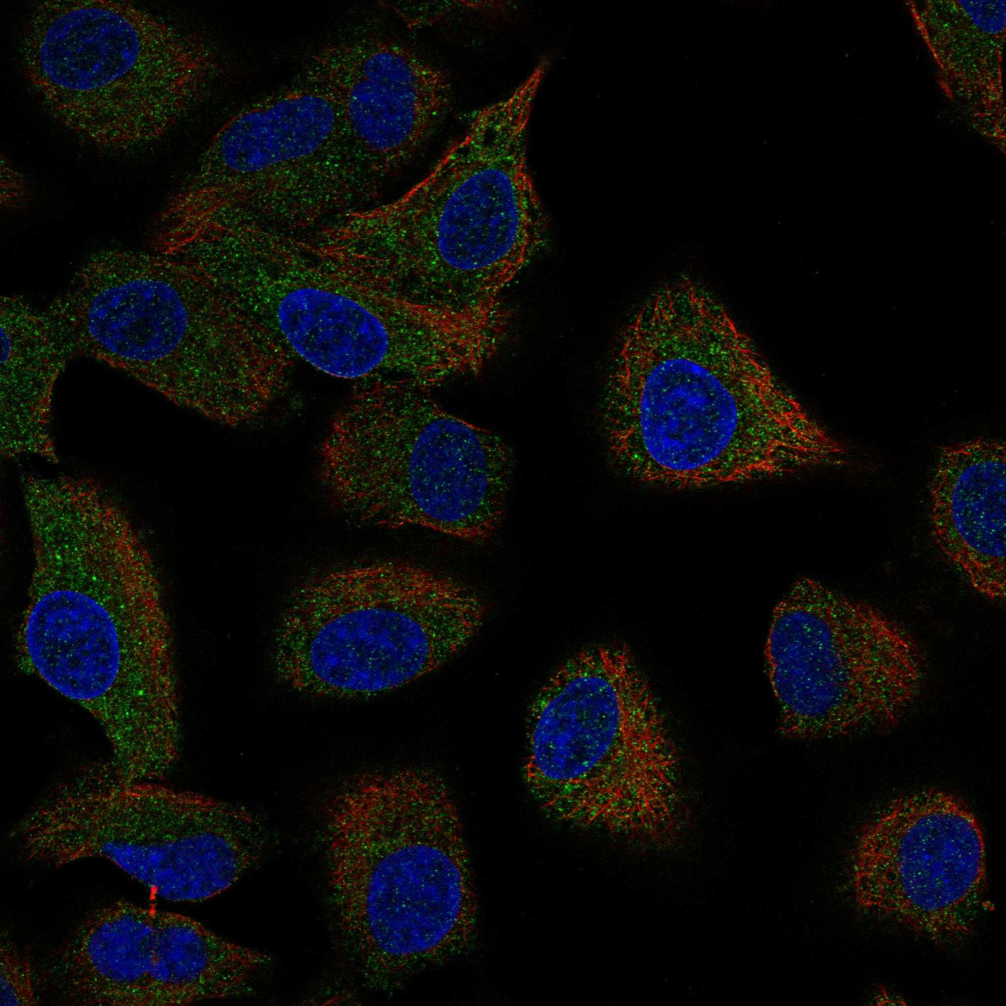
Comprehensive Human Protein Atlas IHC scoring per tissue (reliability: Enhanced — High consistency between antibody staining and RNA expression data. Pending external verification.). Rows are taken directly from the HPA tissue chart — click any row's HPA link to view the source.
| Tissue | Cell type | Level | Evidence | Source |
|---|---|---|---|---|
| Appendix | Enterocytes | High | Protein (IHC) | HPA → |
| Bronchus | Ciliated cells (cell body) | High | Protein (IHC) | HPA → |
| Cervix | Glandular cells | High | Protein (IHC) | HPA → |
| Colon | Goblet cells | High | Protein (IHC) | HPA → |
| Duodenum | Enterocytes | High | Protein (IHC) | HPA → |
| Tissue | Cell type | Level | Evidence | Source |
|---|---|---|---|---|
| Adipose tissue | Adipocytes | Not detected | Protein (IHC) | HPA → |
| Adrenal gland | Glandular cells | Not detected | Protein (IHC) | HPA → |
| Bone marrow | Hematopoietic cells | Not detected | Protein (IHC) | HPA → |
| Caudate | Glial cells | Not detected | Protein (IHC) | HPA → |
| Cerebellum | Cells in granular layer | Not detected | Protein (IHC) | HPA → |
Troubleshoot AGR3 staining in paraffin sections by checking retrieval, cell type, compartment, controls, and scoring before interpreting signal.
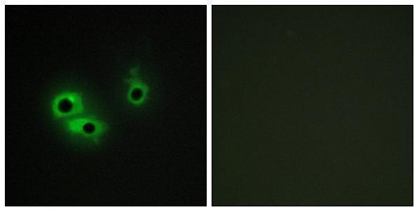
A05950-1 has IHC data in paraffin-embedded human breast carcinoma tissue and IF data in COS7 cells (catalog image captions); listed reactivity is Human and Mouse (catalog: reactivity).
A05950-1 will render with an IHC figure from paraffin-embedded human breast carcinoma tissue (A05950-1 IHC caption). It is listed for IHC, IF, ICC and ELISA, with Human and Mouse reactivity; its IF image shows COS7 cells (A05950-1 catalog).
Which to pick: Choose A05950-1 for tissue IHC: its own image shows paraffin-embedded human breast carcinoma tissue, and its IHC dilution is 1:100–1:300 (A05950-1 IHC caption; datasheet: IHC dilution). The fixative is unreported (A05950-1 IHC caption). The same rabbit polyclonal antibody is listed for IF/ICC at 1:200–1:1000 and for Human and Mouse reactivity, although its pictured IF example is COS7 cells (A05950-1 catalog and IF caption).