This website uses cookies to ensure you get the best experience on our website.


- Table of Contents
Real validated AIRE Western blot protocols, expected-band and isoform facts, troubleshooting for weak or shifted signal, and recommended anti-AIRE WB antibodies. Everything you need to plan the experiment before you commit precious samples.
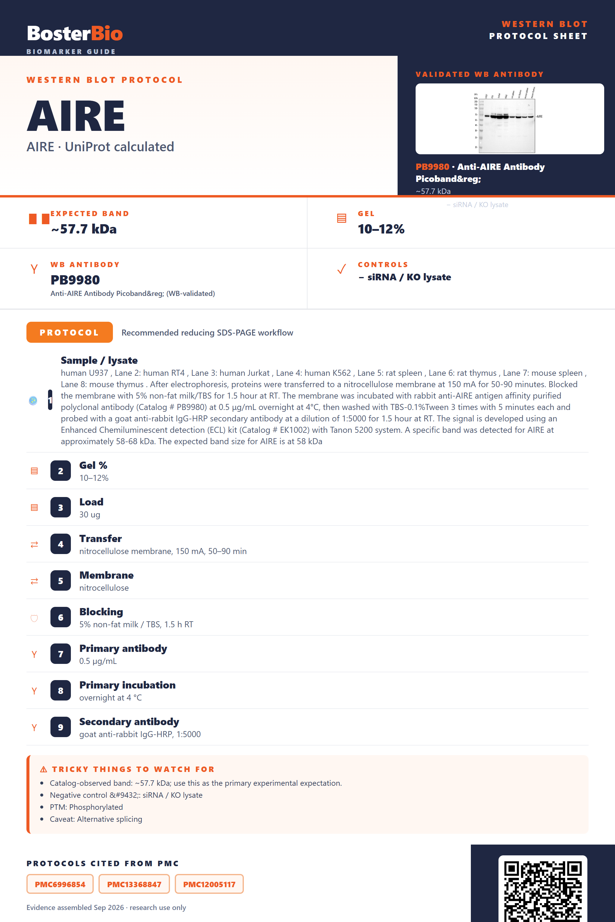
Expected bands, validated protocols, controls and antibodies — the at-a-glance facts below, then the full design guide.
| Expected band | ~57.7 kDa | |
| Gel | 10–12% | |
| Negative control | siRNA / KO lysate |
| PTM | Phosphorylated | |
| Caveat | Alternative splicing | |
| Regulation | M2 macrophage marker | |
| Isoform | 4 isoform(s) |
Literature-validated Western blot parameters for AIRE — gel percentage, transfer, blocking, antibody incubation and detection, extracted from published methods.
| Sample / lysate | human U937 , Lane 2: human RT4 , Lane 3: human Jurkat , Lane 4: human K562 , Lane 5: rat spleen , Lane 6: rat thymus , Lane 7: mouse spleen , Lane 8: mouse thymus . After electrophoresis, proteins were transferred to a nitrocellulose membrane at 150 mA for 50-90 minutes. Blocked the membrane with 5% non-fat milk/TBS for 1.5 hour at RT. The membrane was incubated with rabbit anti-AIRE antigen affinity purified polyclonal antibody (Catalog # PB9980) at 0.5 μg/mL overnight at 4°C, then washed with TBS-0.1%Tween 3 times with 5 minutes each and probed with a goat anti-rabbit IgG-HRP secondary antibody at a dilution of 1:5000 for 1.5 hour at RT. The signal is developed using an Enhanced Chemiluminescent detection (ECL) kit (Catalog # EK1002) with Tanon 5200 system. A specific band was detected for AIRE at approximately 58-68 kDa. The expected band size for AIRE is at 58 kDa |
| Gel % | 10–12% |
| Load | 30 ug |
| Transfer | nitrocellulose membrane, 150 mA, 50–90 min |
| Membrane | nitrocellulose |
| Blocking | 5% non-fat milk / TBS, 1.5 h RT |
| Primary antibody | 0.5 µg/mL |
| Primary incubation | overnight at 4 °C |
| Secondary antibody | goat anti-rabbit IgG-HRP, 1:5000 |
| Wash | TBS-0.1% Tween, 3 × 5 min |
| Detection | ECL |
| Exposure / imaging | Tanon 5200 |
| Observed band | 58–68 kDa |
AIRE runs as a single ~58 kDa monomer band by predicted mass, with occasional higher-MW homodimer or homotetramer bands if oligomers are not fully denatured.
| single band near 58 kDa in nuclear or whole-cell lysate | matches the predicted mass of full-length, unmodified AIRE monomer, which has no signal peptide, propeptide, or glycosylation |
| faint or absent band in cytoplasm-only fraction | AIRE is predominantly nuclear, concentrated in nuclear body-like dots, with only a partial cytoplasmic pool, so non-nuclear fractions give weaker signal |
| additional bands at slightly different apparent sizes | four annotated splice isoforms of AIRE can each contribute a band of distinct length relative to the canonical sequence |
| higher-molecular-weight band near 2x or 4x the monomer size | AIRE self-associates into homodimers and homotetramers; this oligomer can persist as a higher-MW band if samples are not fully denatured, even though no inter-chain disulfide is annotated |
| single sharp band with no smearing | no annotated glycosylation sites, so the protein backbone runs at a constant, unsmeared mass |
| Predicted mass (UniProt) | sets the baseline ~57.7 kDa monomer band expected on a reducing SDS-PAGE gel |
| Absence of glycosylation sites | keeps the band sharp and close to predicted mass rather than smeared or shifted upward |
| No signal peptide or propeptide | means there is no cleaved mature form, so the full-length sequence is what runs on the gel |
| Homodimer/homotetramer self-association (non-disulfide) | can produce higher-MW bands at roughly 2x or 4x the monomer size if lysis or reducing conditions do not fully dissociate the oligomer |
| Four annotated splice isoforms | different isoforms may run at distinct apparent sizes, producing extra bands relative to the canonical sequence |
| Situation | Likely cause | Next action |
|---|---|---|
| No band in lysate | AIRE forms nuclear body-like punctate and filamentous structures and is chromatin/CREBBP-associated, so standard soluble lysis buffers can leave it in the insoluble or nuclear pellet fraction | use a nuclear extraction or lysis protocol with sonication and include the pellet/nuclear fraction rather than only the soluble supernatant |
| Band higher than expected | AIRE self-associates into homodimers and homotetramers through non-disulfide interactions, so incomplete denaturation or reduction can leave residual oligomer intact | boil samples fully in fresh reducing sample buffer and confirm the gel is run under denaturing, not native, conditions |
| Multiple bands | four annotated splice isoforms of AIRE can each contribute a band of slightly different apparent size | compare against isoform-specific positive controls or use an antibody targeting a region common to all isoforms to confirm identity |
| Weak or no signal | AIRE expression is largely restricted to thymic medullary epithelial cells and a few other tissues, so bulk lysates from non-expressing lines show little protein | use thymus tissue or an AIRE-expressing mTEC line as positive control and increase total protein loaded |
| Broad smear instead of sharp band | over-loading or incomplete lysis of chromatin-associated nuclear material can trail into a smear even though AIRE itself has no glycosylation to cause heterogeneity | reduce loading amount, ensure complete sample denaturation, and add a nuclease digestion step to clear DNA-associated smearing |

Comprehensive Human Protein Atlas IHC scoring per tissue. Rows are taken directly from the HPA tissue chart — click any row's HPA link to view the source.
| Tissue | Cell type | Level | Evidence | Source |
|---|
| Tissue | Cell type | Level | Evidence | Source |
|---|
Deeper troubleshooting and optimisation questions for AIRE, answered from its protein features.
BosterBio's AIRE antibodies are among the best-performing WB antibodies on the market — well cited, thoroughly validated, and orthogonally cross-validated against negative tissues and complementary methods.
For AIRE Western blotting, we recommend antibodies selected for top performance, extensive literature citation, and rigorous validation, including orthogonal cross-checks against negative-control tissues and complementary detection methods, ensuring confident, reproducible band identification.
Which to pick: Only one Boster AIRE antibody is catalogued, PB9980, which includes an authentic western blot validation image demonstrating specific reactivity, making it the clear choice for AIRE WB experiments.