AK1 / Adenylate kinase isoenzyme 1 · IHC design guide

Design Immunohistochemistry for AK1

Plan AK1 paraffin IHC around its variable cytoplasmic staining, with skeletal muscle myocytes as a high-staining control (HPA tissue IHC). The IHC-validated antibody has a recommended concentration of 2–5 μg/ml (datasheet PB10033).

Evidence assembled Oct 2026 · For research use; verify linked source records and product datasheet before use
Immunohistochemistry protocol sheet for AK1 (IHC for AK1): expected localisation Cytoplasmic staining in tissue cells (HPA tissue IHC), antibody PB10033, validated IHC image, and IHC protocol steps
Printable AK1 IHC protocol sheet — expected localisation Cytoplasmic staining in tissue cells (HPA tissue IHC), antibody PB10033, controls and protocol steps. Open the full AK1 IHC guide →

AK1 Immunohistochemistry Experimental Design Guide

Expected localisation, validated protocols, controls and antibodies — the at-a-glance facts below, then the full design guide.

Must know before staining
Expected localisation Cytoplasmic staining in tissue cells (HPA tissue IHC)
Staining pattern Variable cytoplasmic staining in myocytes and ciliated cells (HPA tissue IHC)
Antigen retrieval EDTA pH 8.0 HIER, heat-mediated (datasheet PB10033)
Positive control ⓘ Choroid plexus+4 more · see all
Negative control ⓘ Adipose tissue+4 more · see all
Important caveats
Reasons your staining may differ from the expected pattern.
Fixation Keep fixation consistent across sections (standard IHC practice; not target-specific)
Caveat Staining varies by cell type within tissues (HPA tissue IHC)
Regulation Expression varies by tissue and cell type (HPA tissue IHC)
Isoform / epitope One 1–194 chain; no annotated isoforms or propeptide (UniProt)
Section 1

Recommended AK1 IHC & IF Protocols

The catalog antibody’s IHC-P protocol is paired with one published AK1 staining protocol for clinical breast cancer lesions (PMC2882958).

Recommended immunohistochemistry (IHC-P) protocol parameters
SampleParaffin-embedded human colon cancer tissue; fixative not specified (datasheet PB10033)
FixationImage fixative and duration unreported (datasheet PB10033); verify before use.
Sectioning4–5 µm sections on charged slides (standard)
DeparaffinisationXylene, graded ethanol series to water (standard)
Antigen retrievalHeat retrieval: EDTA pH 8.0 (datasheet PB10033); 20 min, 95–100 °C (standard)
Peroxidase block3% H2O2, 10 min, room temperature (standard)
Blocking10% goat serum (datasheet PB10033)
Primary antibodyRabbit anti-AK1, 2-5μg/ml (datasheet PB10033)
Primary incubationOvernight at 4 °C (datasheet PB10033)
DetectionHRP-conjugated secondary, DAB chromogen (datasheet PB10033)
CounterstainHematoxylin, blue, dehydrate and mount (standard)
Expected resultAK1-positive staining in ependymal cells of choroid plexus (HPA tissue IHC: High). HPA tissue profile: Cytoplasmic expression at variable levels in several different cell types, most abundant in skeletal muscle, fallopian tube, choroid plexus and efferent ducts. No signal in the no-primary control.
💡Decision noteStart with heat-mediated EDTA retrieval at pH 8.0 (datasheet: PB10033); the published breast tissue protocol used retrieval solution at 95°C for 40 min (PMC2882958).
Section 2

What Is the Expected AK1 Staining Pattern?

AK1 is cytoplasmic (UniProt P00568; HPA tissue IHC) and has no transmembrane segment (UniProt P00568 topology). In paraffin sections, expect variable cytoplasmic staining across cell types, with prominent signal in skeletal muscle myocytes, choroid plexus ependymal cells, and ciliated cells of the fallopian tube and efferent ducts (HPA tissue IHC). HPA rates the tissue pattern Enhanced, while reporting medium consistency between antibody staining and RNA expression (HPA tissue IHC).

What am I looking at on my slide?
Strong staining fills the cell body of skeletal muscle myocytes or choroid plexus ependymal cells (HPA tissue IHC).This fits the reported High protein expression in those cells (HPA tissue IHC). Assess cytoplasmic signal in the identified cells, rather than judging the section only by its overall colour (general IHC practice).
Signal appears predominantly nuclear or restricted to cell borders in otherwise positive cells (HPA tissue IHC).That distribution conflicts with the cytoplasmic tissue pattern (HPA tissue IHC) and cytoplasmic annotation (UniProt P00568). Review staining specificity and section morphology (general IHC practice). Ciliary signal alone needs context: nasopharyngeal cilia axonemes are reported High (HPA tissue IHC).
Strong staining appears in adipocytes or bone marrow hematopoietic cells (HPA tissue IHC).Those cell populations were Not detected in the HPA tissue survey (HPA tissue IHC). Check cell identification and consider cross-reactivity or endogenous chromogenic activity (general IHC practice). A negative HPA observation is a comparator, not proof that every specimen must be blank.
Brown deposit spreads across stroma, empty spaces, or most cell types without clear cellular boundaries.Treat this as background until the pattern can be resolved (general IHC practice). It does not match the cell-type-dependent cytoplasmic distribution reported for AK1 (HPA tissue IHC). Inspect no-primary and tissue controls alongside the section (general IHC practice).
No specific signal is visible in skeletal muscle myocytes selected as a positive comparator (HPA tissue IHC).HPA reports High expression in these cells (HPA tissue IHC), so first investigate assay performance and tissue quality (general IHC practice). A failed comparator cannot establish that an unknown sample lacks AK1; review controls before scoring it negative (general IHC practice).
💡Expected AK1 appearanceCall a result positive when identifiable high-expressing cells show predominantly cytoplasmic chromogenic signal (HPA tissue IHC; UniProt P00568); isolated nuclear staining or widespread noncellular deposit warrants investigation (general IHC practice).
How each factor affects the staining
Which tissue pattern should guide scoring?Expression varies by cell type: myocytes, choroid plexus ependymal cells, and several ciliated populations are High, while thyroid glandular cells are Medium (HPA tissue IHC). Score the relevant cells within each section (general IHC practice).
How strong is the IHC evidence?HPA assigns Enhanced IHC status to HPA006456 and CAB009893 (HPA antibodies). Its tissue profile also notes medium staining-to-RNA consistency (HPA tissue IHC). Use the reported pattern as a benchmark, with matched controls (general IHC practice).
Does topology predict a membrane rim?AK1 is annotated as cytoplasmic and lacks a transmembrane segment (UniProt P00568). A membrane-only rim is therefore outside the expected dominant pattern; check specificity before accepting it (general IHC practice).
What should IF/ICC show?The separate ICC-IF record places AK1 mainly in the cytosol and additionally in the annulus (HPA subcellular). HPA006456 has Approved ICC status (HPA antibodies). These observations support localisation interpretation, but do not supply an IF/ICC protocol.
Why is my staining missing, weak or wrong?
SituationLikely causeNext action
A known-positive myocyte or ependymal-cell comparator is blank (HPA tissue IHC).The expected High signal is absent (HPA tissue IHC); assay failure or compromised specimen quality is possible (general IHC practice).Check positive-control performance, reagent delivery, retrieval conditions, and detection steps before interpreting the test section (general IHC practice).
All structures carry a similar brown haze, including spaces without cells.Nonspecific detection or endogenous chromogenic activity may obscure the cell-specific pattern (general IHC practice).Compare a no-primary control; review blocking, washing, detection exposure, and endogenous-activity controls as appropriate (general IHC practice).
Adipocytes stain strongly where a low-signal comparator was expected (HPA tissue IHC).HPA reports adipocytes as Not detected (HPA tissue IHC); misidentified cells or cross-reactivity are possible (general IHC practice).Confirm cell identity on the counterstain and compare controls before calling the signal AK1-specific (general IHC practice).
Signal is concentrated in nuclei or outlines membranes.The dominant pattern conflicts with cytoplasmic AK1 localisation (UniProt P00568; HPA tissue IHC).Review morphology and controls, then assess whether the validated cytoplasmic pattern appears in a positive comparator (general IHC practice).
Cilia stain while adjacent cell bodies show a different intensity.HPA reports High signal in nasopharyngeal cilia axonemes and in fallopian-tube ciliated-cell bodies (HPA tissue IHC).Identify the tissue and ciliated-cell compartment before scoring; compare with the location reported for that tissue (HPA tissue IHC).
A test tissue is negative despite a functioning positive comparator.AK1 staining varies among cell types, and several HPA populations are Not detected (HPA tissue IHC).Score the identified cell population against its tissue-specific HPA entry and retain the working positive control as evidence of assay performance (HPA tissue IHC; general IHC practice).

Sample controls for AK1 IHC & IF

🧪Run choroid plexus first and look for staining in ependymal cells (HPA: High in choroid plexus ependymal cells). Run adipose tissue as the negative tissue, focusing on adipocytes (HPA: Not detected in adipose tissue adipocytes); on the positive slide, unstained neighboring cells can serve as a background reference, but no specific internal negative cell type is identified in the supplied HPA rows.
Positive control tissue: Choroid plexus (Ependymal cells, HPA High)
Negative control tissue: Adipose tissue (HPA Not detected)
ICC-IF cell lines (HPA subcellular resource): HPA ICC-IF images show AK1 in A-431, U-251MG, U2OS, ASC52telo, Sperm, with annotated localisation: Cytosol (approved) (HPA subcellular).
Technical controls: Include a no-primary, secondary-only control, a concentration-matched rabbit isotype control (caption: rabbit anti-AK1 primary), and a biological specificity control using AK1 knockout material or a validated peptide block. Quench endogenous peroxidase for chromogenic detection and check for background in the vascular choroid plexus (caption: peroxidase secondary and DAB chromogen; standard IHC practice).
⚠️Feasibility: No target-specific fixation window or fixation effect is reported, and the selected PB10033 paraffin-section caption does not state a fixative (caption: fixative unreported). The caption used heat retrieval in EDTA at pH 8.0, but does not establish whether retrieval is required (caption: heat-mediated EDTA retrieval). The supplied evidence does not establish whether frozen sections or IF/ICC are easier; assess vascular background when scoring choroid plexus (HPA: choroid plexus ependymal cells High; standard IHC practice).

HPA tissue IHC evidence for AK1

Comprehensive Human Protein Atlas IHC scoring per tissue (reliability: Enhanced — Medium consistency between antibody staining and RNA expression data. Protein expression in respiratory epithelia can be correlated with lung RNA-seq data.). Rows are taken directly from the HPA tissue chart — click any row's HPA link to view the source.

Positive expression · recommended positive controls

TissueCell typeLevelEvidenceSource
Choroid plexus Ependymal cells High Protein (IHC) HPA →
Efferent ducts Ciliated cells High Protein (IHC) HPA →
Fallopian tube Ciliated cells (cell body) High Protein (IHC) HPA →
Lung Alveolar cells type II High Protein (IHC) HPA →
Nasopharynx Ciliated cells (cilia axoneme) High Protein (IHC) HPA →

Undetected expression · recommended negative controls

TissueCell typeLevelEvidenceSource
Adipose tissue Adipocytes Not detected Protein (IHC) HPA →
Appendix Glandular cells Not detected Protein (IHC) HPA →
Bone marrow Hematopoietic cells Not detected Protein (IHC) HPA →
Breast Adipocytes Not detected Protein (IHC) HPA →
Cerebellum Cells in granular layer Not detected Protein (IHC) HPA →
Section 3

Advanced AK1 IHC Tips

Troubleshoot AK1 staining in paraffin sections by checking retrieval, cytoplasmic localisation, cell identity, and detection controls before comparing signal intensity.

Which retrieval conditions should I start with for AK1 in paraffin sections?
Start with heat-mediated retrieval in EDTA, pH 8.0, for paraffin-section AK1 IHC (datasheet PB10033). The selected image used this retrieval before staining a human colon cancer section with 2 μg/ml catalog antibody overnight at 4°C (caption PB10033). If staining is weak, first check heating, cooling, section adhesion, and reagent preparation using matched control sections, changing one variable at a time (standard IHC practice). Treat another retrieval buffer as a fallback to test against EDTA pH 8.0, since an alternative buffer is not documented for this selected image (caption PB10033).
How should I troubleshoot weak AK1 staining when fixation history is uncertain?
Target-specific fixation sensitivity is unknown: the selected paraffin-section caption does not state a fixative or fixation duration (caption PB10033). Record the available processing history and compare sections with similar handling before attributing weak staining to AK1 abundance (standard IHC practice). Keep the documented EDTA, pH 8.0 retrieval and 2 μg/ml primary antibody conditions consistent while assessing variation between blocks (caption PB10033). If staining differs, assess morphology, section integrity, and retrieval performance with appropriate controls; those observations can identify a processing problem without establishing an AK1-specific fixation effect (standard IHC practice).
What AK1 staining pattern should I expect within positive cells?
Expect predominantly cytoplasmic staining in tissue sections because AK1 is annotated in the cytoplasm and has no transmembrane segment (UniProt P00568 subcellular location and topology). In cell imaging, the main approved location is cytosol, with an additional approved annulus location (HPA subcellular). Interpret intensity within identified cells: HPA reports high staining in skeletal-muscle myocytes and choroid-plexus ependymal cells, among other cell types (HPA tissue IHC). A dominant nuclear or membrane-rim pattern should prompt a check of controls, counterstain, and cell boundaries before it is scored as AK1 (UniProt P00568 subcellular location; standard IHC practice).
Could an AK1 isoform or modified epitope explain patchy staining?
The supplied record annotates a single 1–194 AK1 chain and 0 isoforms, so it provides no annotated isoform explanation for patchy tissue staining (UniProt P00568 processing and isoforms). It lists N-acetylmethionine at residue 1 and phosphoserine at residue 38, but the selected caption does not identify the antibody epitope (UniProt P00568 modified residues; caption PB10033). Do not assign an observed difference to either modification without epitope and modification-specific evidence (standard IHC interpretation). Compare cell identity, morphology, retrieval, and exposure to chromogen across matched sections before proposing an epitope-based mechanism (standard IHC practice).
How can I assess AK1 localisation by IF alongside the IHC result?
For a separate IF experiment, multiplex AK1 with a marker identifying the expected cell type, such as a validated ependymal-cell marker when examining choroid plexus (HPA tissue IHC; standard IF practice). Choose fluorophores after checking the specimen's autofluorescence, and include single-channel controls to assess spectral bleed-through (standard IF practice). AK1 is cytoplasmic, lacks a transmembrane segment, and is mainly cytosolic, so assess whether permeabilisation gives antibody access to this intracellular location (UniProt P00568 topology; HPA subcellular; standard IF practice). The selected antibody caption documents paraffin-section chromogenic IHC, so establish IF performance with IF controls rather than assuming the IHC conditions transfer (caption PB10033; standard IF practice).
How do I distinguish diffuse AK1 signal from chromogenic background?
AK1 can give cytoplasmic signal at variable levels across cell types, so diffuse DAB alone does not establish specific staining (HPA tissue IHC; standard IHC interpretation). Compare a no-primary control, a known positive section, and morphology-matched cells while keeping development conditions consistent (standard IHC practice). The selected image used 10% goat-serum block, a peroxidase-conjugated secondary, and DAB development; it does not report an endogenous-peroxidase block (caption PB10033). If control sections show DAB deposition, review peroxidase blocking, secondary binding, washing, and chromogen development as general workflow variables before increasing the primary dilution (standard IHC practice).
What should I score when comparing AK1 across tissue sections? ⚠ ANSWER MARKED FOR VERIFICATION
Score cytoplasmic AK1 in a defined cell population, recording either an H-score or the percentage of positive cells at prespecified intensity thresholds (UniProt P00568 subcellular location; standard IHC practice). Normalise to the number of evaluable cells in that population, and report the sampled tissue area or fields so sections with different cellularity remain comparable (standard IHC practice). Keep retrieval, antibody concentration, DAB development, and image settings consistent across the comparison (standard IHC practice). Separate cell types when possible: HPA reports high staining in lung alveolar type II cells but no detected staining in appendix glandular cells, making a whole-section average hard to interpret (HPA tissue IHC).
Which findings argue against a true AK1-positive IHC result?
A credible result follows cell morphology and is predominantly cytoplasmic, consistent with AK1's annotated location and HPA tissue staining profile (UniProt P00568 subcellular location; HPA tissue IHC). Strong nuclear-only or membrane-rim signal warrants review before assignment to AK1, given its cytoplasmic location and lack of a transmembrane segment (UniProt P00568 subcellular location and topology). Check whether apparent positives occur chiefly at section edges, in necrotic regions, or in no-primary controls; these patterns can indicate staining artefact or endogenous enzyme activity (standard IHC practice). Use cell identity and matched controls when interpreting unexpected staining, since HPA reports variable expression between cell types and only medium consistency between staining and RNA expression (HPA tissue IHC).
Boster reagents

Best AK1 / Adenylate kinase isoenzyme 1 IHC Antibodies

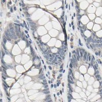
PB10033 has IHC images from paraffin sections of human colon cancer, human lung adenocarcinoma, mouse heart and rat heart (catalog image captions); IF/ICC data are unreported (catalog applications and images).

Real IHC data IHC analysis of AK1 using anti-AK1 antibody (PB10033). AK1 was detected in a paraffin-embedded section of human colon cancer tissue. Heat mediated antigen retrieval was performed in EDTA buffer (pH 8.0, epitope retrieval solution). The tissue section was blocked with 10% goat serum. The tissue section was then incubated with 2 μg/ml rabbit anti-AK1 Antibody (PB10033) overnight at 4°C. Peroxidase Conjugated Goat Anti-rabbit IgG was used as secondary antibody and incubated for 30 minutes at 37°C. The tissue section was developed using HRP Conjugated Rabbit IgG Super Vision Assay Kit (Catalog # SV0002) with DAB as the chromogen.
Anti-Adenylate Kinase 1/AK1 Antibody ®
Cat # PB10033

PB10033 is listed for IHC in human, mouse and rat (catalog applications and reactivity). Its IHC images show paraffin sections of human colon cancer, human lung adenocarcinoma, mouse heart and rat heart (catalog image captions).

Which to pick: Choose PB10033 for paraffin-section tissue IHC: its images use EDTA retrieval at pH 8.0 and 2 μg/ml primary antibody, while the listed IHC range is 2–5 μg/ml (PB10033 image captions; catalog dilution). For cross-species IHC, PB10033 lists human, mouse and rat reactivity and has IHC images for each species (catalog reactivity; PB10033 image captions). No SKU here lists IF/ICC validation or a reported clone type, and the paraffin-section captions do not report the fixative (catalog applications and clone field; PB10033 image captions).

Each figure is that product's own IHC / IF validation image from its datasheet.

References

  1. UniProt Consortium. UniProt entry P00568 (KAD1_HUMAN, Adenylate kinase isoenzyme 1).
  2. Human Protein Atlas. AK1 tissue IHC expression (reliability: Enhanced).
  3. Human Protein Atlas. AK1 subcellular location (ICC-IF): Mainly localized to the cytosol. In addition localized to the annulus..
  4. Human Protein Atlas. AK1 antibody validation summary (2 antibodies).
  5. Genome-wide analysis of mRNA and microRNA expression in colorectal cancer and adjacent normal mucosa. The journal of pathology. Clinical research 2022 — PMC9161315.
  6. Proteome-wide profiling of the MCF10AT breast cancer progression model. PloS one 2010 — PMC2882958.
  7. Inhibition of Sirtuin 2 with Sulfobenzoic Acid Derivative AK1 is Non-Toxic and Potentially Neuroprotective in a Mouse Model of Frontotemporal Dementia. Frontiers in pharmacology 2012 — PMC3298895.
  8. 4EBP1/eIF4E and p70S6K/RPS6 axes play critical and distinct roles in hepatocarcinogenesis driven by AKT and N-Ras proto-oncogenes in mice. Hepatology (Baltimore, Md.) 2015 — PMC4280310.
  9. PubMed PMID:183954 — UniProt-cited evidence.
  10. PubMed PMID:2542324 — UniProt-cited evidence.
  11. PubMed PMID:15489334 — UniProt-cited evidence.