This website uses cookies to ensure you get the best experience on our website.


- Table of Contents
Plan AKR1B10 staining in paraffin sections using the cytoplasmic pattern reported in gastrointestinal and gallbladder tissue (HPA tissue IHC). This guide covers controls, staining interpretation, and the potential effect of secretion on tissue localisation (HPA tissue IHC; UniProt).

Expected localisation, validated protocols, controls and antibodies — the at-a-glance facts below, then the full design guide.
| Expected localisation | Cytoplasmic tissue staining (HPA tissue IHC); lysosomal secretion (UniProt) | |
| Staining pattern | Enterocytes and glandular cells show cytoplasmic staining (HPA tissue IHC) | |
| Antigen retrieval | Citrate pH 6 HIER, heat-mediated (datasheet A02976) | |
| Positive control | Small intestine+4 more · see all | |
| Negative control | Adipose tissue+4 more · see all |
| Fixation | Keep fixation consistent across sections. (standard IHC practice; not target-specific) | |
| Caveat | Secretion may separate protein staining from RNA location (HPA tissue IHC) | |
| Regulation | Regulation not specified (UniProt) | |
| Isoform / epitope | No isoforms annotated; no transmembrane epitope split (UniProt) |
The catalog antibody’s IHC-P protocol is paired with 4 published AKR1B10 IHC protocols covering liver, endometrial, and esophageal tissues (PMC5011670; PMC6201704; PMC8305663; PMC11663324).
| Sample | Paraffin-embedded human intestinal cancer tissues; fixative not specified (datasheet A02976) |
| Fixation | Image fixative and duration unreported (datasheet A02976); verify before use. |
| Sectioning | 4–5 µm sections on charged slides (standard) |
| Deparaffinisation | Xylene, graded ethanol series to water (standard) |
| Antigen retrieval | Heat retrieval: Citrate pH 6, 20 min (datasheet A02976) |
| Peroxidase block | 3% H2O2, 10 min, room temperature (standard) |
| Blocking | 10% goat serum (datasheet A02976) |
| Primary antibody | Rabbit anti-AKR1B10, 0.5-1μg/ml (datasheet A02976) |
| Primary incubation | Overnight at 4 °C (datasheet A02976) |
| Detection | Streptavidin-biotin complex (SABC), DAB chromogen (datasheet A02976) |
| Counterstain | Hematoxylin, blue, dehydrate and mount (standard) |
| Expected result | AKR1B10-positive staining in enterocytes - Microvilli of small intestine (HPA tissue IHC: High). HPA tissue profile: Cytoplasmic expression in gastrointestinal tract and gall bladder. No signal in the no-primary control. |
AKR1B10 should show predominantly cytoplasmic staining in gastrointestinal epithelial cells, especially small intestinal enterocytes and stomach glandular cells (HPA tissue IHC: High). HPA rates the tissue profile Enhanced, while cautioning that the staining evidence includes antibodies targeting proteins from multiple genes (HPA tissue IHC). A membrane-spanning pattern is not expected from the protein sequence (UniProt O60218: no transmembrane segment); supported ICC-IF data also place AKR1B10 mainly in the cytosol (HPA subcellular).
| Strong cytoplasmic signal in small intestinal enterocytes or stomach glandular cells (HPA tissue IHC: High). | This fits the principal positive tissue pattern. Compare epithelial cells with neighboring structures on the same slide, and assess whether the signal is cell-associated rather than a uniform deposit (HPA tissue IHC; general IHC practice). |
| Predominantly nuclear signal, with little epithelial cytoplasmic staining (HPA tissue IHC: cytoplasmic profile). | Treat this as a localization mismatch requiring validation. HPA reports cytoplasmic tissue staining and supported cytosolic ICC-IF localization; UniProt also lists lysosomal localization and secretion, so unusual signal should be checked rather than assigned to AKR1B10 by appearance alone (HPA tissue IHC; HPA subcellular; UniProt O60218). |
| Strong staining in adipocytes or respiratory epithelial cells, while expected gastrointestinal epithelium is weak (HPA tissue IHC). | These cell types are listed as not detected in the HPA tissue examples. Consider cross-reactivity or endogenous detection activity, then compare a matched negative detection control and a known-positive section (HPA tissue IHC; general IHC practice). |
| Even haze across epithelium, stroma, and empty areas of the section (general IHC practice). | Diffuse background has little anatomical selectivity and cannot establish AKR1B10 expression. Review blocking, wash steps, primary antibody concentration, and detection controls before interpreting cell-specific signal (general IHC practice). |
| No epithelial signal in small intestine or stomach (HPA tissue IHC: High). | First question assay performance, because these are strong HPA positive examples. Check tissue preservation, retrieval and detection using the antibody's validated IHC conditions; a failed positive control makes a negative study section uninterpretable (HPA tissue IHC; general IHC practice). |
| Cellular localization and secretion (UniProt O60218; HPA subcellular) | UniProt places AKR1B10 in lysosomes and describes lysosome-mediated secretion; HPA ICC-IF mainly supports cytosol, with additional plasma membrane localization. Interpret intracellular staining in the context of the tissue IHC cytoplasmic profile; secretion can complicate a direct RNA-to-protein comparison (UniProt O60218; HPA subcellular; HPA tissue IHC). |
| Tissue reference and antibody specificity (HPA tissue IHC; HPA antibodies) | HPA lists high staining in small intestine and stomach, medium staining in colon and gallbladder, and Enhanced overall tissue reliability. Its cross-gene antibody caution limits target assignment from a matching stain alone; antibody-level IHC statuses differ: CAB070163 Enhanced and HPA020280 Supported (HPA tissue IHC; HPA antibodies). |
| Adrenal discrepancy (UniProt O60218; HPA tissue IHC) | UniProt describes high adrenal expression, but HPA lists adrenal glandular cells as not detected by tissue IHC. Treat adrenal tissue as an uncertain staining reference and favor the HPA high-staining gastrointestinal examples for an IHC positive control (UniProt O60218; HPA tissue IHC). |
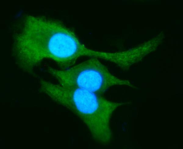
| IF/ICC Q: What pattern should an IF comparison show? (HPA subcellular) | A: Mainly cytosolic signal, with possible additional plasma membrane localization in the HPA ICC-IF record. The subcellular summary cautions that its antibodies target proteins from multiple genes; use this as localization context, not proof that an IHC signal is AKR1B10-specific (HPA subcellular). |
| Situation | Likely cause | Next action |
|---|---|---|
| Known-positive gastrointestinal epithelium is blank (HPA tissue IHC: High). | A failed staining workflow is possible; absence on this section alone does not establish absent protein (general IHC practice). | Review the IHC-validated antibody's stated retrieval and dilution, reagent activity, and detection controls; repeat with a known-positive section in the same run (general IHC practice). |
| Nuclear staining dominates (HPA tissue IHC: cytoplasmic profile). | The distribution conflicts with HPA tissue and supported cytosolic ICC-IF observations (HPA tissue IHC; HPA subcellular). | Compare a known-positive section and negative detection control; review antibody specificity before scoring nuclei as positive (general IHC practice; HPA tissue IHC cross-gene caution). |
| Broad staining appears in HPA not-detected cell types (HPA tissue IHC). | Cross-reactivity or endogenous detection activity may mimic a positive chromogenic signal (general IHC practice). | Check a negative detection control and compare cell types within the section; avoid assigning AKR1B10 from staining in an unexpected compartment alone (general IHC practice; HPA tissue IHC). |
| A diffuse chromogenic veil obscures epithelial boundaries (general IHC practice). | Nonspecific antibody binding, inadequate washes, or detection background are possible general IHC causes (general IHC practice). | Review blocking and washes, and compare the negative detection control before adjusting the antibody concentration within validated IHC conditions (general IHC practice). |
| Adrenal tissue is negative despite the UniProt expression summary (UniProt O60218). | UniProt reports high adrenal expression, whereas HPA records adrenal glandular cells as not detected by IHC (UniProt O60218; HPA tissue IHC). | Use HPA high-staining small intestine or stomach as the assay control; report the adrenal result with the source discrepancy rather than calling the run failed (HPA tissue IHC; UniProt O60218). |
| Membrane-edge or extracellular signal accompanies epithelial staining (HPA subcellular; UniProt O60218). | HPA ICC-IF reports additional plasma membrane localization, and UniProt describes secretion; these observations do not establish the identity of a tissue deposit (HPA subcellular; UniProt O60218). | Score the expected epithelial cytoplasmic pattern separately, check background controls, and avoid treating an isolated edge or deposit as definitive positivity (HPA tissue IHC; general IHC practice). |




Comprehensive Human Protein Atlas IHC scoring per tissue (reliability: Enhanced — High consistency between antibody staining and RNA expression data. Caution, targets protein from more than one gene. At least one protein variant secreted, tissue location of RNA and protein might differ and correlation is complex.). Rows are taken directly from the HPA tissue chart — click any row's HPA link to view the source.
| Tissue | Cell type | Level | Evidence | Source |
|---|---|---|---|---|
| Small intestine | Enterocytes - Microvilli | High | Protein (IHC) | HPA → |
| Stomach | Glandular cells | High | Protein (IHC) | HPA → |
| Appendix | Enterocytes - Microvilli | Medium | Protein (IHC) | HPA → |
| Colon | Enterocytes - Microvilli | Medium | Protein (IHC) | HPA → |
| Duodenum | Enterocytes | Medium | Protein (IHC) | HPA → |
| Tissue | Cell type | Level | Evidence | Source |
|---|---|---|---|---|
| Adipose tissue | Adipocytes | Not detected | Protein (IHC) | HPA → |
| Adrenal gland | Glandular cells | Not detected | Protein (IHC) | HPA → |
| Bone marrow | Hematopoietic cells | Not detected | Protein (IHC) | HPA → |
| Breast | Adipocytes | Not detected | Protein (IHC) | HPA → |
| Bronchus | Respiratory epithelial cells | Not detected | Protein (IHC) | HPA → |
Troubleshoot AKR1B10 staining in paraffin sections by checking retrieval, tissue controls, cellular localisation and detection background before scoring (A02976 tissue-IHC caption; HPA tissue IHC).
A02976 has real IHC images from human paraffin-embedded intestinal and liver cancer tissue and an IF image from A549 cells (A02976 image captions).
A02976 is listed for human IHC, IF and ICC (A02976 catalog: applications and reactivity). Its IHC images show paraffin-embedded intestinal and liver cancer tissue, and its IF image shows A549 cells (A02976 image captions).
Which to pick: For tissue IHC, choose A02976 for paraffin sections; its IHC captions document citrate pH 6 retrieval, but do not report the fixative (A02976 IHC image captions). For IF/ICC, choose A02976; both applications are listed, and its IF image shows A549 cells (A02976 catalog: applications; A02976 IF image caption). No cross-species choice is supported because A02976 lists Human reactivity only (A02976 catalog: reactivity).