This website uses cookies to ensure you get the best experience on our website.
- Table of Contents
1 Citations 6 Q&As
Facts about Aldo-keto reductase family 1 member C2.

Has a top bile-binding ability. .
| Human | |
|---|---|
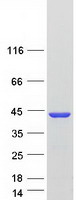
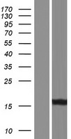
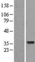
| Gene Name: | AKR1C2 |
| Uniprot: | P52895 |
| Entrez: | 1646 |

| Belongs to: |
|---|
| aldo/keto reductase family |

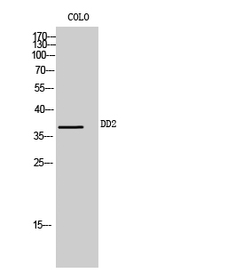
aldo-keto reductase family 1 member C2; aldo-keto reductase family 1, member C2 (dihydrodiol dehydrogenase 2; bile acidbinding protein; 3-alpha hydroxysteroid dehydrogenase, type III); BABP; Chlordecone reductase homolog HAKRD; DD; DD-2; DD2DD/BABP; DDH2FLJ53800; Dihydrodiol dehydrogenase 2; Dihydrodiol dehydrogenase/bile acid-binding protein; EC 1.1.1,3-alpha-HSD3; EC 1.1.1.213; EC 1.3.1.20; HAKRDAKR1C-pseudo; HBAB; MCDR2; pseudo-chlordecone reductase; Trans-1,2-dihydrobenzene-1,2-diol dehydrogenase; type II dihydrodiol dehydrogenase; Type III 3-alpha-hydroxysteroid dehydrogenase
Mass (kDA):
36.735 kDA

| Human | |
|---|---|
| Location: | 10p15.1 |
| Sequence: | 10; NC_000010.11 (4987775..5018033, complement) |
Expressed in fetal testes. Expressed in fetal and adult adrenal glands.
Cytoplasm.


PMID: 8274401 by Qin K.-N., et al. Molecular cloning of multiple cDNAs encoding human enzymes structurally related to 3 alpha-hydroxysteroid dehydrogenase.
PMID: 8011662 by Ciaccio P.J., et al. cDNA and deduced amino acid sequences of a human colon dihydrodiol dehydrogenase.