This website uses cookies to ensure you get the best experience on our website.


- Table of Contents
Plan paraffin-section ALDH1A3 IHC around cytoplasmic staining in prostate glandular cells (HPA tissue IHC). This guide covers fixation consistency, the catalog antibody’s 2–5 μg/mL IHC range (datasheet A03030), and interpretation of presumed off-target staining (HPA tissue IHC).

Expected localisation, validated protocols, controls and antibodies — the at-a-glance facts below, then the full design guide.
| Expected localisation | Cytoplasmic tissue staining (HPA tissue IHC) | |
| Staining pattern | Cytoplasmic staining in prostate glandular cells (HPA tissue IHC) | |
| Antigen retrieval | EDTA pH 8.0 HIER, heat-mediated (datasheet A03030) | |
| Positive control | Prostate+4 more · see all | |
| Negative control | Adrenal gland+4 more · see all |
| Fixation | Keep fixation consistent across sections (standard IHC practice; not target-specific) | |
| Caveat | Presumed off-target staining was observed (HPA tissue IHC) | |
| Regulation | RNA enhanced: prostate, urinary bladder (HPA tissue RNA) | |
| Isoform / epitope | No annotated isoforms; mature chain 2–512 (UniProt) |
The catalog antibody’s IHC-P protocol (datasheet A03030) is accompanied by 3 published IHC protocols (PMC12215419; PMC4430273; PMC6769487).
| Sample | Paraffin-embedded human prostate cancer tissue; fixative not specified (datasheet A03030) |
| Fixation | Image fixative and duration unreported (datasheet A03030); verify before use. |
| Sectioning | 4–5 µm sections on charged slides (standard) |
| Deparaffinisation | Xylene, graded ethanol series to water (standard) |
| Antigen retrieval | Heat retrieval: EDTA pH 8.0 (datasheet A03030); 20 min, 95–100 °C (standard) |
| Peroxidase block | 3% H2O2, 10 min, room temperature (standard) |
| Blocking | 10% goat serum (datasheet A03030) |
| Primary antibody | Rabbit anti-ALDH1A3, 2-5μg/ml (datasheet A03030) |
| Primary incubation | Overnight at 4 °C (datasheet A03030) |
| Detection | HRP-conjugated secondary, DAB chromogen (datasheet A03030) |
| Counterstain | Hematoxylin, blue, dehydrate and mount (standard) |
| Expected result | ALDH1A3-positive staining in glandular cells of prostate (HPA tissue IHC: High). HPA tissue profile: Cytoplasmic expression in several different tissue types including prostate. No signal in the no-primary control. |
ALDH1A3 is a cytoplasmic protein without a transmembrane segment (UniProt P47895: subcellular location and topology). In paraffin section IHC, expect cytoplasmic staining in prostate glandular cells and testis peritubular cells, with staining also reported in several other tissue types (HPA: tissue IHC). HPA rates the tissue profile Enhanced but notes medium agreement with RNA data and presumed off target binding that was disregarded (HPA: tissue IHC reliability).
| Distinct cytoplasmic chromogen in prostate glandular cells, with a stronger pattern than nearby unstained structures. | This fits a useful positive control: HPA reports High staining in prostate glandular cells, and UniProt places ALDH1A3 in the cytoplasm (HPA: prostate tissue IHC; UniProt P47895: subcellular location). Judge the named cells rather than treating every cell on the section as an expected positive. |
| Predominantly nuclear or membranous staining in the IHC section, with little convincing cytoplasmic signal. | That differs from the expected tissue IHC pattern (HPA: cytoplasmic tissue profile; UniProt P47895: cytoplasm and no transmembrane segment). Check whether the signal persists in a no primary control and compare it with a documented positive tissue before assigning it to ALDH1A3. Nuclear ICC IF evidence requires separate interpretation (HPA: subcellular ICC IF). |
| Strong chromogen in cells listed as Not detected, such as adrenal gland glandular cells or heart cardiomyocytes. | Treat the mismatch as a specificity warning, rather than proof that those cells express ALDH1A3 (HPA: adrenal gland and heart muscle, Not detected). Possible explanations include cross reactivity or endogenous detection activity; inspect a no primary control and the staining distribution. HPA itself notes presumed off target binding in its tissue assessment (HPA: tissue IHC reliability). |
| A broad, pale haze covers cells and extracellular areas without a clear cytoplasmic boundary. | This is difficult to score as cell specific ALDH1A3 staining. Compare a no primary section, inspect blocking and washing, and check chromogen development using standard IHC practice. The relevant reference is a cell associated cytoplasmic pattern, not uniform field staining (HPA: tissue IHC profile; UniProt P47895: cytoplasm). |
| No visible signal in prostate glandular cells or testis peritubular cells on a processed positive control. | These are High staining reference cell populations, so a blank result calls for a run check before biological interpretation (HPA: prostate and testis tissue IHC). Confirm tissue identity and control performance, then review retrieval, primary antibody application and detection as general IHC steps. The supplied sources do not establish ALDH1A3 specific fixation sensitivity. |
| Reference tissue and cell population | Prostate glandular cells and testis peritubular cells are High; bronchial respiratory epithelium, endometrial stroma, gallbladder and stomach glandular cells, and bladder urothelium are Medium (HPA: tissue IHC). Select the precise cell population when scoring. A Medium tissue may be less decisive than a High tissue as a run control (HPA: tissue IHC levels). |
| Antibody validation and specificity | The HPA tissue profile has Enhanced reliability, while its description reports medium staining to RNA consistency and presumed off target binding that was disregarded (HPA: tissue IHC reliability). HPA046271 has Enhanced IHC status; HPA064749 has no listed IHC status (HPA: antibody validation). Match the antibody used to its own validation record before relying on an HPA pattern. |
| Topology and protein processing | UniProt places ALDH1A3 in the cytoplasm, annotates no transmembrane segment or signal peptide, and lists a chain spanning residues 2–512 (UniProt P47895: location, topology and processing). These annotations support a cytoplasmic expectation; they do not identify the antibody epitope or predict antigen retrieval behavior. |
| ICC IF question: Does nuclear fluorescence overturn the IHC expectation? | HPA reports nucleoplasm as the main supported ICC IF location and cytosol as an additional supported location, whereas its tissue IHC profile is cytoplasmic (HPA: subcellular ICC IF; HPA: tissue IHC). Interpret fluorescence in the context of its own assay and controls; do not transfer an ICC IF compartment call directly to paraffin section IHC. |
| Endogenous chromogenic activity | Endogenous enzyme activity can imitate a positive chromogen signal in IHC as a general detection issue (standard IHC practice). A no primary control and detection appropriate blocking help identify that contribution. The supplied HPA and UniProt records do not assign a particular endogenous enzyme artifact to ALDH1A3. |
| Situation | Likely cause | Next action |
|---|---|---|
| The positive control is blank in prostate glandular cells. | A staining run problem is plausible because this cell population is High in HPA tissue IHC (HPA: prostate tissue IHC). Tissue identity or an omitted workflow step can also explain the result (standard IHC practice). | Verify the section and antibody identity, then inspect retrieval, primary incubation and detection records. Repeat alongside a confirmed positive section and a no primary control; do not infer an ALDH1A3 specific fixation effect from these sources. |
| Only faint staining appears in a tissue expected to be positive. | Several listed positive populations are Medium rather than High (HPA: tissue IHC), so apparent weakness depends on which cells are being compared. Run performance may also affect visibility (standard IHC practice). | Compare the named cell population with a High reference, preferably prostate glandular cells or testis peritubular cells (HPA: tissue IHC). Review control performance before changing scoring thresholds or general IHC conditions. |
| Staining is strongest in nuclei on the paraffin section. | A nuclear dominant IHC pattern conflicts with the cytoplasmic tissue profile (HPA: tissue IHC; UniProt P47895: cytoplasm). Supported nucleoplasmic localization comes from ICC IF and does not by itself validate the section pattern (HPA: subcellular ICC IF). | Check no primary and positive tissue controls, then reassess the chromogen against cell boundaries. Record the assay distinction when reporting any nuclear signal; seek independent specificity evidence before calling it ALDH1A3 IHC. |
| The whole section shows diffuse brown background. | Poor separation of specific stain from nonspecific chromogen or endogenous detection activity can obscure localization (standard IHC practice). The target reference is cytoplasmic staining in identified cells (HPA: tissue IHC). | Inspect a no primary section; review blocking, washes and chromogen development using standard IHC practice. Score only a resolved cellular pattern that matches the expected compartment and cell type. |
| A nominally negative cell population stains strongly. | HPA lists adrenal gland glandular cells, heart cardiomyocytes and several other populations as Not detected, and notes presumed off target binding in its tissue assessment (HPA: tissue IHC). Cross reactivity or detection background is possible. | Confirm the exact cell type and compare no primary and positive controls. Treat reproducible unexpected staining as unresolved until antibody specificity is supported; do not convert an HPA Not detected reference into a definitive biological absence. |
| Results from two antibodies disagree across the same tissue. | Validation is antibody specific: HPA046271 is Enhanced for IHC, while HPA064749 has no listed IHC status (HPA: antibody validation). Different staining therefore cannot be assumed equally supported. | Record which antibody produced each pattern and compare its own validation status. Recheck tissue identity and controls, then prioritize the pattern supported by the IHC validation record while reporting any remaining disagreement. |
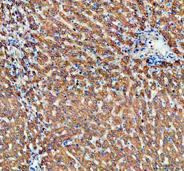
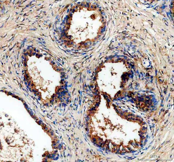
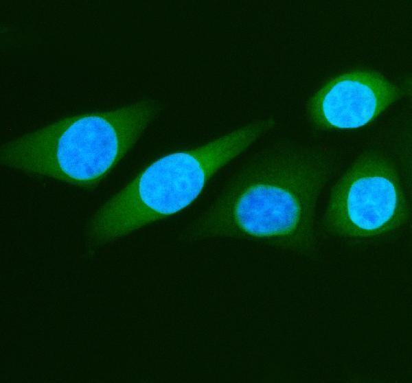


Comprehensive Human Protein Atlas IHC scoring per tissue (reliability: Enhanced — Medium consistency between antibody staining and RNA expression data. Presumed off target binding observed and disregarded.). Rows are taken directly from the HPA tissue chart — click any row's HPA link to view the source.
| Tissue | Cell type | Level | Evidence | Source |
|---|---|---|---|---|
| Prostate | Glandular cells | High | Protein (IHC) | HPA → |
| Testis | Peritubular cells | High | Protein (IHC) | HPA → |
| Bronchus | Respiratory epithelial cells | Medium | Protein (IHC) | HPA → |
| Endometrium | Cells in endometrial stroma | Medium | Protein (IHC) | HPA → |
| Gallbladder | Glandular cells | Medium | Protein (IHC) | HPA → |
| Tissue | Cell type | Level | Evidence | Source |
|---|---|---|---|---|
| Adrenal gland | Glandular cells | Not detected | Protein (IHC) | HPA → |
| Appendix | Glandular cells | Not detected | Protein (IHC) | HPA → |
| Caudate | Glial cells | Not detected | Protein (IHC) | HPA → |
| Heart muscle | Cardiomyocytes | Not detected | Protein (IHC) | HPA → |
| Lymph node | Germinal center cells | Not detected | Protein (IHC) | HPA → |
Troubleshoot ALDH1A3 staining in paraffin sections by checking retrieval, cellular distribution, controls and scoring before interpreting chromogenic signal.
A03030 has IHC data from human prostate cancer and rat lung paraffin sections, plus IF data from SiHa cells (IHC and IF image captions).
A03030 is listed for IHC and IF/ICC, with Human, Mouse and Rat reactivity (catalog: applications and reactivity). Its IHC images show human prostate cancer and rat lung paraffin sections; its IF image shows SiHa cells (catalog: image captions).
Which to pick: For tissue IHC, choose A03030: its paraffin-section captions document EDTA pH 8.0 retrieval and 2 μg/ml primary antibody (A03030 IHC captions). For IF/ICC, A03030 has a SiHa-cell IF image at 5 μg/ml (A03030 IF caption). For cross-species IHC, images document human and rat tissues; Mouse is listed under general reactivity without a mouse IHC image, and the paraffin-section captions do not report a fixative (catalog: reactivity; A03030 IHC captions).