This website uses cookies to ensure you get the best experience on our website.


- Table of Contents
Plan chromogenic ANXA2 IHC in paraffin sections using catalog antibody PA1348 (datasheet: PA1348 IHC). Compare membranous and extracellular staining with tissue observations, while accounting for secreted variants that can weaken RNA–protein agreement (HPA tissue IHC).

Expected localisation, validated protocols, controls and antibodies — the at-a-glance facts below, then the full design guide.
| Expected localisation | Membranous and extracellular tissue staining (HPA tissue IHC) | |
| Staining pattern | Membranous/extracellular; high in lung macrophages (HPA tissue IHC) | |
| Antigen retrieval | EDTA pH 8.0 HIER, heat-mediated (datasheet PA1348) | |
| Positive control | Bronchus+4 more · see all | |
| Negative control | Adrenal gland+4 more · see all |
| Fixation | Keep fixation consistent across sections (standard IHC practice; not target-specific) | |
| Caveat | Secreted variants can weaken tissue RNA–protein agreement (HPA tissue IHC) | |
| Regulation | No expression regulator annotated (UniProt) | |
| Isoform / epitope | 2 isoforms; epitope differences unspecified (UniProt) |
The catalog antibody has a datasheet IHC-P protocol (datasheet: PA1348). The published options below cover parathyroid adenoma, cervical tissue, and endometrium (PMC11279064; PMC10137006; PMC6694070).
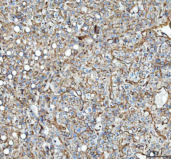
| Sample | Paraffin-embedded human liver cancer tissue; fixative not specified (datasheet PA1348) |
| Fixation | Image fixative and duration unreported (datasheet PA1348); verify before use. |
| Sectioning | 4–5 µm sections on charged slides (standard) |
| Deparaffinisation | Xylene, graded ethanol series to water (standard) |
| Antigen retrieval | Heat retrieval: EDTA pH 8.0 (datasheet PA1348); 20 min, 95–100 °C (standard) |
| Peroxidase block | 3% H2O2, 10 min, room temperature (standard) |
| Blocking | 10% goat serum (datasheet PA1348) |
| Primary antibody | Rabbit anti-ANXA2, 2-5μg/ml (datasheet PA1348) |
| Primary incubation | Overnight at 4 °C (datasheet PA1348) |
| Detection | HRP-conjugated secondary, DAB chromogen (datasheet PA1348) |
| Counterstain | Hematoxylin, blue, dehydrate and mount (standard) |
| Expected result | ANXA2-positive staining in respiratory epithelial cells of bronchus (HPA tissue IHC: High). HPA tissue profile: Ubiquitous membranous and extracellular expression. No signal in the no-primary control. |
In paraffin-section IHC, expect ANXA2 mainly at cell borders and extracellular structures (HPA: ubiquitous membranous and extracellular expression). High staining is reported in selected epithelial and glandular cells, cholangiocytes, and lung macrophages (HPA: tissue IHC). ANXA2 has no transmembrane segment; membrane-associated staining does not imply an integral membrane protein (UniProt P07355 topology). HPA rates the tissue pattern Enhanced, while noting that secretion can complicate RNA–protein comparisons (HPA: reliability description).
| Strong staining outlines esophageal squamous cells, bronchial respiratory epithelium, or liver cholangiocytes. | These are supported positive cell populations (HPA: High in esophageal squamous epithelial cells, bronchial respiratory epithelial cells, and liver cholangiocytes). Assess the named cells and their borders, rather than assigning one score to the whole section (general IHC practice). |
| The dominant signal is confined to nuclei, with little membrane or extracellular staining. | A nuclear-only pattern conflicts with the reported tissue profile and approved ICC location (HPA: membranous and extracellular tissue profile; plasma membrane with additional cytosol in ICC). Consider artefact or nonspecific detection; check the positive control and secondary-only control before interpreting it as ANXA2 (general IHC practice). |
| Adrenal glandular cells, cardiomyocytes, or skeletal myocytes stain strongly. | Those cell populations are reported as Not detected (HPA: adrenal glandular cells, heart cardiomyocytes, skeletal muscle myocytes). Check the cell identity and controls; cross-reactivity or endogenous detection activity may explain staining in these cells (general IHC practice). A negative cell population does not make every structure in its tissue negative. |
| Brown signal spreads fairly evenly across cells and empty spaces, obscuring borders. | The expected profile is membranous and extracellular, so extracellular signal alone is not proof of background (HPA: tissue profile; UniProt P07355 subcellular location). A uniform haze that also appears in a secondary-only control suggests nonspecific detection; reassess blocking, washes, and chromogen development (general IHC practice). |
| A correctly identified positive cell population has no visible signal. | Compare with a reported High population, such as lung macrophages or cervical glandular cells (HPA: tissue IHC). A blank result there makes the run difficult to interpret; verify tissue identity, antibody conditions, retrieval, and detection with a run control (general IHC practice). HPA staining is a reference pattern, not a guarantee for every specimen. |
| Tissue and cell selection | High calls apply to specified cells, including lung macrophages and gallbladder glandular cells (HPA: tissue IHC). Salivary glandular cells are Low, while selected adrenal glandular cells are Not detected (HPA: tissue IHC). Record the cell type as well as the tissue when comparing signal. |
| Membrane and extracellular location | The tissue profile includes both compartments (HPA: tissue IHC). UniProt lists extracellular space, matrix, and basement membrane, as well as movement from cytoplasm to cell surface (UniProt P07355 subcellular location). Interpret extracellular deposits alongside cellular staining and control slides. |
| Antibody validation | Three listed antibodies have Enhanced IHC status: HPA046964, HPA061798, and CAB004311 (HPA: antibody validation). This supports the reported pattern across validation evidence; it does not establish identical staining in every specimen or with an unlisted reagent. |
| RNA and protein comparison | Esophagus is tissue enhanced at RNA level (HPA: RNA specificity). HPA cautions that at least one secreted protein variant can make tissue RNA and protein locations differ (HPA: reliability description). Use observed cell-specific protein staining to judge this IHC section, rather than RNA abundance alone. |
| IF/ICC Q&A: where should signal appear? | Mainly at the plasma membrane, with additional cytosolic signal (HPA: approved ICC-IF locations). This is an ICC-IF location reference, not an IHC intensity standard; the paraffin-section tissue calls come from HPA tissue IHC. |
| Situation | Likely cause | Next action |
|---|---|---|
| No staining in a reported High cell population. | The run may have failed, or the sampled section may lack the identified cells (general IHC practice; HPA: cell-specific High calls). | Confirm cell identity, then review the run control, retrieval, antibody dilution, detection reagents, and development (general IHC practice). |
| Strong nuclear-only signal. | This does not match the reported dominant locations (HPA: tissue membranous/extracellular profile; approved ICC plasma membrane). | Check the positive and secondary-only controls, then reassess detection conditions before scoring it as ANXA2 (general IHC practice). |
| Strong signal in adrenal glandular cells or cardiomyocytes. | These cells are reported as Not detected (HPA: tissue IHC); nonspecific binding or endogenous detection activity is possible (general IHC practice). | Verify morphology and compare secondary-only and detection controls; reassess blocking or detection chemistry if control staining persists (general IHC practice). |
| Diffuse brown haze masks cell borders. | Background from detection or development is possible (general IHC practice); genuine extracellular ANXA2 is also reported (HPA: tissue profile). | Compare with a secondary-only control and inspect whether signal follows anatomical structures; review washes and development (general IHC practice). |
| Only weak signal in salivary glandular cells. | That cell population is reported Low (HPA: salivary glandular cells). | Compare with a reported High cell population on a suitable control section before changing staining conditions (HPA: tissue IHC; general IHC practice). |
| IHC and IF/ICC show different apparent distributions. | The evidence describes tissue IHC as membranous and extracellular, while ICC-IF also reports cytosol (HPA: tissue profile; approved ICC-IF locations). | Evaluate each preparation against its own HPA location reference and its controls; do not transfer an ICC-IF intensity call to paraffin-section IHC (general IHC practice). |
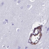


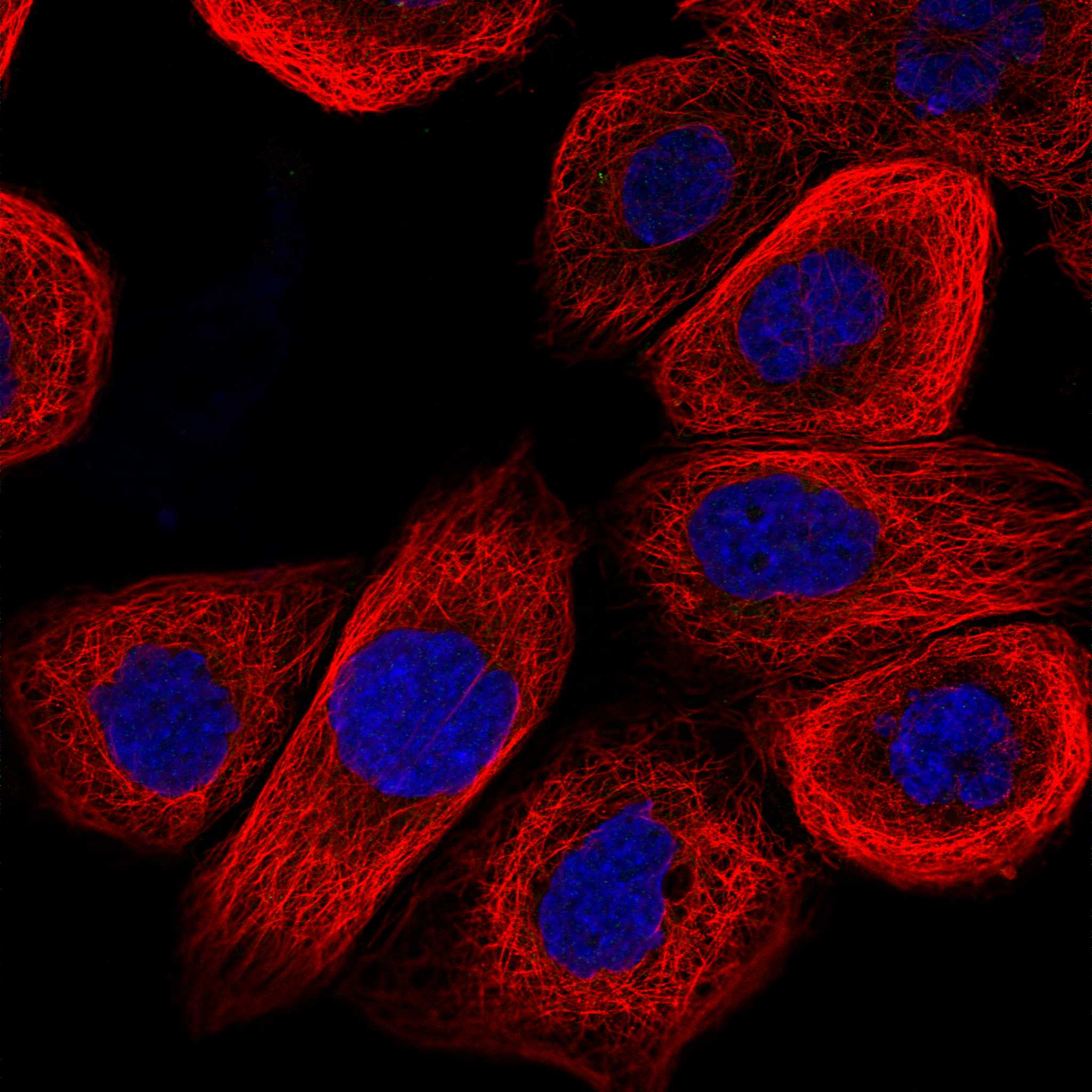

Comprehensive Human Protein Atlas IHC scoring per tissue (reliability: Enhanced — At least one protein variant secreted, tissue location of RNA and protein might differ and correlation is complex. Antibody staining in cells/structures not annotated, view images.). Rows are taken directly from the HPA tissue chart — click any row's HPA link to view the source.
| Tissue | Cell type | Level | Evidence | Source |
|---|---|---|---|---|
| Bronchus | Respiratory epithelial cells | High | Protein (IHC) | HPA → |
| Cervix | Glandular cells | High | Protein (IHC) | HPA → |
| Endometrium | Glandular cells | High | Protein (IHC) | HPA → |
| Esophagus | Squamous epithelial cells | High | Protein (IHC) | HPA → |
| Gallbladder | Glandular cells | High | Protein (IHC) | HPA → |
| Tissue | Cell type | Level | Evidence | Source |
|---|---|---|---|---|
| Adrenal gland | Glandular cells | Not detected | Protein (IHC) | HPA → |
| Caudate | Glial cells | Not detected | Protein (IHC) | HPA → |
| Cerebellum | Cells in granular layer | Not detected | Protein (IHC) | HPA → |
| Epididymis | Glandular cells | Not detected | Protein (IHC) | HPA → |
| Heart muscle | Cardiomyocytes | Not detected | Protein (IHC) | HPA → |
Troubleshoot ANXA2 staining in paraffin sections by checking retrieval, cell compartment, controls and scoring before interpreting a chromogenic signal.
PA1348 has a human liver cancer paraffin-section IHC image (catalog image caption); M00868 lists IHC and IF/ICC applications with human, mouse, and rat reactivity (catalog applications/reactivity).
PA1348 is the SKU with a rendered card and its own IHC figure (cards payload). Its image shows chromogenic ANXA2 detection in a paraffin-embedded human liver cancer section (PA1348 image caption).
Which to pick: Choose PA1348 for paraffin-section tissue IHC: its own image documents EDTA retrieval, 2 μg/mL primary antibody, and DAB detection; the fixative is unreported (PA1348 image caption). For IF/ICC, M00868 lists those applications and is rabbit monoclonal, but has no IF image in the payload (M00868 applications/host/clone/image alts). Both list human, mouse, and rat reactivity for cross-species planning; the pictured IHC example is human only (catalog reactivity; PA1348 image caption).