Click image to see more details
Product Info Summary
| SKU: | PA1348 |
|---|---|
| Size: | 100 μg/vial |
| Reactive Species: | Human, Mouse, Rat |
| Host: | Rabbit |
| Application: | Flow Cytometry, IHC, WB |
Customers Who Bought This Also Bought

Product info
Product Name
Anti-Annexin A2/ANXA2 Antibody Picoband®
SKU/Catalog Number
PA1348
BA0641 is an alternative SKU for this antibody, used in previous lots.
Size
100 μg/vial
Form
Lyophilized
Description
ANXA2 is a multi-compartment annexin that supports dynamic membrane-related processes; at the cell surface, ANXA2 (often as an ANXA2•S100A10 complex) promotes fibrinolysis by organizing plasmin generation, linking it to vascular homeostasis and inflammatory remodeling interfaces (context dependent). Assay context: antibody tested for flow cytometry, IHC, and WB in human/mouse/rat. Commonly contextualized with protease-inhibitor balance markers like SERPINA1 and ECM remodeling markers like MMP1 when interpreting net proteolytic environments (putative).
Storage & Handling
Store at -20˚C for one year from date of receipt. After reconstitution, at 4˚C for one month. It can also be aliquotted and stored frozen at -20˚C for six months. Avoid repeated freeze-thaw cycles.
Cite This Product
Anti-Annexin A2/ANXA2 Antibody Picoband® (Boster Biological Technology, Pleasanton CA, USA, Catalog # PA1348)
Host
Rabbit
Contents
Each vial contains 4 mg Trehalose, 0.9 mg NaCl and 0.2 mg Na2HPO4.
Clonality
Polyclonal
Isotype
Rabbit IgG
Immunogen
A synthetic peptide corresponding to a sequence in the middle region of human Annexin A2, different from the rat sequence by one amino acid.
Cross-reactivity
No cross-reactivity with other proteins
Reactive Species
PA1348 is reactive to ANXA2 in Human, Mouse, Rat
Observed Molecular Weight
36 kDa
Calculated molecular weight
38.6 kDa
Background of ANXA2
Annexin A2 also known as annexin II is a protein that in humans is encoded by the ANXA2 gene. The ANXA2 gene has three pseudogenes located on chromosomes 4, 9 and 10, respectively. Multiple alternatively spliced transcript variants encoding different isoforms have been found for this gene. This protein is a member of the annexin family. Members of this calcium-dependent phospholipid-binding protein family play a role in the regulation of cellular growth and in signal transduction pathways. This protein functions as an autocrine factor which heightens osteoclast formation and bone resorption. Richard et al. (1994) presented an integration of the physical, expression, and genetic maps of human chromosome 15. They placed the ANXA2 gene in their region IV, i.e., 15q21-q22, thus confirming the previous localization.
Antibody Validation
Boster validates all antibodies on WB, IHC, ICC, Immunofluorescence, and ELISA with known positive control and negative samples to ensure specificity and high affinity, including thorough antibody incubations.
Application & Images
Applications
PA1348 is guaranteed for Flow Cytometry, IHC, WB Boster Guarantee
Recommend Dilution
| Application | Dilution | Species |
|---|---|---|
| Western blot | 0.1-0.5μg/ml | Human, Mouse, Rat |
| Immunohistochemistry (Paraffin-embedded Section) | 2-5μg/ml | Human |
| Flow Cytometry(Fixed) | 1-3 μg/1x106 cells | Human |
Tested application
Suggested blocking solution with 5% non-fat milk or BSA; (*)Recommended protein loading: 20-40 µg per lane
Use TE buffer pH 9.0 for antigen retrieval; (*) citrate buffer pH 6.0 is an alternative.
Validation Images & Assay Conditions
Click image to see more details
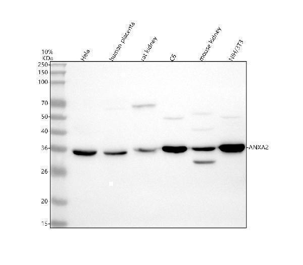
Western blot analysis of ANXA2 using anti-ANXA2 antibody (PA1348).
Electrophoresis was performed on a 10% SDS-PAGE gel at 80V (Stacking gel) / 120V (Resolving gel) for 2 hours. The sample well of each lane was loaded with 30 ug of sample under reducing conditions.
Lane 1: human Hela whole cell lysates,
Lane 2: human placenta tissue lysates,
Lane 3: rat kidney tissue lysates,
Lane 4: rat C6 whole cell lysates,
Lane 5: mouse kidney tissue lysates,
Lane 6: mouse NIH/3T3 whole cell lysates.
After electrophoresis, proteins were transferred to a nitrocellulose membrane at 150 mA for 50-90 minutes. Blocked the membrane with 5% non-fat milk/TBS for 1.5 hour at RT. The membrane was incubated with rabbit anti-ANXA2 antigen affinity purified polyclonal antibody (PA1348) at 0.5 μg/mL overnight at 4°C, then washed with TBS-0.1%Tween 3 times with 5 minutes each and probed with a goat anti-rabbit IgG-HRP secondary antibody (Catalog # BA1054) at a dilution of 1:5000 for 1.5 hour at RT. The signal is developed using an ECL Plus Western Blotting Substrate (Catalog # AR1196-200) with Tanon 5200 system. A specific band was detected for ANXA2 at approximately 36 kDa. The expected band size for ANXA2 is at 39 kDa.

Click image to see more details
IHC analysis of ANXA2 using anti-ANXA2 antibody (PA1348).
ANXA2 was detected in a paraffin-embedded section of human liver cancer tissue. Heat mediated antigen retrieval was performed in EDTA buffer (pH 8.0, epitope retrieval solution). The tissue section was blocked with 10% goat serum. The tissue section was then incubated with 2 μg/ml rabbit anti-ANXA2 Antibody (PA1348) overnight at 4°C. Peroxidase Conjugated Goat Anti-rabbit IgG was used as secondary antibody and incubated for 30 minutes at 37°C. The tissue section was developed using HRP Conjugated Rabbit IgG Super Vision Assay Kit (Catalog # SV0002) with DAB as the chromogen.

Click image to see more details
Flow Cytometry analysis of Hela cells using anti-ANXA2 antibody (PA1348).
Overlay histogram showing Hela cells stained with PA1348 (Blue line). The cells were fixed with 4% paraformaldehyde and blocked with 10% normal goat serum. And then incubated with rabbit anti-ANXA2 Antibody (PA1348, 1 μg/1x106 cells) for 30 min at 20°C. DyLight®488 conjugated goat anti-rabbit IgG (BA1127, 5-10 μg/1x106 cells) was used as secondary antibody for 30 minutes at 20°C. Isotype control antibody (Green line) was rabbit IgG (1 μg/1x106) used under the same conditions. Unlabelled sample without incubation with primary antibody and secondary antibody (Red line) was used as a blank control.
Specific Publications For Anti-Annexin A2/ANXA2 Antibody Picoband® (PA1348)
Loading publications
Recommended Resources
Here are featured tools and databases that you might find useful.
- Boster's Pathways Library
- Protein Databases
- Bioscience Research Protocol Resources
- Data Processing & Analysis Software
- Photo Editing Software
- Scientific Literature Resources
- Research Paper Management Tools
- Molecular Biology Software
- Primer Design Tools
- Bioinformatics Tools
- Phylogenetic Tree Analysis
Customer Reviews
Have you used Anti-Annexin A2/ANXA2 Antibody Picoband®?
Share your experimental results or join a short interview to earn up to $1,000 in product credits or other rewards.
0 Reviews For Anti-Annexin A2/ANXA2 Antibody Picoband®
Customer Q&As
Have a question?
Find answers in Q&As, reviews.
Can't find your answer?
Submit your question
3 Customer Q&As for Anti-Annexin A2/ANXA2 Antibody Picoband®
Question
My question regarding product PA1348, anti-Annexin A2/ANXA2 antibody. I was wondering if it would be possible to conjugate this antibody with biotin. I would need it to be without BSA or sodium azide. I am planning on using a buffer exchange of sodium azide with PBS only. Would there be problems for me to conjugate the antibody and store it in -20 degrees in small aliquots?
Verified Customer
Verified customer
Asked: 2019-05-10
Answer
We do not advise storing this antibody with PBS buffer only in -20 degrees. If you want to store it in -20 degrees it is best to add some cryoprotectant like glycerol. If you want carrier free PA1348 anti-Annexin A2/ANXA2 antibody, we can provide it to you in a special formula with trehalose and/or glycerol. These molecules will not interfere with conjugation chemistry and provide a good level of protection for the antibody from degradation. Please be sure to specify this in your purchase order.
Boster Scientific Support
Answered: 2019-05-10
Question
We are currently using anti-Annexin A2/ANXA2 antibody PA1348 for rat tissue, and we are satisfied with the WB results. The species of reactivity given in the datasheet says human, mouse, rat. Is it possible that the antibody can work on bovine tissues as well?
O. Wu
Verified customer
Asked: 2019-04-02
Answer
The anti-Annexin A2/ANXA2 antibody (PA1348) has not been tested for cross reactivity specifically with bovine tissues, though there is a good chance of cross reactivity. We have an innovator award program that if you test this antibody and show it works in bovine you can get your next antibody for free. Please contact me if I can help you with anything.
Boster Scientific Support
Answered: 2019-04-02
Question
Would anti-Annexin A2/ANXA2 antibody PA1348 work on monkey IHC with umbilical vein endothelial cell?
Verified Customer
Verified customer
Asked: 2018-11-21
Answer
Our lab technicians have not validated anti-Annexin A2/ANXA2 antibody PA1348 on monkey. You can run a BLAST between monkey and the immunogen sequence of anti-Annexin A2/ANXA2 antibody PA1348 to see if they may cross-react. If the sequence homology is close, then you can perform a pilot test. Keep in mind that since we have not validated monkey samples, this use of the antibody is not covered by our guarantee. However we have an innovator award program that if you test this antibody and show it works in monkey umbilical vein endothelial cell in IHC, you can get your next antibody for free.
Boster Scientific Support
Answered: 2018-11-21
