This website uses cookies to ensure you get the best experience on our website.


- Table of Contents
Plan ANXA7 paraffin IHC using the reported cytoplasmic and membranous tissue pattern (HPA tissue IHC). Start with the catalog antibody's 0.5–1 μg/mL IHC range (datasheet PA2076), and interpret staining alongside HPA's low consistency with RNA expression (HPA tissue IHC).

Expected localisation, validated protocols, controls and antibodies — the at-a-glance facts below, then the full design guide.
| Expected localisation | Cytoplasmic and membranous tissue staining (HPA tissue IHC) | |
| Staining pattern | Broad cytoplasmic and membranous staining; glandular cells positive (HPA tissue IHC) | |
| Antigen retrieval | EDTA pH 8.0 HIER, heat-mediated (datasheet PA2076) | |
| Positive control | Adrenal gland+4 more · see all | |
| Negative control | Adipose tissue+1 more · see all |
| Fixation | Keep fixation consistent across sections (standard IHC practice; not target-specific) | |
| Caveat | Antibody staining and RNA levels show low consistency (HPA tissue IHC) | |
| Regulation | Isoform abundance differs by tissue (UniProt) | |
| Isoform / epitope | 2 isoforms; epitope coverage is unknown (UniProt) |
The catalog antibody’s IHC-P protocol uses EDTA pH 8.0 retrieval (datasheet: PA2076). Two published ANXA7 IHC protocols provide additional paraffin-section conditions (PMC6188866; PMC11659777).
| Sample | Paraffin-embedded Human Mammary Cancer tissue; fixative not specified (datasheet PA2076) |
| Fixation | Image fixative and duration unreported (datasheet PA2076); verify before use. |
| Sectioning | 4–5 µm sections on charged slides (standard) |
| Deparaffinisation | Xylene, graded ethanol series to water (standard) |
| Antigen retrieval | Heat retrieval: EDTA pH 8.0 (datasheet PA2076); 20 min, 95–100 °C (standard) |
| Peroxidase block | 3% H2O2, 10 min, room temperature (standard) |
| Blocking | 10% goat serum (datasheet PA2076) |
| Primary antibody | Rabbit anti-ANXA7, 0.5-1μg/ml (datasheet PA2076) |
| Primary incubation | Overnight at 4 °C (datasheet PA2076) |
| Detection | Streptavidin-biotin complex (SABC), DAB chromogen (datasheet PA2076) |
| Counterstain | Hematoxylin, blue, dehydrate and mount (standard) |
| Expected result | ANXA7-positive staining in glandular cells of adrenal gland (HPA tissue IHC: Medium). HPA tissue profile: Ubiquitous cytoplasmic and membranous expression. No signal in the no-primary control. |
ANXA7 should appear mainly in cytoplasm and along cell membranes, with medium staining reported in several glandular and respiratory epithelial cells and in hematopoietic and lymphoid tissue (HPA: ubiquitous cytoplasmic and membranous expression; listed medium signals). It has no transmembrane segment, so membrane staining need not form a continuous surface outline (UniProt P20073: no transmembrane segment). HPA rates its tissue IHC Approved while noting low consistency with RNA expression (HPA: reliability description).
| Cytoplasmic staining, with some membranous accentuation, in adrenal or breast glandular cells. | This fits the reported compartment and cell-level pattern; HPA lists medium staining in those cells (HPA: tissue IHC). Judge intensity against a matched control section, since slide preparation and detection affect its appearance (general IHC practice). |
| Predominantly nuclear staining, with little cytoplasmic or membranous signal. | That distribution does not match HPA’s reported cytoplasmic and membranous pattern (HPA: tissue IHC). Treat it as unconfirmed; inspect the negative control and repeat with adjusted staining conditions before assigning ANXA7 to nuclei (general IHC practice). |
| Strong adipocyte or small-intestinal glandular-cell staining. | HPA reports ANXA7 as not detected in those respective cells (HPA: tissue IHC). Check morphology and background; unexpected signal may reflect cross-reactivity or endogenous detection activity, but this pattern alone does not establish its cause (general IHC practice). |
| Broad, hazy colour across cells, stroma and empty areas. | Signal without a recognizable cellular pattern is difficult to score as ANXA7. Compare a no-primary control and check wash, blocking and detection steps for background (general IHC practice); HPA’s expected pattern is cytoplasmic and membranous (HPA: tissue IHC). |
| No staining in a technically adequate section of adrenal gland or breast. | Their glandular cells are reported at medium intensity, so complete absence warrants a control and workflow check (HPA: tissue IHC). It does not by itself prove the specimen lacks ANXA7; confirm the tissue and cell type before interpreting a negative result (general IHC practice). |
| Compartment and topology | HPA describes cytoplasmic and membranous staining (HPA: tissue IHC). ANXA7 has no transmembrane segment (UniProt P20073: topology); membrane association should not be read as proof of membrane insertion. |
| Isoforms and tissue context | Isoform 1 is expressed in brain, heart and skeletal muscle; isoform 2 is more abundant in liver, lung, kidney, spleen, fibroblasts and placenta (UniProt P20073: tissue specificity). The supplied IHC record does not identify which isoform an antibody detects. |
| Strength of the tissue evidence | The catalog antibody CAB004312 is IHC Approved, while HPA reports low consistency between antibody staining and RNA data (HPA: antibodies; tissue IHC reliability). Use the listed cell-level observations as reference patterns, with that limitation. |
| Antigen retrieval | No ANXA7-specific retrieval condition or fixation sensitivity is supplied (provided UniProt/HPA records). If optimizing paraffin-section IHC, compare retrieval settings using the same known-positive tissue and controls (general IHC practice). |
| IF/ICC Q: is a localisation pattern validated here? | A: HPA lists no main subcellular location, no cell lines with ICC-IF images and no ICC status for CAB004312 (HPA: subcellular record; antibodies). These data do not establish an IF pattern or supply an IF protocol. |
| Situation | Likely cause | Next action |
|---|---|---|
| Expected positive glandular cells are blank. | The cause cannot be assigned from a blank section; tissue identification, retrieval, primary staining or detection may need checking (general IHC practice). | Confirm the cell type and run a matched positive-control section through the full IHC workflow; compare with HPA’s medium glandular-cell examples (HPA: tissue IHC). |
| Only nuclei show strong colour. | A nuclear-dominant pattern differs from HPA’s cytoplasmic and membranous description (HPA: tissue IHC); its source remains uncertain. | Review morphology and a no-primary control, then repeat under adjusted staining conditions before scoring it as ANXA7 (general IHC practice). |
| Adipocytes or small-intestinal glandular cells look positive. | Those cells are listed as not detected (HPA: tissue IHC). Cross-reactivity or endogenous detection activity is possible, but neither is established by the image alone. | Compare adjacent expected-positive cells and a no-primary control; address control staining before interpreting the unexpected cells (general IHC practice). |
| Diffuse colour obscures cell boundaries. | Background from blocking, washing or detection can prevent assessment of cellular localisation (general IHC practice). | Inspect a no-primary control, improve the relevant background step and rescore only clearly identifiable cytoplasmic or membranous signal (general IHC practice; HPA: tissue IHC pattern). |
| One positive tissue stains while another does not. | HPA lists cell-specific levels and low consistency with RNA expression; its profile does not promise equal staining across tissues (HPA: tissue IHC). | Compare the same named cell types, specimen quality and matched controls; report each tissue’s observed result instead of imposing one intensity threshold (general IHC practice). |
| IF shows puncta or a compartment unlike the IHC reference. | HPA supplies no main ICC-IF location or cell-line images and gives CAB004312 no ICC status (HPA: subcellular record; antibodies). | Treat the IF localisation as provisional and evaluate it with controls on its separate guide page; use the HPA IHC pattern only as tissue context (HPA: tissue IHC; general IF practice). |
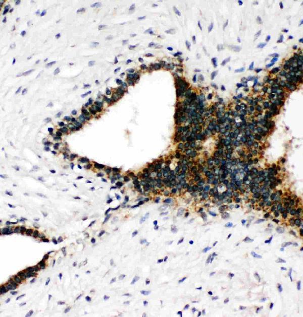
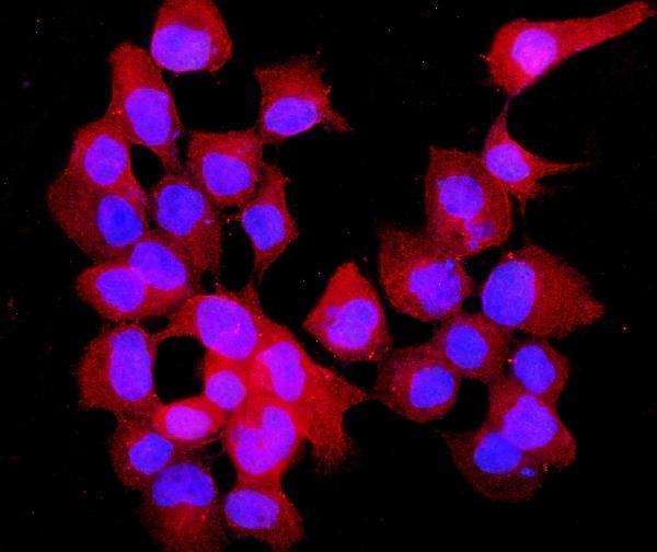

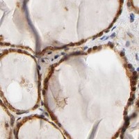


Comprehensive Human Protein Atlas IHC scoring per tissue (reliability: Approved — Low consistency between antibody staining and RNA expression data.). Rows are taken directly from the HPA tissue chart — click any row's HPA link to view the source.
| Tissue | Cell type | Level | Evidence | Source |
|---|---|---|---|---|
| Adrenal gland | Glandular cells | Medium | Protein (IHC) | HPA → |
| Appendix | Lymphoid tissue | Medium | Protein (IHC) | HPA → |
| Bone marrow | Hematopoietic cells | Medium | Protein (IHC) | HPA → |
| Breast | Glandular cells | Medium | Protein (IHC) | HPA → |
| Bronchus | Respiratory epithelial cells | Medium | Protein (IHC) | HPA → |
Troubleshoot ANXA7 staining by keeping the validated paraffin-section conditions traceable and assessing signal in the relevant cells and compartments.
PA2076 has IHC data from human mammary cancer paraffin sections and IF data from A431 cells (PA2076 image captions); the catalog lists human reactivity (PA2076 catalog).
PA2076 is the only SKU with a rendered card and its own IHC figure, showing human mammary cancer paraffin sections (PA2076 IHC caption). Its IF figure shows A431 cells (PA2076 IF caption).
Which to pick: Choose PA2076 for human tissue IHC because its own figure documents paraffin sections, EDTA retrieval at pH 8.0, and 1 μg/ml primary antibody (PA2076 IHC caption); the fixative is unreported (PA2076 IHC caption). For IF/ICC, PA2076 has an A431 cell IF image at 5 μg/ml (PA2076 IF caption). For mouse or rat IHC, consider rabbit monoclonals M04889-1 or M04889-2; both list IHC and human, mouse, and rat reactivity, but neither has an IHC image in the payload (M04889-1 and M04889-2 catalog).