AP2A2 / AP-2 complex subunit alpha-2 · IHC design guide

Design Immunohistochemistry for AP2A2

Plan chromogenic AP2A2 IHC in paraffin sections with the catalog antibody's 2–5 μg/ml range (datasheet A08527-1). Expect variable cytoplasmic staining, sometimes granular, across tissues (HPA tissue IHC).

Evidence assembled Oct 2026 · For research use; verify linked source records and product datasheet before use
Immunohistochemistry protocol sheet for AP2A2 (IHC for AP2A2): expected localisation Cytoplasmic tissue staining, sometimes granular (HPA tissue IHC), antibody A08527-1, validated IHC image, and IHC protocol steps
Printable AP2A2 IHC protocol sheet — expected localisation Cytoplasmic tissue staining, sometimes granular (HPA tissue IHC), antibody A08527-1, controls and protocol steps. Open the full AP2A2 IHC guide →

AP2A2 Immunohistochemistry Experimental Design Guide

Expected localisation, validated protocols, controls and antibodies — the at-a-glance facts below, then the full design guide.

Must know before staining
Expected localisation Cytoplasmic tissue staining, sometimes granular (HPA tissue IHC)
Staining pattern Variable cytoplasmic staining; lung macrophages stain highly (HPA tissue IHC)
Antigen retrieval EDTA pH 8.0 HIER, heat-mediated (datasheet A08527-1)
Positive control ⓘ Adrenal gland+4 more · see all
Negative control ⓘ Skeletal muscle
Important caveats
Reasons your staining may differ from the expected pattern.
Fixation Matched tissue-IHC evidence does not establish the fixation claim. Validate the specimen-specific method before use. (selected-SKU IHC image A08527-1)
Caveat High lung macrophage staining can skew tissue scoring (HPA tissue IHC)
Regulation Brain-enhanced RNA expression (HPA tissue RNA)
Isoform / epitope 3 isoforms; epitope differences unspecified (UniProt)
Section 1

Recommended AP2A2 IHC & IF Protocols

The catalog antibody protocol is paired with one published AP2A2 IHC protocol using human brain sections (PMC8204704).

Recommended immunohistochemistry (IHC-P) protocol parameters
SampleParaffin-embedded human lymphoma tissue; fixative not specified (datasheet A08527-1)
FixationImage fixative and duration unreported (datasheet A08527-1); verify before use.
Sectioning4–5 µm sections on charged slides (standard)
DeparaffinisationXylene, graded ethanol series to water (standard)
Antigen retrievalHeat retrieval: EDTA pH 8.0 (datasheet A08527-1); 20 min, 95–100 °C (standard)
Peroxidase block3% H2O2, 10 min, room temperature (standard)
Blocking10% goat serum (datasheet A08527-1)
Primary antibodyRabbit anti-AP2A2, 2-5 μg/ml (datasheet A08527-1)
Primary incubationOvernight at 4 °C (datasheet A08527-1)
DetectionStreptavidin-biotin complex (SABC), DAB chromogen (datasheet A08527-1)
CounterstainHematoxylin, blue, dehydrate and mount (standard)
Expected resultAP2A2-positive staining in glandular cells of adrenal gland (HPA tissue IHC: High). HPA tissue profile: Cytoplasmic expression at variable levels in most tissues, sometimes with a granular pattern. No signal in the no-primary control.
💡Decision noteStart with heat-mediated EDTA retrieval at pH 8.0 (datasheet: A08527-1); the published IHC method does not specify retrieval (PMC8204704).
Section 2

What Is the Expected AP2A2 Staining Pattern?

AP2A2 is associated with the cell membrane and coated pits and has no transmembrane segment (UniProt O94973 topology and subcellular location). In tissue IHC, expect variable cytoplasmic staining, sometimes granular, across many cell types (HPA: tissue IHC). HPA rates the tissue pattern Supported, with medium consistency between staining and RNA expression (HPA: reliability).

What am I looking at on my slide?
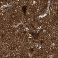
Cytoplasmic staining, sometimes granular, in cerebral cortex neurons or lung macrophages.This fits the reported tissue pattern; both cell populations have High staining (HPA: tissue IHC). Assess the named cells rather than assigning one intensity to an entire section.
Predominantly crisp nuclear staining, with little cytoplasmic signal, in a tissue IHC section.This does not match the reported dominant tissue IHC pattern (HPA: cytoplasmic expression). Review specificity and controls before calling it AP2A2; nucleoplasm is an additional ICC-IF location (HPA: subcellular), so context matters.
Strong staining in skeletal muscle myocytes, especially if the no-primary control also stains.HPA reports AP2A2 as Not detected in these myocytes (HPA: skeletal muscle). Cross-reactivity or endogenous detection activity is possible; the control result helps distinguish detection background from primary-antibody staining (general IHC practice).
Diffuse chromogen across cells, stroma, or the whole section without a clear cell pattern.The distribution is less convincing than variable cellular cytoplasm (HPA: tissue IHC). Check the no-primary control and staining conditions for nonspecific background (general IHC practice); do not score diffuse haze as a positive cell population.
No cellular staining in cerebral cortex neurons or lung macrophages.Both are reported High in tissue IHC (HPA: cerebral cortex; HPA: lung). A blank result warrants a run-control and detection check before concluding that AP2A2 is absent in the specimen (general IHC practice).
💡Expected AP2A2 appearanceCall a positive result when identifiable cells show cytoplasmic staining, sometimes granular, with readily visible signal in reported High populations such as cerebral cortex neurons or lung macrophages (HPA: tissue IHC); isolated nuclear color or diffuse section-wide haze needs control review (HPA: tissue IHC; general IHC practice).
How each factor affects the staining
Cell population chosen for interpretationHigh staining is reported in cerebral cortex neurons, lung macrophages, and several other named populations; hepatocytes and smooth muscle cells are Low, while skeletal myocytes are Not detected (HPA: tissue IHC). Compare like cell types when judging intensity.
Subcellular patternTissue IHC is described as variably cytoplasmic and sometimes granular (HPA: tissue IHC). UniProt places AP2A2 at the cell membrane and coated pits and lists no transmembrane segment (UniProt O94973); a continuous surface outline is not required to recognize the HPA tissue pattern.
Antibody evidenceCAB017514 has Supported IHC status, whereas HPA043040 has no IHC status in the supplied antibody record (HPA: antibodies). HPA's overall tissue reliability is Supported with medium staining–RNA consistency (HPA: reliability); neither label validates every cell in a new specimen.
IF/ICC: what localisation should I compare?HPA reports vesicles as the supported main ICC-IF location and nucleoplasm as an approved additional location (HPA: subcellular). Use those as IF/ICC interpretation context; the tissue IHC description remains cytoplasmic, sometimes granular (HPA: tissue IHC).
Why is my staining missing, weak or wrong?
SituationLikely causeNext action
Reported High cells are blank while the counterstain is visible.The target staining run may have failed; High tissue IHC staining is reported for cerebral cortex neurons and lung macrophages (HPA: tissue IHC).Check a known-positive control section, primary-antibody step, detection reagents, and chromogen development before interpreting the specimen as negative (general IHC practice).
Skeletal myocytes show strong color.That result conflicts with Not detected staining in skeletal myocytes (HPA: tissue IHC); nonspecific binding or detection background may contribute (general IHC practice).Review a no-primary control and the cellular distribution; reassess antibody specificity if color persists only when the primary is included (general IHC practice).
Color appears throughout the section, including areas without discernible cells.Diffuse deposit does not match the reported variable cellular cytoplasmic pattern (HPA: tissue IHC); nonspecific or endogenous detection signal is possible (general IHC practice).Inspect no-primary and reagent controls, then review blocking, washing, and chromogen development for the detection system used (general IHC practice).
Only nuclei appear positive in tissue IHC.The dominant tissue IHC pattern is cytoplasmic (HPA: tissue IHC), although nucleoplasm is an additional ICC-IF location (HPA: subcellular).Compare cytoplasm and nuclei in a reported High population and check controls; do not equate an IF nucleoplasmic observation with a nuclear-only IHC call.
Hepatocytes look faint beside strongly stained lung macrophages.These are reported Low and High, respectively, in tissue IHC (HPA: liver hepatocytes; HPA: lung macrophages).Score each named cell population against its reported level and the run controls; faint hepatocyte staining alone does not establish technical failure.
Granules are visible but there is no continuous plasma-membrane rim.Granular cytoplasmic staining is within the reported tissue pattern (HPA: tissue IHC); AP2A2 is associated with coated pits and lacks a transmembrane segment (UniProt O94973).Judge whether the granules belong to identifiable cells and are absent from background controls; do not require an uninterrupted membrane outline (HPA: tissue IHC; general IHC practice).

Sample controls for AP2A2 IHC & IF

🧪Run adrenal gland first: glandular cells must stain (HPA: High in adrenal gland glandular cells). Run skeletal muscle as the negative tissue (HPA: Myocytes not detected); on the adrenal slide, cells without specific staining should show only counterstain, but no cell population there is established as an internal biological negative by the supplied HPA rows.
Positive control tissue: Adrenal gland (Glandular cells, HPA High)
Negative control tissue: Skeletal muscle (HPA Not detected)
ICC-IF cell lines (HPA subcellular resource): HPA ICC-IF images show AP2A2 in A-431, U-251MG, U2OS, HEK293, NIH 3T3, with annotated localisation: Vesicles (supported) (HPA subcellular).
Technical controls: Include a no-primary (secondary-only) control, a concentration-matched rabbit IgG isotype control matched to the primary antibody’s clonality if known, and AP2A2-knockout material as a biological specificity control (caption: rabbit anti-AP2A2; standard IHC practice). For the caption’s biotin-based DAB method, block endogenous peroxidase and assess endogenous biotin background on the adrenal section (caption: SABC with DAB; standard IHC practice).
⚠️Feasibility: A target-specific fixation window or fixation effect is unreported, and the selected-SKU tissue-IHC caption does not state a fixative (A08527-1 caption: fixative not stated). That paraffin-section example uses heat retrieval in EDTA at pH 8.0, but the evidence does not establish whether retrieval is required (A08527-1 caption). Neither frozen sections nor IF can be judged easier from a matched protocol; ICC-IF images support vesicular localization, while endogenous peroxidase or biotin may complicate interpretation of adrenal SABC/DAB staining (HPA: vesicles supported; caption: SABC with DAB; standard IHC practice).

HPA tissue IHC evidence for AP2A2

Comprehensive Human Protein Atlas IHC scoring per tissue (reliability: Supported — Medium consistency between antibody staining and RNA expression data.). Rows are taken directly from the HPA tissue chart — click any row's HPA link to view the source.

Positive expression · recommended positive controls

TissueCell typeLevelEvidenceSource
Adrenal gland Glandular cells High Protein (IHC) HPA →
Bone marrow Hematopoietic cells High Protein (IHC) HPA →
Bronchus Respiratory epithelial cells High Protein (IHC) HPA →
Caudate Glial cells High Protein (IHC) HPA →
Cerebellum GLUC cells - cytoplasm/membrane High Protein (IHC) HPA →

Undetected expression · recommended negative controls

TissueCell typeLevelEvidenceSource
Skeletal muscle Myocytes Not detected Protein (IHC) HPA →
Section 3

Advanced AP2A2 IHC Tips

Troubleshoot AP2A2 staining in paraffin sections by checking retrieval, compartment, controls and scoring before interpreting differences between samples.

Which retrieval condition should I try first for weak AP2A2 staining?
Start with heat-mediated antigen retrieval in EDTA at pH 8.0 (datasheet A08527-1). The catalog antibody detected AP2A2 in a paraffin section of human lymphoma tissue after this retrieval, followed by 2 μg/ml primary antibody overnight at 4°C (datasheet A08527-1). If staining is weak, first check heating consistency and whether sections stayed covered by buffer throughout retrieval (standard IHC practice). Then compare a modestly longer retrieval with the original condition on adjacent sections, watching for tissue damage or stronger background (standard IHC practice). Keep detection and development conditions identical so the retrieval comparison is interpretable (standard IHC practice).
How can I troubleshoot fixation when AP2A2 staining varies between sections?
Target-specific sensitivity of AP2A2 to fixation is unknown from the supplied evidence; the selected tissue caption identifies a paraffin section but does not state its fixative (datasheet A08527-1). Record each sample’s fixative, fixation duration and processing history before comparing staining (standard IHC practice). Run sections from differently processed samples together with the same EDTA pH 8.0 retrieval and 2 μg/ml primary antibody condition (datasheet A08527-1). Include a consistently processed control section in each run to detect changes in staining performance (standard IHC practice). Do not attribute a weak section to AP2A2-specific fixation effects without a direct comparison under controlled conditions (standard IHC practice).
Should AP2A2 appear at the membrane or throughout the cytoplasm?
Expect a cytoplasmic signal that may be granular in tissue sections (HPA: cytoplasmic expression at variable levels, sometimes granular). AP2A2 is associated with the cell membrane and coated pits, and has no transmembrane segment (UniProt O94973: subcellular location and topology). HPA also reports mainly vesicular localisation in cell imaging, with an additional nucleoplasmic location (HPA: subcellular summary). Assess the staining pattern within intact cells at high magnification before calling diffuse chromogen signal specific (standard IHC practice). Because AP-2 can disengage shortly before vesicle internalisation, absence of a continuous membrane rim alone should not exclude AP2A2 staining (UniProt O94973: subcellular note).
Could an AP2A2 isoform or hidden epitope explain inconsistent staining?
AP2A2 has 3 annotated isoforms, so check the catalog antibody’s immunogen against their sequences before assuming equal recognition (UniProt O94973: isoforms). The supplied evidence does not identify this antibody’s epitope or establish isoform-specific staining (datasheet A08527-1: selected tissue caption). If an epitope lies in a region absent from one isoform, that isoform would not be expected to stain with an antibody against that region (standard antibody interpretation). Compare adjacent sections using the established EDTA pH 8.0 retrieval before changing more than one staining variable (datasheet A08527-1; standard IHC practice). Interpret differing patterns cautiously until epitope coverage and antibody specificity are established (standard IHC practice).
How should I check an AP2A2 pattern by multiplex IF?
Use IF as a separate confirmation experiment while retaining chromogenic paraffin IHC as this page’s primary workflow (datasheet A08527-1: tissue IHC caption). In cerebral cortex, pair AP2A2 with a neuronal marker to identify the expected cell population (HPA: high staining in cerebral cortex neuronal cells). Choose a red or far-red fluorophore after checking tissue autofluorescence in an unstained section, and use single-stain controls to assess channel bleed-through (standard IF practice). AP2A2 has no transmembrane segment and associates with membrane coated pits, so plan permeabilisation to expose its cytosolic epitope while preserving punctate structures (UniProt O94973: topology and location; standard IF practice). Confirm any IF pattern against the cell-resolved chromogenic result, allowing for differences between detection methods (standard IHC/IF practice).
How do I reduce diffuse or granular AP2A2 IHC background?
First distinguish cell-associated granular staining from diffuse deposit outside intact cells; granular cytoplasmic staining can be part of the reported AP2A2 tissue pattern (HPA: tissue IHC profile). The selected lymphoma-section workflow used 10% goat serum blocking and 2 μg/ml primary antibody overnight at 4°C (datasheet A08527-1). Run a no-primary control, review wash consistency and shorten chromogen development if the background also appears without primary antibody (standard IHC practice). Block endogenous peroxidase when using a peroxidase-based chromogenic workflow, particularly before judging brown DAB signal (standard IHC practice). Compare adjustments on adjacent sections so changes in background can be separated from changes in tissue expression (standard IHC practice).
What should I score when comparing AP2A2 across paraffin sections? ⚠ ANSWER MARKED FOR VERIFICATION
Define the cell population and compartment before scoring because reported tissue staining is cytoplasmic and can be granular (HPA: tissue IHC profile). For comparable sections, record the percentage of positive cells and staining intensity to calculate an H-score, or count positive cells per mm² within a predefined tissue area (standard IHC practice). Normalise cell counts to the analysed viable tissue area and compare the same cell type across specimens (standard IHC practice). Use the same retrieval, antibody concentration, imaging settings and chromogen development conditions for every comparison; the catalog condition includes EDTA pH 8.0 and 2 μg/ml primary antibody (datasheet A08527-1). Report excluded damaged or necrotic regions and keep scoring blinded where feasible (standard IHC practice).
How can I distinguish genuine AP2A2 staining from artefact?
Look for cell-associated cytoplasmic or granular staining, consistent with the tissue profile, while recognising AP2A2’s membrane and coated-pit association (HPA: tissue IHC profile; UniProt O94973: subcellular location). Check whether positive cells fit the specimen: lung macrophages show high staining, whereas skeletal-muscle myocytes were not detected in the supplied HPA profile (HPA: lung macrophages high; skeletal-muscle myocytes not detected). Discount signal confined to section edges, folds or necrotic areas, and examine a no-primary control for endogenous enzyme or detection background (standard IHC practice). Unexpected strong nuclear-only staining needs independent confirmation; cell imaging lists nucleoplasm as an additional location, not its main location (HPA: subcellular summary). Use concordant morphology, controls and replicate sections before assigning biological meaning to a pattern (standard IHC practice).
Boster reagents

Best AP2A2 / AP-2 complex subunit alpha-2 IHC Antibodies

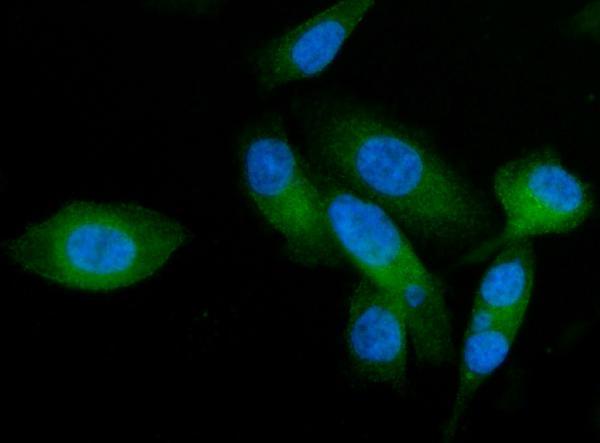
The catalog antibody A08527-1 has IHC images from human paraffin sections and an IF image from PC-3 cells (catalog image captions). Listed reactivity includes human, mouse and rat (datasheet: reactivity).

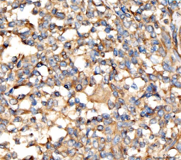
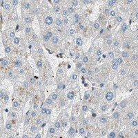
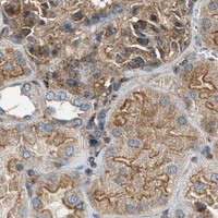
Real IHC data IHC analysis of AP2A2 using anti-AP2A2 antibody (A08527-1). AP2A2 was detected in a paraffin-embedded section of human lymphoma tissue. Heat mediated antigen retrieval was performed in EDTA buffer (pH 8.0, epitope retrieval solution). The tissue section was blocked with 10% goat serum. The tissue section was then incubated with 2 μg/ml rabbit anti-AP2A2 Antibody (A08527-1) overnight at 4°C. Biotinylated goat anti-rabbit IgG was used as secondary antibody and incubated for 30 minutes at 37°C. The tissue section was developed using Strepavidin-Biotin-Complex (SABC) (Catalog # SA1022) with DAB as the chromogen.
Anti-AP2A2 Antibody ®
Cat # A08527-1

A08527-1 will render with an IHC figure from a human lymphoma paraffin section (catalog IHC image caption). Its other IHC captions show human breast, liver and stomach cancer sections, and its IF caption shows PC-3 cells (catalog image captions).

Which to pick: Choose A08527-1 for paraffin-section IHC: its own figure shows human lymphoma tissue stained at 2 μg/ml after EDTA retrieval at pH 8.0 (catalog IHC image caption); the fixative is unreported (catalog IHC image caption). The same SKU is listed for IF/ICC at 5 μg/ml and has a PC-3 IF image (datasheet: applications and dilution; catalog IF image caption). For mouse or rat work, A08527-1 has listed reactivity, but the supplied IHC and IF images show human samples only; clonality is unreported (datasheet: reactivity and clone; catalog image captions).

Each figure is that product's own IHC / IF validation image from its datasheet.

References

  1. UniProt Consortium. UniProt entry O94973 (AP2A2_HUMAN, AP-2 complex subunit alpha-2).
  2. Human Protein Atlas. AP2A2 tissue IHC expression (reliability: Supported).
  3. Human Protein Atlas. AP2A2 subcellular location (ICC-IF): Mainly localized to vesicles. In addition localized to the nucleoplasm..
  4. Human Protein Atlas. AP2A2 antibody validation summary (2 antibodies).
  5. Alzheimer Disease Pathology-Associated Polymorphism in a Complex Variable Number of Tandem Repeat Region Within the MUC6 Gene, Near the AP2A2 Gene. Journal of neuropathology and experimental neurology 2020 — PMC8204704.
  6. Alpha adaptins show isoform-specific association with neurofibrillary tangles in Alzheimer's disease. Neuropathology and applied neurobiology 2022 — PMC8810620.
  7. PubMed PMID:10048485 — UniProt-cited evidence.
  8. PubMed PMID:12168954 — UniProt-cited evidence.
  9. PubMed PMID:14702039 — UniProt-cited evidence.