This website uses cookies to ensure you get the best experience on our website.


- Table of Contents
Plan AP2M1 paraffin-section IHC around the cytoplasmic staining reported in most tissues (HPA tissue IHC). This guide highlights control-tissue selection, fixation consistency and the uncertainty in the reported tissue staining (HPA tissue IHC).

Expected localisation, validated protocols, controls and antibodies — the at-a-glance facts below, then the full design guide.
| Expected localisation | Cytoplasmic tissue staining (HPA tissue IHC); membrane and coated-pit association (UniProt) | |
| Staining pattern | Cytoplasmic staining in most tissues (HPA tissue IHC) | |
| Antigen retrieval | EDTA pH 8.0 HIER, heat-mediated (datasheet A06179-3) | |
| Positive control | Appendix+4 more · see all | |
| Negative control | Adipose tissue+4 more · see all |
| Fixation | Keep fixation consistent across sections (standard IHC practice; not target-specific) | |
| Caveat | Tissue staining reliability is uncertain (HPA tissue IHC) | |
| Regulation | Low tissue specificity (HPA tissue RNA) | |
| Isoform / epitope | 2 isoforms; epitope coverage is unspecified (UniProt) |
The catalog antibody has a datasheet IHC-P protocol (datasheet: A06179-3). One published chromogenic AP2M1 IHC protocol is available for colorectal tissue (PMC10900655).
| Sample | Paraffin-embedded human kidney tissue; fixative not specified (datasheet A06179-3) |
| Fixation | Image fixative and duration unreported (datasheet A06179-3); verify before use. |
| Sectioning | 4–5 µm sections on charged slides (standard) |
| Deparaffinisation | Xylene, graded ethanol series to water (standard) |
| Antigen retrieval | Heat retrieval: EDTA pH 8.0 (datasheet A06179-3); 20 min, 95–100 °C (standard) |
| Peroxidase block | 3% H2O2, 10 min, room temperature (standard) |
| Blocking | 10% goat serum (datasheet A06179-3) |
| Primary antibody | Rabbit anti-AP2M1, 2-5 μg/ml (datasheet A06179-3) |
| Primary incubation | Overnight at 4 °C (datasheet A06179-3) |
| Detection | HRP-conjugated secondary, DAB chromogen (datasheet A06179-3) |
| Counterstain | Hematoxylin, blue, dehydrate and mount (standard) |
| Expected result | AP2M1-positive staining in glandular cells of appendix (HPA tissue IHC: Medium). HPA tissue profile: Cytoplasmic expression in most tissues. No signal in the no-primary control. |
AP2M1 is an AP-2 adaptor associated with the cell membrane and coated pits; it has no transmembrane segment (UniProt Q96CW1). In paraffin-section IHC, expect mainly cytoplasmic staining in expressing cells, including neuronal and glandular cells (HPA: cytoplasmic expression in most tissues; medium in selected neuronal and glandular cells). Interpret modest signals cautiously because HPA rates tissue IHC reliability as Uncertain (HPA: Uncertain).
| Cytoplasmic staining in neuronal cells of the cerebral cortex or caudate, or in cerebellar granular-layer cells (HPA: Medium in each). | This matches reported tissue IHC. AP2M1 acts in membrane traffic at coated pits (UniProt Q96CW1), but a chromogenic cytoplasmic pattern need not resolve individual pits. Compare staining with the surrounding cells and a matched control; HPA calls the overall tissue pattern Uncertain (HPA: Uncertain). |
| Cytoplasmic staining in appendix, cervix, epididymis, or gallbladder glandular cells (HPA: Medium in each). | These are additional reported positive cell populations (HPA: Medium). Score the specified cells rather than treating every structure in the section as an expected positive. The HPA levels describe observed staining, not a guaranteed intensity for another antibody or laboratory (HPA: tissue IHC; HPA: Uncertain). |
| Predominantly nuclear staining, with little cytoplasmic signal, in an otherwise positive cell population. | A nuclear-dominant pattern conflicts with HPA tissue cytoplasmic staining and its supported ICC-IF plasma-membrane location (HPA: tissue IHC; HPA: subcellular). Treat it as a possible artefact or off-target signal and review controls and morphology before assigning AP2M1 positivity; the evidence does not establish nuclear localisation. |
| Strong staining in adipocytes or esophageal squamous epithelial cells (HPA: Not detected in these cells). | This differs from the reported tissue pattern and warrants checking cross-reactivity or endogenous chromogenic activity (HPA: tissue IHC; general IHC practice). Do not call a single stained cell definitive proof of either problem: HPA rates its tissue IHC reliability Uncertain, and the result may depend on the antibody (HPA: Uncertain). |
| Diffuse colour across cells, stroma, or an entire section, without a discernible cellular pattern. | Diffuse background cannot establish AP2M1 localisation; assess the no-primary control, blocking and detection steps (general IHC practice). A meaningful result should be interpretable in cells against their surroundings, with the cytoplasmic tissue pattern as the reference (HPA: tissue IHC). |
| Tissue and cell selection (HPA: tissue IHC). | HPA reports medium staining in selected neuronal and glandular populations, low staining in several other cell populations, and no detection in specified cells such as adipocytes (HPA: tissue IHC). Choose a reported positive population for an interpretive control, while allowing for the Uncertain reliability rating (HPA: Uncertain). |
| Compartment and membrane association (UniProt Q96CW1; HPA: subcellular). | AP2M1 associates with the cell membrane and coated pits without spanning the membrane (UniProt Q96CW1). HPA tissue IHC is described as cytoplasmic, whereas ICC-IF supports a plasma-membrane location (HPA: tissue IHC; HPA: subcellular). These observations guide interpretation at each method's resolution; they do not imply that every pit will be visible in IHC. |
| IHC antibody evidence (HPA: antibodies). | HPA lists antibody HPA036849 as Uncertain for IHC and HPA069870 as Supported for ICC, with no IHC status reported for HPA069870 (HPA: antibodies). Do not transfer an ICC validation label to paraffin-section IHC; interpret the catalog antibody against its own IHC controls (general IHC practice). |
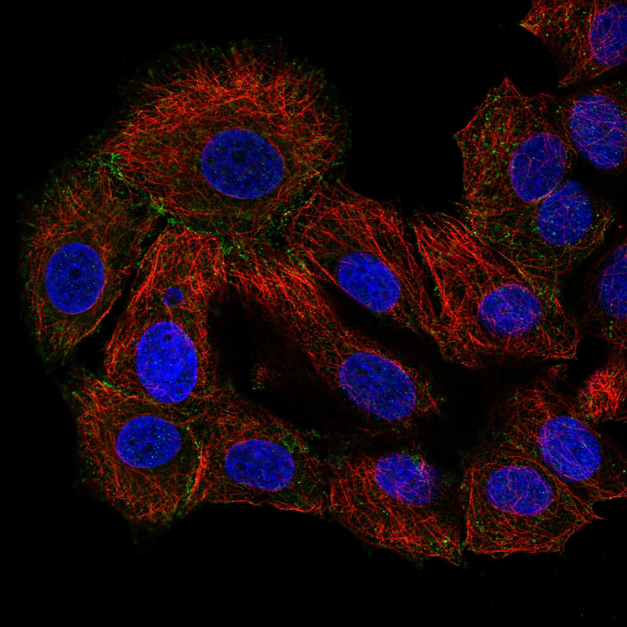
| IF/ICC Q: What pattern is reported? (HPA: subcellular). | A: Plasma-membrane localisation is supported in ICC-IF images from MCF-7 and U-251MG (HPA: subcellular). That observation informs compartment comparison, but this IHC section gives no IF/ICC protocol or claim that its image validation establishes the IHC pattern (HPA: antibodies). |
| Situation | Likely cause | Next action |
|---|---|---|
| No signal in a reported medium-staining population, such as cerebral-cortex neuronal cells (HPA: Medium). | The run may have lost detectable signal, or this antibody and preparation may differ from HPA's observations; HPA tissue IHC reliability is Uncertain (HPA: Uncertain; general IHC practice). | Check a same-run reported positive control, primary-antibody and detection steps, and the selected cell population before concluding absence (HPA: tissue IHC; general IHC practice). |
| Colour appears in the no-primary control. | Primary-independent detection or endogenous activity can contribute to chromogenic staining (general IHC practice). | Review the detection and blocking controls, then compare the control with the tissue slide at the same exposure to the chromogen (general IHC practice). Do not score matching background as AP2M1. |
| Most structures show diffuse stain that obscures individual cells. | Excess background from staining or detection can prevent cell-level interpretation (general IHC practice). | Review blocking, washes, antibody concentration and chromogen development using matched controls (general IHC practice). Rescore only when the reported cytoplasmic pattern can be distinguished in cells (HPA: tissue IHC). |
| Signal is chiefly nuclear in a putative positive cell population. | This disagrees with the reported cytoplasmic tissue pattern and supported ICC-IF plasma-membrane localisation (HPA: tissue IHC; HPA: subcellular). | Verify cell boundaries and counterstain, inspect the no-primary control, and repeat with a validated IHC reagent if needed (general IHC practice). Keep the compartment discrepancy explicit when reporting the result. |
| Adipocytes or esophageal squamous epithelial cells stain strongly (HPA: Not detected). | Off-target binding or endogenous detection activity is possible; HPA's Uncertain tissue-IHC rating also limits a categorical negative call (HPA: tissue IHC; HPA: Uncertain; general IHC practice). | Compare a reported medium-staining cell population and the no-primary control in the same run, then assess whether the unexpected stain tracks tissue structures or background (HPA: tissue IHC; general IHC practice). |
| Only faint staining appears in a reported medium-staining cell population (HPA: tissue IHC). | HPA's level is an observed category, not a fixed intensity target for this antibody or run, and its tissue IHC reliability is Uncertain (HPA: tissue IHC; HPA: Uncertain). | Confirm that cells and controls are identifiable, review detection and antibody conditions, and score the observed compartment and cells rather than forcing a Medium grade (general IHC practice; HPA: tissue IHC). |
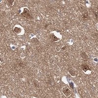
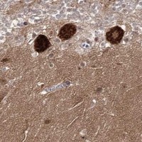
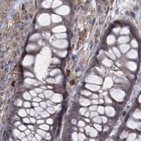
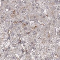

Comprehensive Human Protein Atlas IHC scoring per tissue (reliability: Uncertain — Medium consistency between antibody staining and RNA expression data.). Rows are taken directly from the HPA tissue chart — click any row's HPA link to view the source.
| Tissue | Cell type | Level | Evidence | Source |
|---|---|---|---|---|
| Appendix | Glandular cells | Medium | Protein (IHC) | HPA → |
| Caudate | Neuronal cells | Medium | Protein (IHC) | HPA → |
| Cerebellum | Cells in granular layer | Medium | Protein (IHC) | HPA → |
| Cerebral cortex | Neuronal cells | Medium | Protein (IHC) | HPA → |
| Cervix | Glandular cells | Medium | Protein (IHC) | HPA → |
| Tissue | Cell type | Level | Evidence | Source |
|---|---|---|---|---|
| Adipose tissue | Adipocytes | Not detected | Protein (IHC) | HPA → |
| Bone marrow | Hematopoietic cells | Not detected | Protein (IHC) | HPA → |
| Esophagus | Squamous epithelial cells | Not detected | Protein (IHC) | HPA → |
| Oral mucosa | Squamous epithelial cells | Not detected | Protein (IHC) | HPA → |
| Ovary | Ovarian stroma cells | Not detected | Protein (IHC) | HPA → |
Troubleshoot AP2M1 staining in paraffin sections by checking retrieval, staining controls, cellular distribution and scoring against the available IHC evidence.
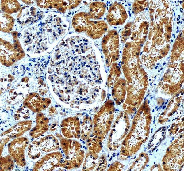
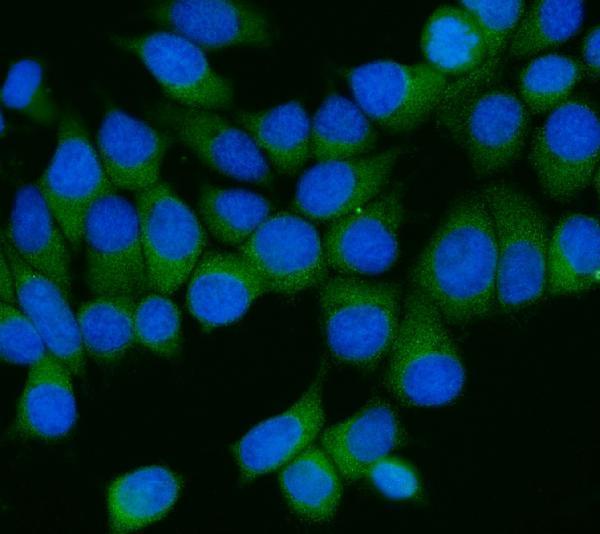
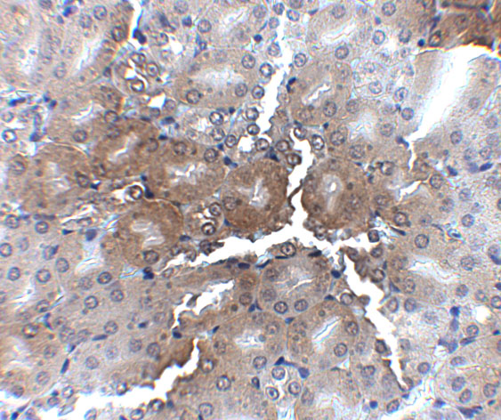
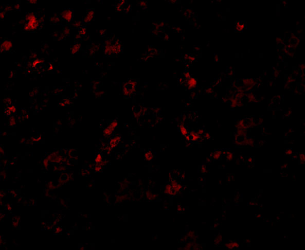
Two anti-AP2M1 antibodies have IHC images from paraffin sections of human or mouse kidney (catalog image captions); IF images show MCF-7 or mouse kidney cells (catalog IF image captions).
A06179-3 shows IHC in a human kidney paraffin section and IF in MCF-7 cells (A06179-3 image captions). A06179-1 shows IHC in mouse kidney tissue and IF in mouse kidney cells (A06179-1 image captions).
Which to pick: For human tissue IHC, choose A06179-3: its paraffin-section caption reports 2 μg/mL, EDTA retrieval at pH 8.0, and DAB detection; the fixative is unreported (A06179-3 IHC caption). For mouse tissue IHC, A06179-1 has a mouse kidney image at 2.5 μg/mL; its caption does not report section processing or fixative (A06179-1 IHC caption). For IF/ICC, A06179-3 lists both applications and shows IF in MCF-7 cells, while A06179-1 lists IF and shows mouse kidney cells; both list human, mouse, and rat reactivity, but their IHC images document only the species stated above (catalog applications, reactivity, and image captions).