This website uses cookies to ensure you get the best experience on our website.
- Table of Contents
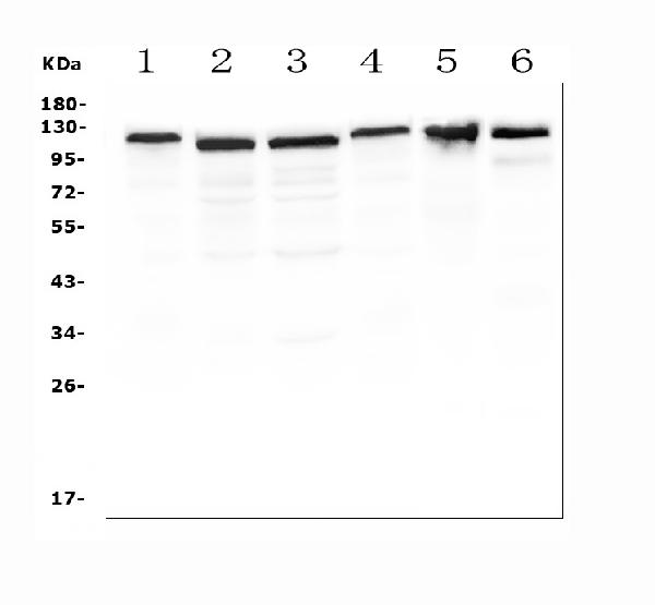
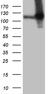
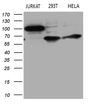
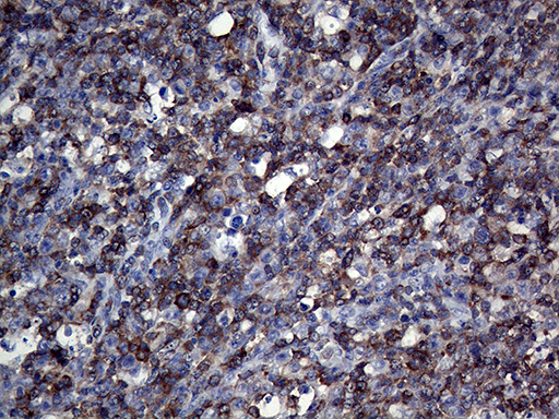
Facts about Amyloid beta A4 precursor protein-binding family B member 1-interacting protein.

Mediates Rap1- induced adhesion. .
| Human | |
|---|---|
| Gene Name: | APBB1IP |
| Uniprot: | Q7Z5R6 |
| Entrez: | 54518 |

| Belongs to: |
|---|
| MRL family |

amyloid beta (A4) precursor protein-binding, family B, member 1 interactingprotein; amyloid beta A4 precursor protein-binding family B member 1-interacting protein; APBB1-interacting protein 1; APBB1IP; INAG1; PREL1; PREL-1; proline rich EVH1 ligand 1; Proline-rich EVH1 ligand 1; Proline-rich protein 73; Rap1-GTP-interacting adapter molecule; Rap1-interacting adaptor molecule; RARP1; Retinoic acid-responsive proline-rich protein 1; RIAM; RIAMRARP-1
Mass (kDA):
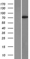
73.183 kDA

| Human | |
|---|---|
| Location: | 10p12.1 |
| Sequence: | 10; NC_000010.11 (26438324..26567879) |
Widely expressed with high expression in thymus, spleen, lymph node, bone marrow and peripheral leukocytes.
Cell membrane; Peripheral membrane protein. Cell projection, lamellipodium. Cell junction, focal adhesion. Cytoplasm, cytoskeleton. Colocalizes with ENA/VASP proteins at lamellipodia tips and focal adhesions, and F-actin at the leading edge. At the membrane surface, associates, via the PH domain, preferentially with the inositol phosphates, PtdIns(5)P and PtdIns(3)P. This binding appears to be necessary for the efficient interaction of the RA domain to Ras-GTPases (By similarity).


PMID: 14530287 by Inagaki T., et al. The retinoic acid-responsive proline-rich protein is identified in promyeloleukemic HL-60 cells.
PMID: 15469846 by Lafuente E.M., et al. RIAM, an Ena/VASP and profilin ligand, interacts with Rap1-GTP and mediates Rap1-induced adhesion.