Click image to see more details
-

-
-
-
-
+3
Product Info Summary
| SKU: | PA1960 |
|---|---|
| Size: | 100 μg/vial |
| Reactive Species: | Human, Mouse, Rat |
| Host: | Rabbit |
| Application: | Flow Cytometry, IF, IHC, IHC-F, ICC, WB |
Customers Who Bought This Also Bought

Product info
Product Name
Anti-RIAM/APBB1IP Antibody Picoband®
SKU/Catalog Number
PA1960
BA3845 is an alternative SKU for this antibody, used in previous lots.
Size
100 μg/vial
Form
Lyophilized
Description
Boster Bio Anti-RIAM/APBB1IP Antibody catalog # PA1960. Tested in Flow Cytometry, IF, IHC, IHC-F, ICC, WB applications. This antibody reacts with Human, Mouse, Rat. The brand Picoband indicates this is a premium antibody that guarantees superior quality, high affinity, and strong signals with minimal background in Western blot applications. Only our best-performing antibodies are designated as Picoband, ensuring unmatched performance.
Storage & Handling
Store at -20˚C for one year from date of receipt. After reconstitution, at 4˚C for one month. It can also be aliquotted and stored frozen at -20˚C for six months. Avoid repeated freeze-thaw cycles.
Cite This Product
Anti-RIAM/APBB1IP Antibody Picoband® (Boster Biological Technology, Pleasanton CA, USA, Catalog # PA1960)
Host
Rabbit
Contents
Each vial contains antibody formulated with stabilizing components, 0.9mg NaCl, 0.2mg Na2HPO4, 0.05mg Thimerosal, 0.05mg NaN3.
*This antibody is supplied in a stabilized formulation.
Compatibility with conjugation reactions depends on the chemistry of the conjugation method used.
For conjugation methods that are not compatible with the stabilizing components present in this formulation, a carrier-free antibody format is required.
Clonality
Polyclonal
Isotype
Rabbit IgG
Immunogen
A synthetic peptide corresponding to a sequence at the C-terminus of human APBB1IP, different from the related rat sequence by one amino acid, and from the related mouse sequence by two amino acids.
Cross-reactivity
No cross-reactivity with other proteins
Reactive Species
PA1960 is reactive to APBB1IP in Human, Mouse, Rat
Observed Molecular Weight
110-120 kDa
Calculated molecular weight
73.2 kDa
Background of APBB1IP
APBB1IP (APBB1-Interacting Protein), also called RIAM or RARP1, is a protein that in humans is encoded by the APBB1IP gene. By genomic sequence analysis, Lafuente et al. (2004) mapped the RIAM gene to chromosome 10p12.1. Using promoter-reporter gene assays, Inagaki et al. (2003) found that RARP1 suppressed transcription from AP1 and SRE sites, but not CRE sites, in all cell lines examined. The proline-rich regions of RARP1 suppressed AP1 transactivation. Lafuente et al. (2004) found that RIAM interacted with profilin and VASP, molecules that regulate actin dynamics, as well as with RAP1-GTP.
Antibody Validation
Boster validates all antibodies on WB, IHC, ICC, Immunofluorescence, and ELISA with known positive control and negative samples to ensure specificity and high affinity, including thorough antibody incubations.
Application & Images
Applications
PA1960 is guaranteed for Flow Cytometry, IF, IHC, IHC-F, ICC, WB Boster Guarantee
Recommend Dilution
| Application | Dilution | Species |
|---|---|---|
| Western blot | 0.1-0.5μg/ml | Human, Mouse, Rat |
| Immunohistochemistry (Paraffin-embedded Section) | 0.5-1μg/ml | Human, Rat |
| Immunohistochemistry (Frozen Section) | 0.5-1μg/ml | Rat |
| Immunocytochemistry | 0.5-1μg/ml | Human |
| Immunocytochemistry/Immunofluorescence | 5μg/ml | Human |
| Flow Cytometry (Fixed) | 1-3μg/1x106 cells | Human |
Tested application
Suggested blocking solution with 5% non-fat milk or BSA; (*)Recommended protein loading: 20-40 µg per lane
Use TE buffer pH 9.0 for antigen retrieval; (*) citrate buffer pH 6.0 is an alternative.
Validation Images & Assay Conditions
Click image to see more details
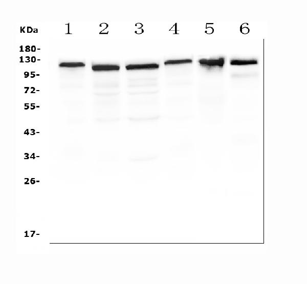
Western blot analysis of APBB1IP using anti-APBB1IP antibody (PA1960 ).
Electrophoresis was performed on a 5-20% SDS-PAGE gel at 70V (Stacking gel) / 90V (Resolving gel) for 2-3 hours. The sample well of each lane was loaded with 50ug of sample under reducing conditions.
Lane 1: human THP-1 whole cell lysates,
Lane 2: Jurkat whole cell lysates,
Lane 3: human Raji whole cell lysates,
Lane 4: U937 whole cell lysates,
Lane 5: rat thymus tissue lysates,
Lane 6: mouse thymus tissue lysates.
After Electrophoresis, proteins were transferred to a Nitrocellulose membrane at 150mA for 50-90 minutes. Blocked the membrane with 5% Non-fat Milk/ TBS for 1.5 hour at RT. The membrane was incubated with rabbit anti-APBB1IP antigen affinity purified polyclonal antibody (Catalog # PA1960 ) at 0.5 μg/mL overnight at 4°C, then washed with TBS-0.1%Tween 3 times with 5 minutes each and probed with a goat anti-rabbit IgG-HRP secondary antibody at a dilution of 1:10000 for 1.5 hour at RT. The signal is developed using an Enhanced Chemiluminescent detection (ECL) kit (Catalog # EK1002) with Tanon 5200 system. A specific band was detected for APBB1IP at approximately 110-120KD. The expected band size for APBB1IP is at 73KD.

Click image to see more details
Anti-RIAM antibody, PA1960, ICC
ICC: JURKAT Cell

Click image to see more details
Anti-RIAM antibody, PA1960, IHC(P)
IHC(P): Human Tonsil Tissue

Click image to see more details
IHC analysis of APBB1IP using anti-APBB1IP antibody (PA1960). APBB1IP was detected in paraffin-embedded section of rat lymphaden. Heat mediated antigen retrieval was performed in citrate buffer (pH6, epitope retrieval solution) for 20 mins. The tissue section was blocked with 10% goat serum. The tissue section was then incubated with 1μg/ml rabbit anti-APBB1IP Antibody (PA1960) overnight at 4°C. Biotinylated goat anti-rabbit IgG was used as secondary antibody and incubated for 30 minutes at 37°C. The tissue section was developed using Strepavidin-Biotin-Complex (SABC)(Catalog # SA1022) with DAB as the chromogen.

Click image to see more details
IHC analysis of APBB1IP using anti-APBB1IP antibody (PA1960). APBB1IP was detected in frozen section of rat spleen. Heat mediated antigen retrieval was performed in citrate buffer (pH6, epitope retrieval solution) for 20 mins. The tissue section was blocked with 10% goat serum. The tissue section was then incubated with μg/ml rabbit anti-APBB1IP Antibody (PA1960) overnight at 4°C. Biotinylated goat anti-rabbit IgG was used as secondary antibody and incubated for 30 minutes at 37°C. The tissue section was developed using Strepavidin-Biotin-Complex (SABC)(Catalog # SA1022) with DAB as the chromogen.

Click image to see more details
IF analysis of APBB1IP using anti-APBB1IP antibody (PA1960).
APBB1IP was detected in immunocytochemical section of A431 cells. Enzyme antigen retrieval was performed using IHC enzyme antigen retrieval reagent (AR0022) for 15 mins. The cells were blocked with 10% goat serum. And then incubated with 5μg/mL rabbit anti-APBB1IP Antibody (PA1960) overnight at 4°C. DyLight®594 Conjugated Goat Anti-Rabbit IgG (BA1142) was used as secondary antibody at 1:100 dilution and incubated for 30 minutes at 37°C. The section was counterstained with DAPI. Visualize using a fluorescence microscope and filter sets appropriate for the label used.

Click image to see more details
Flow Cytometry analysis of SiHa cells using anti-APBB1IP antibody (PA1960).
Overlay histogram showing SiHa cells stained with PA1960 (Blue line). To facilitate intracellular staining, cells were fixed with 4% paraformaldehyde and permeabilized with permeabilization buffer. The cells were blocked with 10% normal goat serum. And then incubated with rabbit anti-APBB1IP Antibody (PA1960, 1μg/1x106 cells) for 30 min at 20°C. DyLight®488 conjugated goat anti-rabbit IgG (BA1127, 5-10μg/1x106 cells) was used as secondary antibody for 30 minutes at 20°C. Isotype control antibody (Green line) was rabbit IgG (1μg/1x106) used under the same conditions. Unlabelled sample without incubation with primary antibody and secondary antibody (Red line) was used as a blank control.
Specific Publications For Anti-RIAM/APBB1IP Antibody Picoband® (PA1960)
Loading publications
Recommended Resources
Here are featured tools and databases that you might find useful.
- Boster's Pathways Library
- Protein Databases
- Bioscience Research Protocol Resources
- Data Processing & Analysis Software
- Photo Editing Software
- Scientific Literature Resources
- Research Paper Management Tools
- Molecular Biology Software
- Primer Design Tools
- Bioinformatics Tools
- Phylogenetic Tree Analysis
Customer Reviews
Have you used Anti-RIAM/APBB1IP Antibody Picoband®?
Share your experimental results or join a short interview to earn up to $1,000 in product credits or other rewards.
0 Reviews For Anti-RIAM/APBB1IP Antibody Picoband®
Customer Q&As
Have a question?
Find answers in Q&As, reviews.
Can't find your answer?
Submit your question
3 Customer Q&As for Anti-RIAM/APBB1IP Antibody Picoband®
Question
I see that the anti-RIAM/APBB1IP antibody PA1960 works with ICC, what is the protocol used to produce the result images on the product page?
Verified Customer
Verified customer
Asked: 2020-03-06
Answer
You can find protocols for ICC on the "support/technical resources" section of our navigation menu. If you have any further questions, please send an email to support@bosterbio.com
Boster Scientific Support
Answered: 2020-03-06
Question
Is this PA1960 anti-RIAM/APBB1IP antibody reactive to the isotypes of APBB1IP?
Verified Customer
Verified customer
Asked: 2019-08-16
Answer
The immunogen of PA1960 anti-RIAM/APBB1IP antibody is A synthetic peptide corresponding to a sequence at the C-terminus of human APBB1IP(647-666aa EQDFMSDLMKALQKKRGNVS), different from the related rat sequence by one amino acid, and from the related mouse sequence by two amino acids. Could you tell me which isotype you are interested in so I can help see if the immunogen is part of this isotype?
Boster Scientific Support
Answered: 2019-08-16
Question
We are currently using anti-RIAM/APBB1IP antibody PA1960 for rat tissue, and we are content with the ICC results. The species of reactivity given in the datasheet says human, mouse, rat. Is it likely that the antibody can work on goat tissues as well?
G. Wu
Verified customer
Asked: 2015-10-12
Answer
The anti-RIAM/APBB1IP antibody (PA1960) has not been tested for cross reactivity specifically with goat tissues, though there is a good chance of cross reactivity. We have an innovator award program that if you test this antibody and show it works in goat you can get your next antibody for free. Please contact me if I can help you with anything.
Boster Scientific Support
Answered: 2015-10-12
