This website uses cookies to ensure you get the best experience on our website.


- Table of Contents
Plan APC chromogenic IHC in paraffin sections using the catalog antibody’s colon section workflow (datasheet M00008). Assess cytoplasmic and membranous staining against cell specific tissue controls (HPA tissue IHC).

Expected localisation, validated protocols, controls and antibodies — the at-a-glance facts below, then the full design guide.
| Expected localisation | Cytoplasmic and membranous tissue staining (HPA tissue IHC) | |
| Staining pattern | Cytoplasmic and membranous; glandular cells often high (HPA tissue IHC) | |
| Antigen retrieval | EDTA pH 8.0 HIER, heat-mediated (datasheet M00008) | |
| Positive control | Appendix+4 more · see all | |
| Negative control | Adipose tissue+4 more · see all |
| Fixation | Keep fixation consistent across sections (standard IHC practice; not target-specific) | |
| Caveat | Staining has medium agreement with RNA expression (HPA tissue IHC) | |
| Regulation | Isoform 1A is brain enriched (UniProt) | |
| Isoform / epitope | 3 isoforms; antibody epitope coverage is unreported (UniProt; datasheet M00008) |
The catalog antibody’s IHC-P protocol (datasheet M00008) is accompanied by published APC staining methods for colorectal carcinoma tissue (PMC10865248) and liver tissue (PMC2757596).
| Sample | Paraffin-embedded human colon tissue; fixative not specified (datasheet M00008) |
| Fixation | Image fixative and duration unreported (datasheet M00008); verify before use. |
| Sectioning | 4–5 µm sections on charged slides (standard) |
| Deparaffinisation | Xylene, graded ethanol series to water (standard) |
| Antigen retrieval | Heat retrieval: EDTA pH 8.0 (datasheet M00008); 20 min, 95–100 °C (standard) |
| Peroxidase block | 3% H2O2, 10 min, room temperature (standard) |
| Blocking | 10% goat serum (datasheet M00008) |
| Primary antibody | Rabbit monoclonal (clone FAD-1) anti-APC, 1:50 (datasheet M00008) |
| Primary incubation | Overnight at 4 °C (datasheet M00008) |
| Detection | HRP-conjugated secondary, DAB chromogen (datasheet M00008) |
| Counterstain | Hematoxylin, blue, dehydrate and mount (standard) |
| Expected result | APC-positive staining in glandular cells of appendix (HPA tissue IHC: High). HPA tissue profile: General cytoplasmic and membranous expression. No signal in the no-primary control. |
In paraffin-section IHC, expect APC staining mainly in the cytoplasm and along cell membranes, including epithelial junctions (HPA: general cytoplasmic and membranous expression; UniProt P25054: adherens junction and cytoplasm). Glandular and respiratory epithelial cells are useful positive populations (HPA: High in appendix glandular cells and bronchus respiratory epithelial cells). APC has no transmembrane segment, so membrane-associated staining does not imply a membrane-spanning protein (UniProt P25054 topology). HPA rates tissue IHC reliability as Supported, with medium consistency against RNA expression (HPA tissue IHC).
| Cytoplasmic staining with a membranous outline in glandular epithelium. | This fits APC's reported tissue pattern (HPA: general cytoplasmic and membranous expression). Appendix, breast, duodenum and gallbladder glandular cells each provide reported High examples (HPA tissue IHC). Judge the pattern within the identified cells; surrounding cells need not share their intensity. |
| Predominantly nuclear staining, with little cytoplasmic or membranous signal. | That is outside the supplied dominant IHC pattern (HPA: general cytoplasmic and membranous expression) and listed UniProt locations (UniProt P25054 subcellular). Treat it as unconfirmed staining; check the same run against a reported positive cell population and the detection controls before assigning it to APC. |
| Strong staining in adipocytes, cardiomyocytes or ovarian follicle cells. | These particular populations were Not detected in the HPA survey (HPA: adipose adipocytes, heart cardiomyocytes, ovary follicle cells). Reproducible signal there merits checks for antibody cross-reactivity or endogenous detection activity. The HPA observations concern those cells, not every cell in each tissue. |
| Diffuse color across tissue, lumens or a section with no primary antibody. | A pattern without cell-specific cytoplasmic or membranous enrichment is less convincing than the reported APC IHC profile (HPA: general cytoplasmic and membranous expression). In a no-primary control, color points to detection background, including possible endogenous activity; this is a general chromogenic IHC control interpretation. |
| No staining in the expected positive cells of a control section. | A blank result in appendix glandular cells or bronchial respiratory epithelium conflicts with their reported High staining (HPA tissue IHC). First verify that the relevant cells are present and the control run worked. HPA's Supported rating still reflects only medium antibody–RNA consistency, so a single negative slide does not establish biological absence (HPA tissue IHC). |
| Tissue and cell selection | Use a named positive cell population when judging the run: appendix glandular cells, bronchial respiratory epithelium, kidney proximal tubule cell bodies and liver cholangiocytes are High (HPA tissue IHC). HPA's Not detected calls are cell-specific and should not be used as whole-organ absence claims (HPA tissue IHC). |
| Compartment and cellular state | UniProt places APC at adherens junctions, the cytoskeleton, lamellipodia and ruffle membranes (UniProt P25054 subcellular). Those locations support some pattern variation, while HPA's tissue-level expectation remains broadly cytoplasmic and membranous (HPA tissue IHC). HGF-associated projection localization is context-dependent, not a required paraffin-section feature (UniProt P25054 subcellular). |
| Isoforms and antibody recognition | UniProt lists isoforms 1A, 2 and 1B; 1A is especially strong in brain, while 1B predominates outside brain (UniProt P25054 tissue specificity). The supplied sources do not identify the catalog antibody's epitope or isoform coverage, so do not infer isoform-specific staining from an IHC result. |
| Evidence and antibody validation | HPA labels tissue IHC Supported with medium staining–RNA consistency (HPA tissue IHC). Its listed rabbit polyclonal HPA013349 has IHC Supported status; HPA013362 has ICC Supported status, with no IHC status supplied (HPA antibodies). These labels support the reported applications but do not establish identical performance for an unspecified catalog antibody. |
| Situation | Likely cause | Next action |
|---|---|---|
| The positive control is blank. | The relevant reported High cell population may be absent, or a general IHC workflow step may have failed (HPA: High in appendix glandular cells). | Confirm the cell population on the section, then review retrieval, primary-antibody use and chromogenic detection against the laboratory's validated IHC workflow (general IHC practice). Target-specific fixation sensitivity is unreported in the supplied sources. |
| All cells have similar diffuse color. | Non-specific staining or detection background may obscure the cell-linked cytoplasmic and membranous pattern (HPA tissue IHC; general IHC practice). | Compare with a no-primary control; review blocking, washes and detection conditions using the assay's established controls (general IHC practice). Avoid interpreting uniform color as APC expression. |
| A reported Not detected cell population stains strongly. | Antibody cross-reactivity or endogenous detection activity is possible; HPA reports adipocytes and cardiomyocytes as Not detected (HPA tissue IHC). | Check a no-primary control and an independently validated APC antibody if available (general IHC practice). Record the exact stained cell type before comparing it with HPA. |
| Only nuclear staining is apparent. | The location disagrees with the supplied cytoplasmic and membranous tissue profile (HPA tissue IHC) and UniProt's listed APC compartments (UniProt P25054 subcellular). | Compare its distribution with a reported High epithelial control, inspect the no-primary control and withhold an APC-positive call until the compartment discrepancy is resolved (general IHC practice). |
| Two tissues give different staining intensities. | Cell populations differ: kidney proximal tubule cell bodies are High, whereas prostate glandular cells are Low (HPA tissue IHC). Isoform expression also varies by tissue (UniProt P25054 tissue specificity). | Score the specified cell populations separately and compare each with its own HPA entry. Do not treat a lower-intensity population as a failed run when a reported High control performs as expected. |
| Q: What if IF/ICC shows a different APC pattern? | HPA reports mainly plasma-membrane localization and additional Golgi localization in ICC-IF (HPA subcellular); imaging format and cells differ from paraffin tissue IHC. | A: Interpret the IF/ICC image against its own controls and the dedicated IF/ICC guide. Do not use the Golgi finding as a required chromogenic tissue-IHC pattern (HPA subcellular; HPA tissue IHC). |


Comprehensive Human Protein Atlas IHC scoring per tissue (reliability: Supported — Medium consistency between antibody staining and RNA expression data.). Rows are taken directly from the HPA tissue chart — click any row's HPA link to view the source.
| Tissue | Cell type | Level | Evidence | Source |
|---|---|---|---|---|
| Appendix | Glandular cells | High | Protein (IHC) | HPA → |
| Breast | Glandular cells | High | Protein (IHC) | HPA → |
| Bronchus | Respiratory epithelial cells | High | Protein (IHC) | HPA → |
| Cerebral cortex | Neuropil | High | Protein (IHC) | HPA → |
| Duodenum | Glandular cells | High | Protein (IHC) | HPA → |
| Tissue | Cell type | Level | Evidence | Source |
|---|---|---|---|---|
| Adipose tissue | Adipocytes | Not detected | Protein (IHC) | HPA → |
| Caudate | Glial cells | Not detected | Protein (IHC) | HPA → |
| Heart muscle | Cardiomyocytes | Not detected | Protein (IHC) | HPA → |
| Hippocampus | Glial cells | Not detected | Protein (IHC) | HPA → |
| Lymph node | Germinal center cells | Not detected | Protein (IHC) | HPA → |
Troubleshoot APC staining in paraffin sections by checking retrieval, cell compartment, antibody epitope, controls, and scoring before interpreting chromogenic signal.
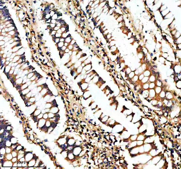
M00008 has a human colon paraffin-section IHC figure (catalog image caption). IF/ICC and human, mouse and rat reactivity are listed, but no IF image is supplied (catalog applications/reactivity; IF image list).
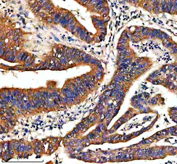
M00008 renders with its human colon paraffin-section IHC figure (catalog image caption). A second caption describes human colon cancer paraffin-section IHC; IF/ICC and mouse/rat reactivity are catalog listings without corresponding images in this payload (catalog image captions; applications/reactivity).
Which to pick: For tissue IHC, choose M00008: its own figure caption documents human colon paraffin-section staining with EDTA retrieval at pH 8.0 and a 1:50 primary dilution (catalog IHC image caption). The fixative is unreported (catalog IHC image caption). For IF/ICC or mouse/rat samples, M00008 is the listed option because it is a rabbit monoclonal clone FAD-1 with those applications and species listed; no IF or mouse/rat IHC image is supplied (catalog host/clone/applications/reactivity; image captions).