recombinant

APC antibody (M00008) performs well in IP, IF, IHC, ICC, WB and shows reactivity with Human, Mouse, Rat.
  • Validated for inter-lot consistency, specificity, and reproducibility
  • Committed to quality since 1993
  • Over 2 Publications
Filter:
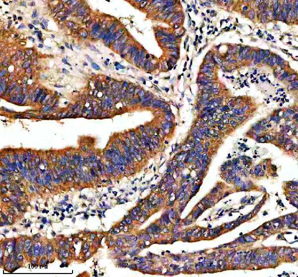
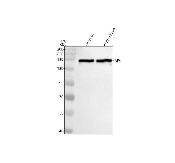
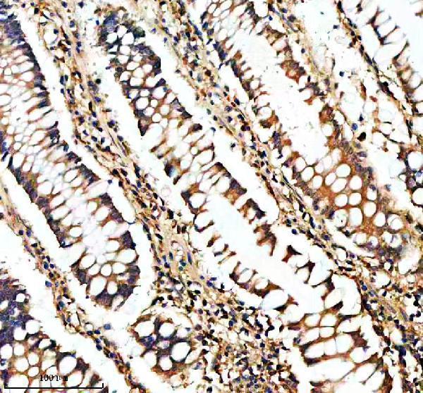
m00008 apc primary antibodies wb testing 1m00008 apc primary antibodies ihc testing 2m00008 apc primary antibodies ihc testing 3

Anti-APC Monoclonal Antibody

APC antibody

Reactive SpeciesHostApplication
Human, Mouse, RatRabbitIP, IF, IHC, ICC, WB

SKU: M00008

Size: 100 μl

Bundle to save: I do not want free secondary ab
Price:
$370/100 μl

In stock. Ships Next Business Day.

Quantity: 1

1
Get A Quote
Guarantee / Product Promise

Tested Applications

APC antibody was positively detected by IHC in:

human colon cancer tissue

Use TE buffer pH 9.0 for antigen retrieval; (*) citrate buffer pH 6.0 is an alternative.

Recommend Dilution

Application
Dilution
Species
WB 1:500-2000
IHC 1:50-200
ICC/IF 1:50-200
IP 1:20

* It is recommended to optimize the amount of this reagent in each experimental system to achieve the best results.

Reactive Data

Filter:
Boster Guarantee:What is this?
WB
IHC
ICC
IF
IP
Human
Recommend
Tested
Recommend
Recommend
Recommend
Mouse
Recommend
Recommend
Recommend
Recommend
Rat
Recommend
Recommend
Recommend
Recommend

Product Information

Product Name
Anti-APC Monoclonal Antibody
View all APC Antibodies
Form
Liquid
Uniprot
Gene ID
Storage & Handling
Store at -20°C for one year. For short term storage and frequent use, store at 4°C for up to one month. Avoid repeated freeze-thaw cycles.
Host
Rabbit
Contents
Rabbit IgG in stabilizing components, phosphate buffered saline, pH 7.4, 150mM NaCl, 0.02% sodium azide and 50% glycerol.
*This antibody is supplied in a stabilized formulation.
Compatibility with conjugation reactions depends on the chemistry of the conjugation method used.
For conjugation methods that are not compatible with the stabilizing components present in this formulation, a carrier-free antibody format is required.
Clonality
Monoclonal
Clone Number
FAD-1
Isotype
Rabbit IgG
Immunogen
A synthesized peptide derived from human APC Tumor suppressor. Promotes rapid degradation of CTNNB1 and participates in Wnt signaling as a negative regulator. APC activity is correlated with its phosphorylation state. Activates the GEF activity of SPATA13 and ARHGEF4. Plays a role in hepatocyte growth factor (HGF) -induced cell migration. Required for MMP9 up-regulation via the JNK signaling pathway in colorectal tumor cells. Acts as a mediator of ERBB2-dependent stabilization of microtubules at the cell cortex.
Reconstitution
Restore with deionized water (or equivalent) for reconstitution volume of 1.0 mL
Observed Molecular Weight
160 kDa
Calculated Molecular Weight

311.6 kDa

Cite This Product
Anti-APC Monoclonal Antibody (Boster Biological Technology, Pleasanton CA, USA, Catalog # M00008)

Protocols

Immunohistochemistry (IHC) protocol for APC antibody

Publications

Application:
microRNA-146a inhibits cancer metastasis by downregulating VEGF through dual pathways in hepatocellular carcinoma
PMID: 25608619
Li M, Tian L, Wang L, Yao H, Zhang J, Lu J, Sun Y, Gao X, Xiao H, Liu M. Plos One. 2013 Oct 23;8(10):E77829. Doi: 10.1371/Journal.Pone.0077829. Ecollection 2013. Down-Regulation Of Mir-129-5P Inhibits Growth And Induces Apoptosis In Laryngeal Squa...
PMID: 24194897

Review

Have you used Anti-APC Monoclonal Antibody?

Submit a review and receive $370 in product credits, a $185 Amazon gift card, or our carefully curated gift bundles.
1 Review For Anti-APC Monoclonal Antibody

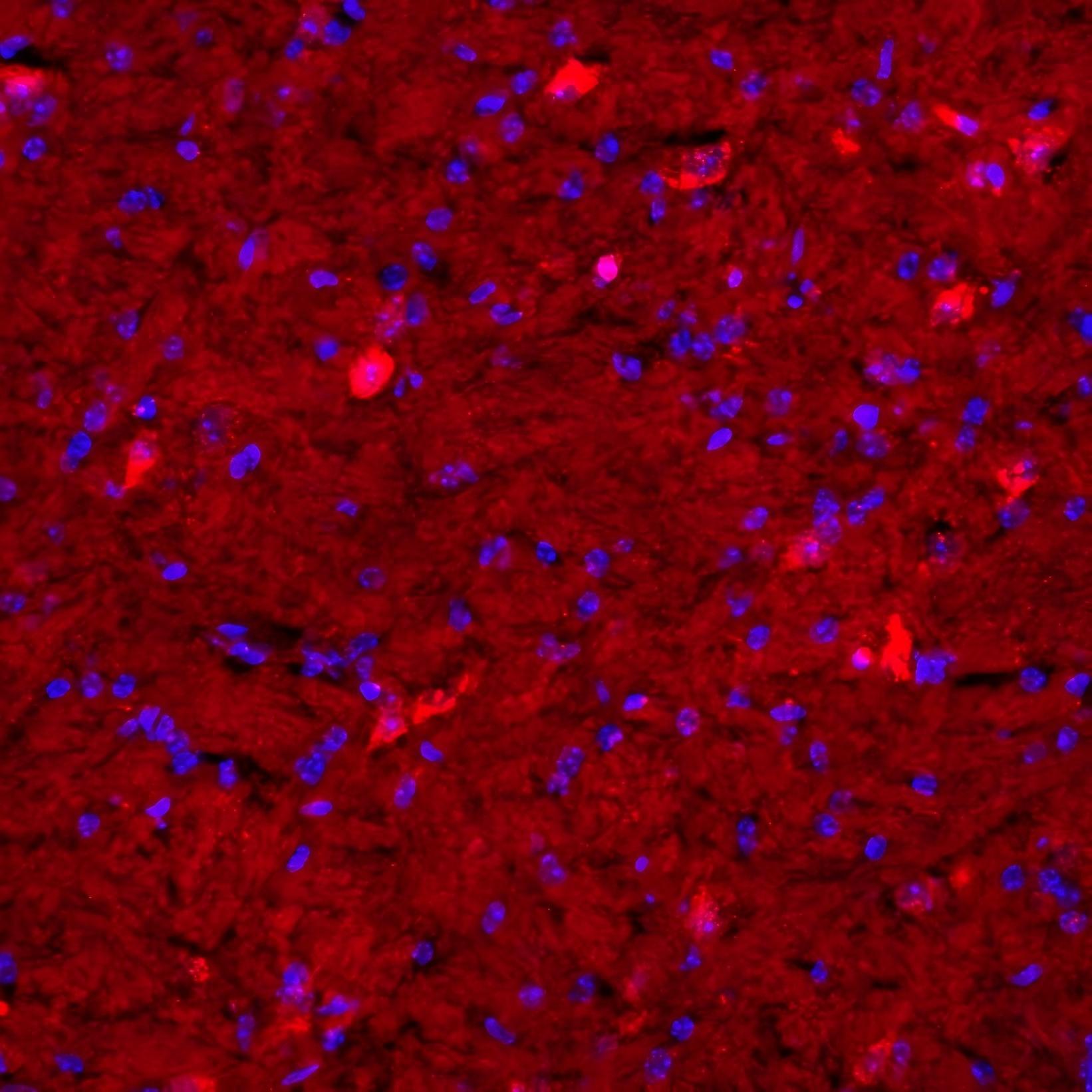
Anti-APC Antibody (M00008, Clone#FAD-1) exhibited high specificity for immunofluorescence detection of APC protein in mouse brain tissue.

SKU M00008
Application Immunofluorescence
Sample mouse brain tissue
Sample Processing Description Mouse heart perfusion was followed by paraformaldehyde fixation for 48 h, brain tissue was frozen and embedded for sectioning, EDTA antigen retrieval was performed for 10 min, and goat serum blocking was performed for 30 min.
Other Reagents EDTA antigen retrieval solution, DAPI staining solution
Primary Antibody APC Monoclonal Antibody
Primary Incubation 1:100, overnight at 4 ℃
Secondary Antibody Fluoro 594-goat anti-mouse (H+L) (Boster BA1141)
Secondary Incubation 37℃, 30 min
Detection Imaging system: OLS5000 laser confocal microscope
Results Summary This image shows the immunofluorescence staining results of mouse brain tissue at 100x magnification. The arrow in the image indicates positive staining for APC protein. APC is a tumor suppressor protein that acts as an antagonist of the Wnt signaling pathway. It participates in cell migration and adhesion, transcriptional activation, and apoptosis. Its expression level is abnormally elevated in apoptotic and tumor cells, and this experimental result verifies this.

Verified customer

Submitted

Product Promise

We promise Anti-APC Monoclonal Antibody performs excellently in relevant applications for IP, IF, IHC, ICC, WB. If it doesn't perform as promised, we will refund the product. Learn more


In stock
Order Product
M00008
Buy one primary antibody get one 0.5ml HRP or Biotin secondary antibody for free.
*Sample sizes are prepared on demand and will take extra lead time. (cannot be conjugated)
$370.00

Troubleshooting

troubleshooting-box-image

Download troubleshooting handbooks for IHC, Western blot and ELISA for FREE.

Download Free PDFs Now

Boster Guarantee

Guaranteed product quality

Guaranteed product quality

We promise all of our products perform as described in datasheets.

This product is for Research Use Only. Not for use in diagnostic procedures.

Update date: Oct 13, 2026