This website uses cookies to ensure you get the best experience on our website.


- Table of Contents
Plan chromogenic paraffin IHC around the ubiquitous nuclear APEX1 staining observed in tissue (HPA tissue IHC). This guide covers fixation, retrieval, antibody titration, controls, and interpretation of possible cytoplasmic signal during regulated nuclear export (UniProt).

Expected localisation, validated protocols, controls and antibodies — the at-a-glance facts below, then the full design guide.
| Expected localisation | Ubiquitous nuclear tissue staining (HPA tissue IHC) | |
| Staining pattern | Nuclear staining across many tissue cell types (HPA tissue IHC) | |
| Antigen retrieval | EDTA pH 8.0 HIER, heat-mediated (datasheet PA1494) | |
| Positive control | Adrenal gland+4 more · see all | |
| Negative control | None in HPA (detected in all 45 tissues); use no-primary + isotype controls |
| Fixation | Keep fixation consistent across sections (standard IHC practice; not target-specific); optimize locally. | |
| Caveat | Nitric oxide can promote cytoplasmic APEX1 staining (UniProt) | |
| Regulation | Nucleolar signal varies with cell cycle (UniProt) | |
| Isoform / epitope | No annotated isoforms; mitochondrial chain lacks aa 2–31 (UniProt) |
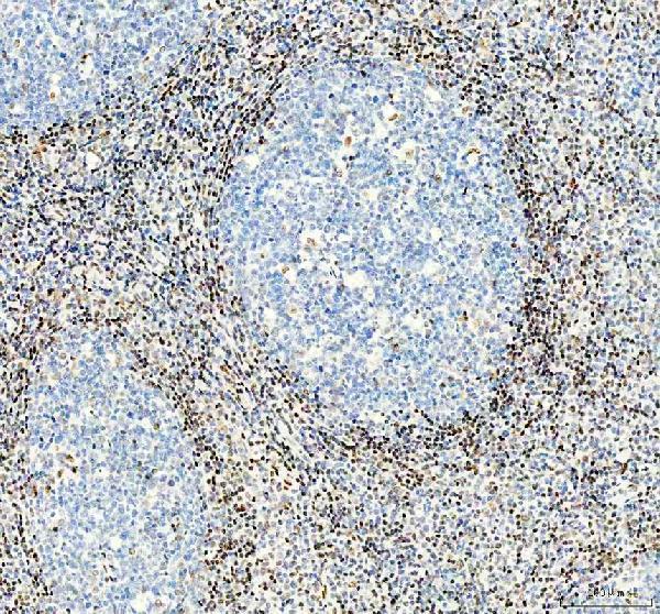
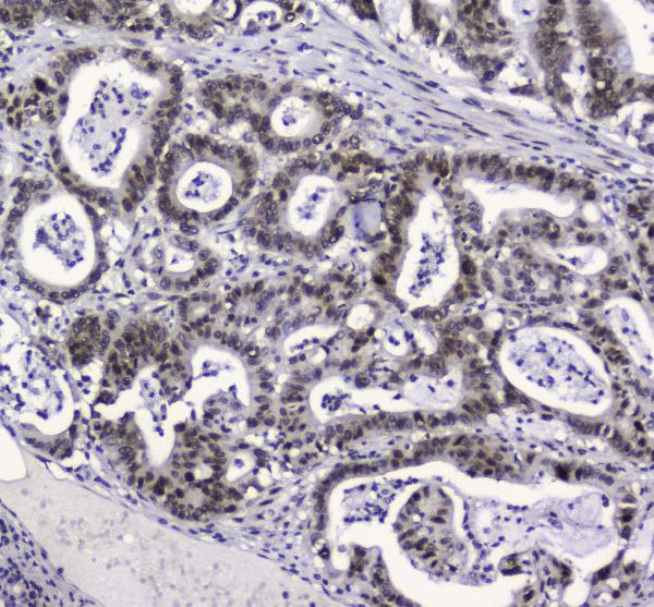
Compare the catalog antibody’s IHC-P protocol (datasheet PA1494) with four published APEX1 tissue IHC examples (PMC12149194; PMC5234431; PMC6723795; PMC4554949).
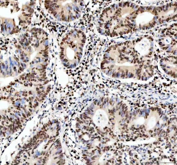
| Sample | Paraffin-embedded human colon adenocarcinoma tissue; fixative not specified (datasheet PA1494) |
| Fixation | Image fixative and duration unreported (datasheet PA1494); verify before use. |
| Sectioning | 4–5 µm sections on charged slides (standard) |
| Deparaffinisation | Xylene, graded ethanol series to water (standard) |
| Antigen retrieval | Heat retrieval: EDTA pH 8.0 (datasheet PA1494); 20 min, 95–100 °C (standard) |
| Peroxidase block | 3% H2O2, 10 min, room temperature (standard) |
| Blocking | 10% goat serum (datasheet PA1494) |
| Primary antibody | Rabbit anti-APEX1, 2-5μg/ml (datasheet PA1494) |
| Primary incubation | Overnight at 4 °C (datasheet PA1494) |
| Detection | HRP-conjugated secondary, DAB chromogen (datasheet PA1494) |
| Counterstain | Hematoxylin, blue, dehydrate and mount (standard) |
| Expected result | APEX1-positive staining in glandular cells of adrenal gland (HPA tissue IHC: High). HPA tissue profile: Ubiquitous nuclear expression. No signal in the no-primary control. |
APEX1 should show widespread nuclear staining in paraffin tissue sections: HPA reports ubiquitous nuclear expression with Supported reliability and high consistency between antibody staining and RNA data (HPA tissue IHC). Expect stronger staining in the listed High cell populations, including adrenal glandular cells and colon endocrine cells (HPA: High). APEX1 has no transmembrane segment; UniProt also lists nucleolar, cytoplasmic, endoplasmic reticulum and mitochondrial localizations (UniProt P27695 topology and subcellular location).
| Nuclear chromogen appears across multiple cell types, with strong signal in an HPA High population. | This fits the reported ubiquitous nuclear profile and provides a useful positive reference (HPA tissue IHC: Supported; HPA: High). Compare nuclei with the counterstain so pigment or tissue deposits are not mistaken for staining (general IHC practice). |
| Strong, widespread cytoplasmic staining dominates while nuclei remain unstained. | That pattern conflicts with the expected tissue IHC profile (HPA tissue IHC: ubiquitous nuclear expression). Cytoplasmic APEX1 can occur under specific conditions, including nitric oxide stimulated nuclear export, so inspect a positive control and detection controls before calling it artefact (UniProt P27695 subcellular location; general IHC practice). |
| The strongest signal is confined to cardiomyocytes or smooth muscle cells, while an HPA High population is negative. | HPA lists cardiomyocytes and smooth muscle cells as Low, so this distribution warrants a specificity check; Low does not mean absent (HPA tissue IHC: Low). Cross-reactivity or endogenous detection activity is possible; compare no-primary and positive tissue controls (general IHC practice). |
| Chromogen is diffuse across cells, stroma or slide areas without a clear nuclear pattern. | This does not support a compartment-specific APEX1 call (HPA tissue IHC: ubiquitous nuclear expression). Check the no-primary control, blocking and detection steps for nonspecific or endogenous signal before changing the biological interpretation (general IHC practice). |
| An HPA High cell population shows no convincing nuclear signal. | Treat the result as an assay failure to investigate, not evidence that APEX1 is absent: HPA reports High staining in specified populations, including endometrial glandular cells (HPA: High). Review the tissue control, antibody conditions, retrieval and detection using the applicable IHC protocol (general IHC practice). |
| Tissue and cell choice | HPA reports Low tissue specificity at the RNA level and ubiquitous nuclear protein staining; its named High and Low cell populations are relative references, not guaranteed positive and negative controls (HPA tissue IHC). |
| Compartment and cell state | Nuclear staining is the tissue IHC expectation (HPA tissue IHC). UniProt also describes nucleolar localization dependent on cell cycle and active rRNA transcription, plus stimulus linked nuclear to cytoplasmic shuttling; do not require either feature in every section (UniProt P27695 subcellular location). |
| Antibody validation | HPA lists IHC as Supported for HPA002564, CAB004294 and CAB047307; HPA000956 has no listed IHC status (HPA antibodies). Validation status supports selecting an IHC reagent, but the section and controls still determine whether a particular stain is interpretable (general IHC practice). |
| Topology and processing | APEX1 has no transmembrane segment or signal sequence, and UniProt lists protein chains beginning at residues 2 and 32 (UniProt P27695 topology and processing). These annotations do not establish an extracellular staining pattern or predict whether a particular antibody recognizes both chains. |
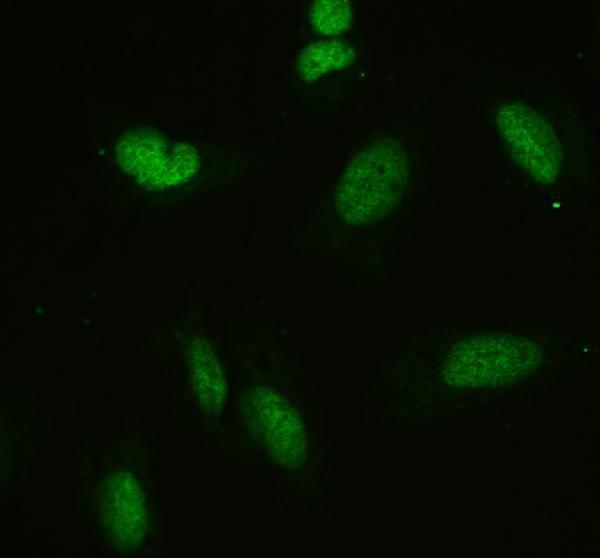
| IF/ICC Q&A: where should signal appear? | Mainly in the nucleoplasm, with an additional centrosome location (HPA subcellular ICC-IF: nucleoplasm Supported; centrosome Approved). This is an IF/ICC observation, not a paraffin IHC requirement; the IF/ICC guide covers its own workflow. |
| Situation | Likely cause | Next action |
|---|---|---|
| The expected nuclear signal is absent in an HPA High cell population. | The section or assay may have failed; HPA High describes an observed cell population, not a guarantee for every specimen (HPA tissue IHC). | Confirm that the cells are present, examine a known-positive section, then review the catalog antibody's IHC-P conditions, retrieval and detection (general IHC practice). |
| Staining is predominantly cytoplasmic and nuclear staining is weak or absent. | This departs from the usual tissue profile, although UniProt documents regulated nuclear-cytoplasmic shuttling (HPA tissue IHC; UniProt P27695 subcellular location). | Check a positive tissue and no-primary control, and assess whether the pattern is cell-specific and reproducible before assigning biological meaning (general IHC practice). |
| Only HPA Low cells stain strongly. | Cell distribution differs from the HPA reference; cross-reactivity or endogenous detection activity may contribute (HPA tissue IHC: Low; general IHC practice). | Compare an HPA High cell population in parallel and inspect no-primary and detection controls; Low is a relative category, not a required blank (HPA tissue IHC; general IHC practice). |
| Background obscures nuclear boundaries. | Nonspecific antibody binding or detection-system background can prevent compartment scoring (general IHC practice). | Inspect no-primary controls, blocking, antibody dilution and wash steps; score APEX1 only where nuclei can be distinguished from background (general IHC practice; HPA tissue IHC: nuclear profile). |
| Signal appears as coarse deposits rather than cellular staining. | Chromogen precipitate or endogenous detection activity may mimic a positive stain (general IHC practice). | Use the counterstain and no-primary control to distinguish deposits from nuclear signal, then review the detection reagents and staining steps (general IHC practice). |
| A paraffin section lacks the centrosome pattern described for IF/ICC. | The additional centrosome location comes from ICC-IF, while the tissue IHC profile is nuclear (HPA subcellular ICC-IF; HPA tissue IHC). | Judge the paraffin result by nuclear staining in appropriate cells; use the separate IF/ICC guide when assessing subcellular IF images (HPA tissue IHC; HPA subcellular ICC-IF). |


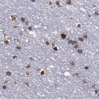

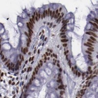


Comprehensive Human Protein Atlas IHC scoring per tissue (reliability: Supported — High consistency between antibody staining and RNA expression data.). Rows are taken directly from the HPA tissue chart — click any row's HPA link to view the source.
| Tissue | Cell type | Level | Evidence | Source |
|---|---|---|---|---|
| Adrenal gland | Glandular cells | High | Protein (IHC) | HPA → |
| Appendix | Endocrine cells | High | Protein (IHC) | HPA → |
| Breast | Adipocytes | High | Protein (IHC) | HPA → |
| Cerebellum | GLUC cells - nucleus | High | Protein (IHC) | HPA → |
| Colon | Endocrine cells | High | Protein (IHC) | HPA → |
| Tissue | Cell type | Level | Evidence | Source |
|---|---|---|---|---|
| None in HPA: APEX1 is detected in all 45 scored tissues. Use a no-primary (secondary-only) and an isotype control instead. | ||||
Use the documented PA1494 paraffin-section conditions as the IHC starting point, then evaluate nuclear staining, compartment shifts, and tissue background against matched controls.
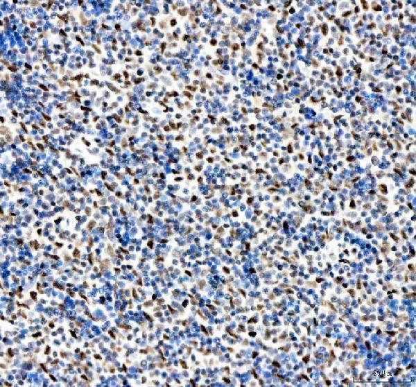
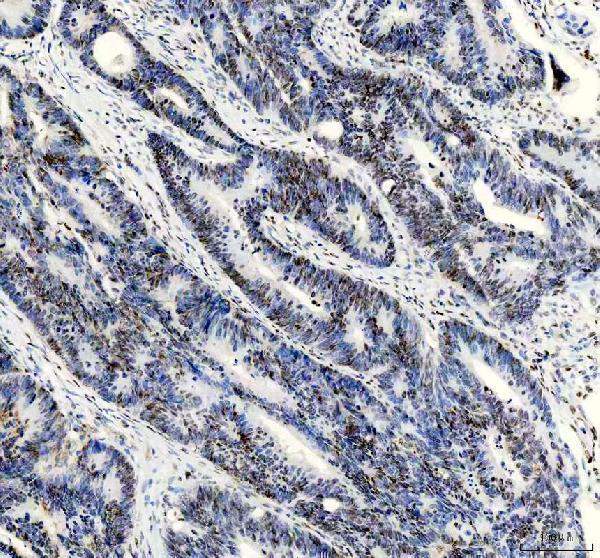
Catalog antibodies have paraffin-section IHC images from human, mouse and rat tissues (IHC captions: PA1494, M00627-3, PB9128); PA1494 and PB9128 also have cell and tissue IF images (IF captions).
PA1494 shows IHC in human colon adenocarcinoma, spleen and prostatic cancer and mouse liver; M00627-3 shows IHC in human colon cancer and tonsil, mouse liver and rat liver (IHC captions: PA1494, M00627-3). PB9128 shows IHC in human lung cancer and mouse and rat brain, while M00627 shows IHC in human intestinal cancer (IHC captions: PB9128, M00627).
Which to pick: For paraffin-section IHC across human, mouse and rat, choose rabbit monoclonal M00627-3 if clonality matters; its IHC captions cover all 3 species and describe EDTA retrieval at pH 8.0 (catalog: clone 17A44; IHC captions: M00627-3). For IF/ICC, choose PA1494 for its human cell and human and rat tissue IF images, or PB9128 for its human cell and tissue IF images (IF captions: PA1494, PB9128). PB9128 also has paraffin-section IHC images across human, mouse and rat with citrate retrieval at pH 6; the IHC captions report paraffin embedding but do not report the fixative (IHC captions: PB9128).