This website uses cookies to ensure you get the best experience on our website.


- Table of Contents
Real validated APLN Western blot protocols, expected-band and isoform facts, troubleshooting for weak or shifted signal, and recommended anti-APLN WB antibodies. Everything you need to plan the experiment before you commit precious samples.
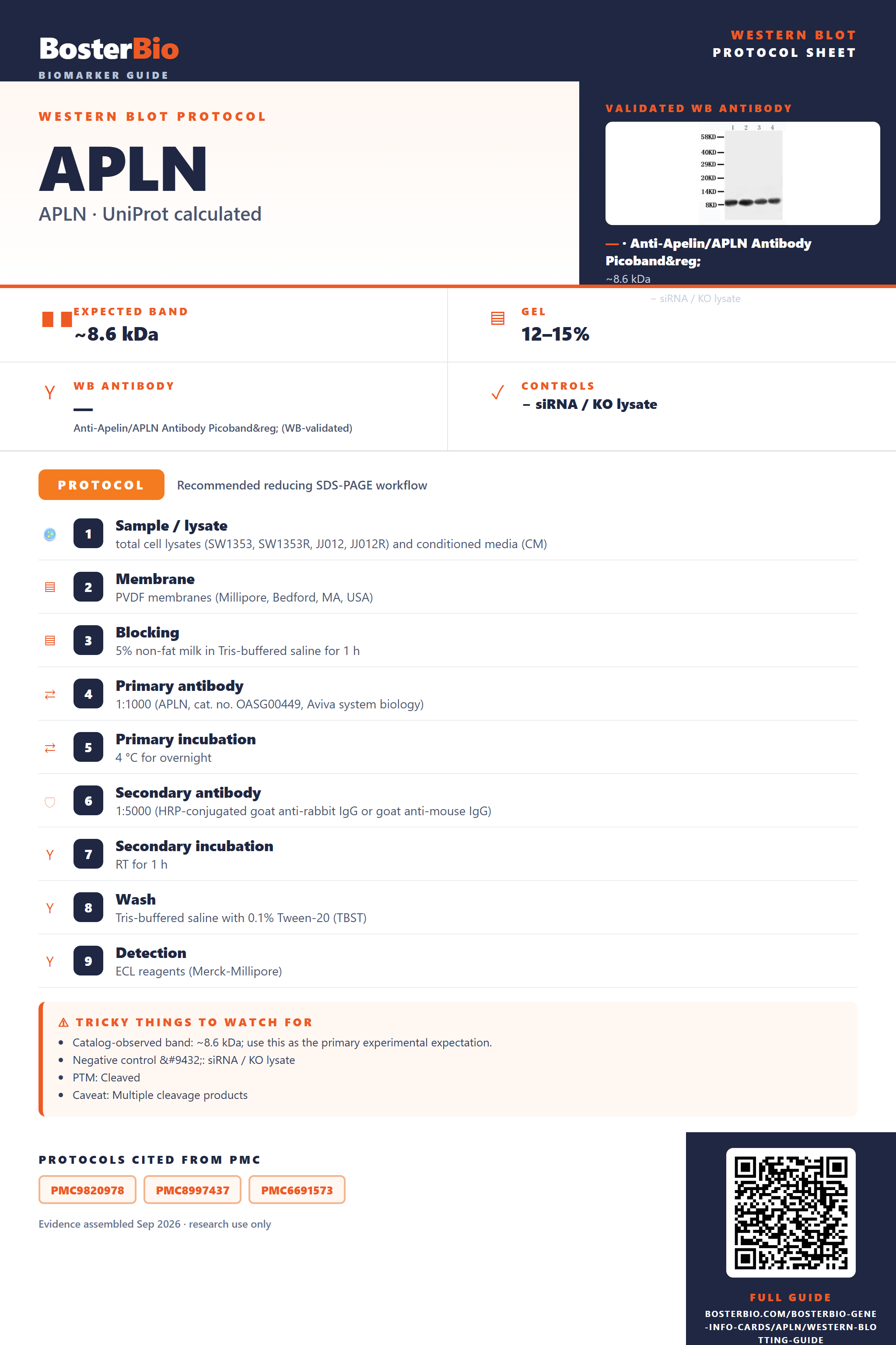
Expected bands, validated protocols, controls and antibodies — the at-a-glance facts below, then the full design guide.
| Expected band | ~8.6 kDa | |
| Gel | 12–15% | |
| Negative control | siRNA / KO lysate |
| PTM | Cleaved | |
| Caveat | Multiple cleavage products | |
| Regulation | LPS-suppressed | |
| Isoform | 1 isoform(s) |
Literature-validated Western blot parameters for APLN — gel percentage, transfer, blocking, antibody incubation and detection, extracted from published methods.
| Sample / lysate | total cell lysates (SW1353, SW1353R, JJ012, JJ012R) and conditioned media (CM) |
| Membrane | PVDF membranes (Millipore, Bedford, MA, USA) |
| Blocking | 5% non-fat milk in Tris-buffered saline for 1 h |
| Primary antibody | 1:1000 (APLN, cat. no. OASG00449, Aviva system biology) |
| Primary incubation | 4 °C for overnight |
| Secondary antibody | 1:5000 (HRP-conjugated goat anti-rabbit IgG or goat anti-mouse IgG) |
| Secondary incubation | RT for 1 h |
| Wash | Tris-buffered saline with 0.1% Tween-20 (TBST) |
| Detection | ECL reagents (Merck-Millipore) |
| Exposure / imaging | autoradiography |
Apelin's 8.6 kDa preproapelin precursor is cleaved at its signal peptide and propeptide to release a much smaller mature secreted peptide.
| Single band near 8-9 kDa | Represents the unprocessed preproapelin precursor before signal peptide and propeptide removal |
| Faint or diffuse band below 5 kDa | Corresponds to the small mature apelin peptide left after signal peptide and propeptide cleavage |
| No detectable band in whole-cell lysate | Apelin is secreted into extracellular space, so intracellular retention is minimal and conditioned media or secretome samples are needed |
| No shift between reducing and non-reducing conditions | No disulfide bonds are annotated for apelin, so band position should not depend on reducing agent |
| No additional bands at other stable molecular weights | Only one isoform is annotated, so multiple distinct bands from alternative splicing are not expected |
| Extra faint bands smaller than the mature peptide | The pair-of-basic-residues cleavage site allows further internal proteolytic processing into shorter bioactive fragments |
| Predicted precursor mass (8.6 kDa, 77 aa) | Sets the baseline size for the full-length unprocessed preproapelin band |
| Signal peptide cleavage (residues 1-22) | Removes the signal sequence upon secretion, shifting the protein below the precursor mass |
| Propeptide cleavage (residues 23-41) | Further removes the propeptide segment, producing a much smaller mature apelin band around 4 kDa |
| Cleavage on pair of basic residues | Enables additional internal proteolytic processing that can generate several shorter bioactive fragments below the mature peptide size |
| Secreted, extracellular localization | Limits detectable signal in whole-cell lysates and favors detection in conditioned media or secretome preparations |
| Single annotated isoform | No alternative splice forms are expected to add extra bands at different masses |
| Situation | Likely cause | Next action |
|---|---|---|
| No band in lysate | Apelin is secreted, so intracellular pools are low and most protein is released into the extracellular space | Test conditioned media, serum, or secretome-enriched fractions instead of only whole-cell lysate |
| Weak or no signal | The mature apelin peptide is very small (around 4 kDa or less), which can run off standard gels or transfer poorly to membrane | Use a high-percentage or peptide-optimized gel, a low molecular weight transfer protocol, and a low-porosity membrane suited to small peptides |
| Band lower than expected | Signal peptide and propeptide removal generates the mature apelin peptide well below the 8.6 kDa precursor mass | Confirm the expected mature peptide size (around 4 kDa) rather than assuming the full precursor mass |
| Fragments below expected size | The pair-of-basic-residues cleavage motif allows further internal proteolytic processing into shorter bioactive apelin fragments | Use fresh samples with protease inhibitors, and note that multiple short fragments can be biologically genuine rather than degradation |
| Multiple bands | Both the unprocessed preproapelin precursor and the cleaved mature peptide can be present in the same sample | Compare band sizes to the precursor (about 8.6 kDa) and mature (about 4 kDa) forms to distinguish processing intermediates from nonspecific bands |
| Band higher than expected | Incomplete processing leaves uncleaved preproapelin near the full 8.6 kDa predicted mass instead of the smaller mature peptide | Check whether the antibody epitope lies within the mature peptide or the propeptide region to determine which form is being detected |

Comprehensive Human Protein Atlas IHC scoring per tissue. Rows are taken directly from the HPA tissue chart — click any row's HPA link to view the source.
| Tissue | Cell type | Level | Evidence | Source |
|---|
| Tissue | Cell type | Level | Evidence | Source |
|---|
Deeper troubleshooting and optimisation questions for APLN, answered from its protein features.
BosterBio's APLN antibodies are among the best-performing WB antibodies on the market — well cited, thoroughly validated, and orthogonally cross-validated against negative tissues and complementary methods.
For APLN Western blots, Boster's PA1501 anti-Apelin antibody is the recommended choice, backed by an actual published blot showing specific, reproducible detection across multiple human cell lysates at a defined working concentration for dependable, well-validated results.
Which to pick: Only one APLN antibody is catalogued: PA1501. It comes with a real WB validation image (U87, MCF-7, and HeLa lysates at 0.5 μg/ml, 40 μg protein/lane), so it's the default and only option to select for this target.