This website uses cookies to ensure you get the best experience on our website.
- Table of Contents
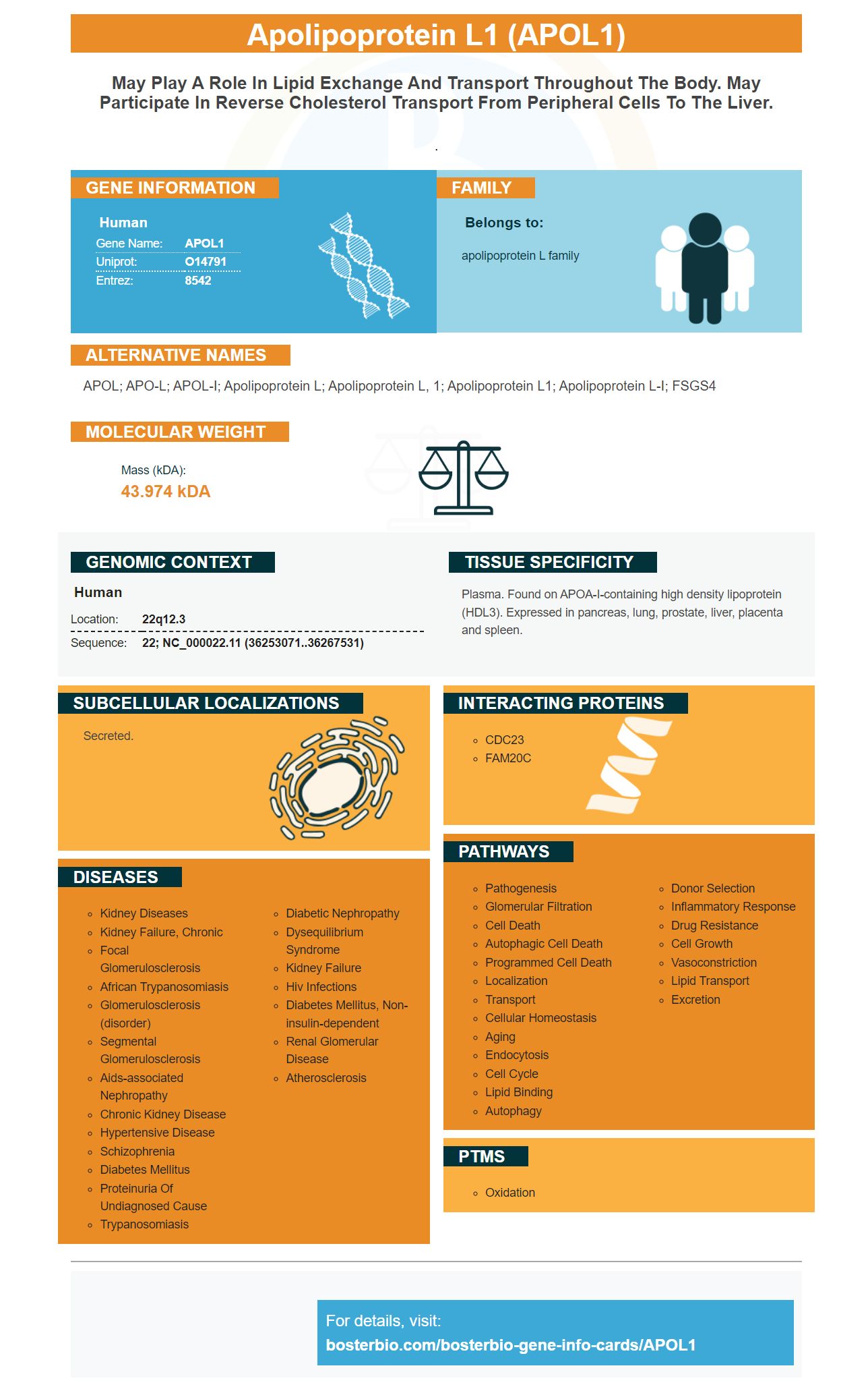
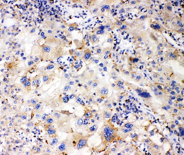
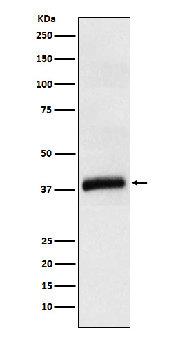
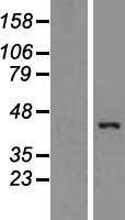
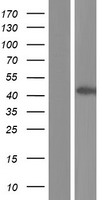
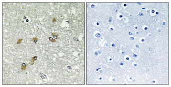
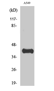
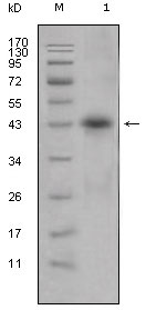
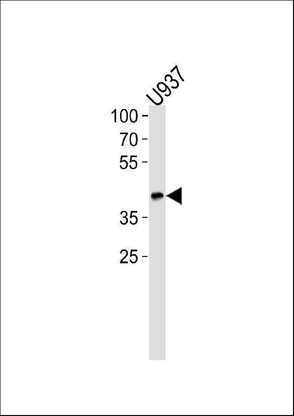
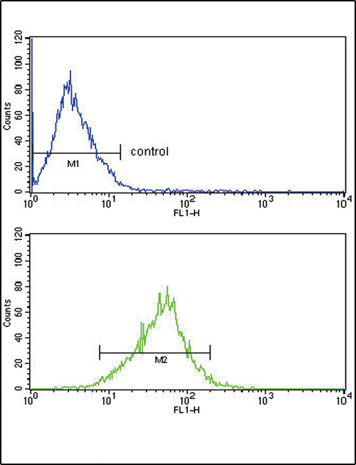
Facts about Apolipoprotein L1.
.
Human | |
---|---|
Gene Name: | APOL1 |
Uniprot: | O14791 |
Entrez: | 8542 |
Belongs to: |
---|
apolipoprotein L family |
APOL; APO-L; APOL-I; Apolipoprotein L; apolipoprotein L, 1; apolipoprotein L1; Apolipoprotein L-I; FSGS4
Mass (kDA):
43.974 kDA
Human | |
---|---|
Location: | 22q12.3 |
Sequence: | 22; NC_000022.11 (36253071..36267531) |
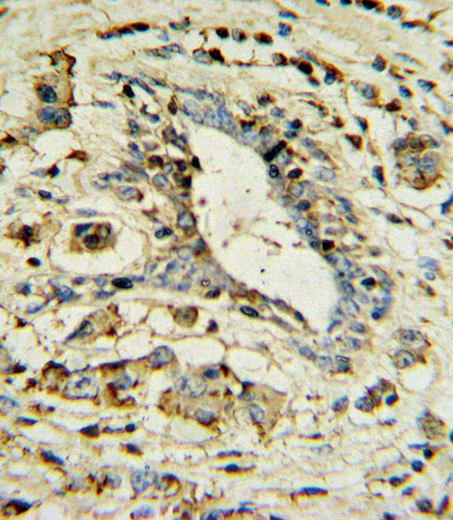
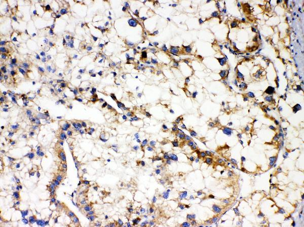
Plasma. Found on APOA-I-containing high density lipoprotein (HDL3). Expressed in pancreas, lung, prostate, liver, placenta and spleen.
Secreted.
Many applications are possible for the APOL1 genetic code in the diagnosis and treatment of disease. This article will discuss the APOL1 genotype and its association with pathologic features. The following articles will help you learn more. Then you'll be well-prepared to find the best uses possible of the APOL1 genome. In the meantime, feel free to contact us for more information.
Although there is not much evidence to support APOL1 mediated kidney disease, this variant protects against renal failure. The expression of APOL1-G1 in hepatocellular carcinoma cells causes cytotoxicity, and the gene can also inhibit p38 MAPK signaling. However, APOL1G2 expression in the kidneys is less efficient than in other organs.
APOL1 variations have been shown to increase the risk for non-diabetic diseases such as kidney disease. The APOL1 G1 version codes for two substitutions of amino acids, while APOL1 G2 has a deletion of six bases. The latter mutation results in the loss 388N and 389Y amino acids residues.
When transiently expressed, the APOL1–G0 gene causes a blockage to autophagy of HEK293 cells. Autophagy inhibitors didn't completely block APOL1–G0's autophagy activity. The APOL1 genotype induced morphological deterioration in Xenopus oocytes, but preventing autophagy-independent morphotoxicity was ineffective.
APOL1 is primarily expressed on HDL particles. Among African populations, it confers resistance against trypanosomiasis. It kills trypanosomes by lysing them. However, some subspecies have developed resistance against APOL1-dependent killing. APOL1G0 may not be as effective in HIV-associated kidney disease treatment.
Although the APOL1–G0 toxin is cytotoxic in mammalian cells it is not yet known how it kills parasites. This toxin has homology with other bacteria-pore-forming chemicals. Its cytotoxicity is dependent on channel activity and is associated with increased cell swelling. It can also trigger trypanolysis.
We have shown that APOL1 mutations are associated in Africa with the endemicity T. b. rhodesiense infections. We genotyped 184 T.b. cases that had been clinically confirmed. We genotyped 184 T.b. clinically confirmed cases and 180 controls to the G1 haplotype. This haplotype contains two nonsynonymous substitutions in APOL1 (rs73885319 and rs60910145) that are situated 128 bp apart. The variant is in near perfect linkage equilibrium.
Patients with the G1 Haplotype did not participate in the study. Because they did not have a full G1 genotype, patients with partial G1 haplotypes were excluded from the study. The complete G1 haplotype was found in the carrier group. It was found that 30 individuals (Raw–G1M) were carriers. One individual was incorrectly labeled G1M/G1.
This study involved 98 African American patients without HIVAN, and focuses on APOL1 risk alleles. Participants were assessed for their age as well as their sex and use of illicit drugs. They also had to be evaluated for kidney function. 29 patients reached ESRD after 310 year of follow-up. Individuals with two APOL1 risks were nearly three times more at risk of ESRD compared to those with zero risk.
PCR/RFLP analysis was used in determining the APOL1 genome. The G1 variant includes two non-synonymous substituts and is near perfect in linkage disequilibrium. The G2 version is a 2 amino acid in-frame delete. Prepared FTA cards discs were used to create templates for PCR. Three primers were used in order to amplify a product with 458 bp.
Retrospective analysis of APOL1 genetics in African populations revealed that there were associations between APOL1 G1 variants and susceptibility two different forms African trypanosomiasis. T.b.rhodesiense can be found in East Africa while T.gambiense can be found in West Africa.
The association between APOL1 G1 and G2 risk variants and human African trypanosomiasis suggests that the two haplotypes have more complex relationships than previously thought. These findings have implications both for individuals and subspecies T.b. gambiense. We also discuss both G1- and G2-related alleles in the selection variants associated with APOL1 Kidney Disease.
This study used TaqMan to genotype the APOL1 genome. The G1 variant is composed of two missense variations and the G2 variant is a six-bp In-frame deletion. These alleles have been called risk alleles. High-risk individuals have at least two risk alleles. Individuals with a single risk allele were classified as low-risk.
Despite being closely associated with preeclampsia during pregnancy, the APOL1 gene of the mother was not found to be associated with the fetal genome. Preeclampsia is associated with higher chances of fetal liver disease in women who have other maternal conditions. The APOL1 gene may help to predict this risk. This study could lead to early intervention to improve the health of fetal kidneys.
Different epigenetic DNA methylation levels are also associated in the APOL1 gene promor. This is evident in AFR carriers born in Africa and the UK. Moreover APOL1 methylation can be correlated with the presence CKD for AFR carriers. A higher methylation level for the APOL1 gene enhancer could also prevent transcriptional silencing. However, this information is not yet accessible for CKD patients and kidney tissue.
PMID: 9325276 by Duchateau P.N., et al. Apolipoprotein L, a new human high density lipoprotein apolipoprotein expressed by the pancreas. Identification, cloning, characterization, and plasma distribution of apolipoprotein L.
PMID: 11290834 by Duchateau P.N., et al. Apolipoprotein L gene family: tissue-specific expression, splicing, promoter regions; discovery of a new gene.