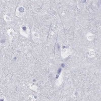
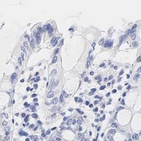
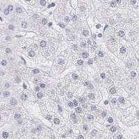
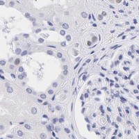
AR / Androgen receptor · IHC design guide

Design Immunohistochemistry for AR

Plan AR staining in paraffin sections around the nuclear pattern reported in reproductive tissues (HPA tissue IHC). The guide covers fixation, controls and interpretation of ligand-dependent nuclear localisation (UniProt).

Evidence assembled Sep 2026 · For research use; verify linked source records and product datasheet before use
Immunohistochemistry protocol sheet for AR (IHC for AR): expected localisation Predominantly nuclear in tissue; cytoplasmic AR is possible (HPA tissue IHC; UniProt), antibody M00542-1, validated IHC image, and IHC protocol steps
Printable AR IHC protocol sheet — expected localisation Predominantly nuclear in tissue; cytoplasmic AR is possible (HPA tissue IHC; UniProt), antibody M00542-1, controls and protocol steps. Open the full AR IHC guide →

AR Immunohistochemistry Experimental Design Guide

Expected localisation, validated protocols, controls and antibodies — the at-a-glance facts below, then the full design guide.

Must know before staining
Expected localisation Predominantly nuclear in tissue; cytoplasmic AR is possible (HPA tissue IHC; UniProt)
Staining pattern Nuclear staining in reproductive glandular cells (HPA tissue IHC)
Antigen retrieval Tris-EDTA pH 9.0 HIER, 95–98 °C, 20 min (rule: nuclear antigen)
Positive control ⓘ Epididymis+4 more · see all
Negative control ⓘ Adipose tissue+4 more · see all
Important caveats
Reasons your staining may differ from the expected pattern.
Fixation Keep formalin fixation consistent; prolonged fixation may mask epitopes (standard IHC practice)
Caveat Ligand binding can shift AR from cytoplasm to nucleus (UniProt)
Regulation Androgen drives nuclear accumulation (UniProt)
Isoform / epitope 4 isoforms; validate antibody epitope coverage (UniProt)
Section 1

Recommended AR IHC & IF Protocols

The catalog antibody’s IHC-P protocol (datasheet) is paired with a published AR method for FFPE breast cancer biopsies (PMC13515587).

Recommended immunohistochemistry (IHC-P) protocol parameters
SampleFFPE tissue sections (standard)
Fixation10% neutral-buffered formalin, 24 h, standard processing (standard)
Sectioning4–5 µm sections on charged slides (standard)
DeparaffinisationXylene, graded ethanol series to water (standard)
Antigen retrievalHeat-induced epitope retrieval in Tris-EDTA buffer, pH 9.0, 20 min at 95–98 °C (standard rule: nuclear antigen)
Peroxidase block3% H2O2, 10 min, room temperature (standard)
Blocking10% normal serum of the secondary host, 30 min, room temperature (standard)
Primary antibodyRabbit monoclonal (clone HD-1) anti-AR, 1:50-1:100 (datasheet M00542-1)
Primary incubationOvernight at 4 °C (standard)
DetectionHRP-polymer secondary, DAB chromogen 5–10 min (standard)
CounterstainHematoxylin, blue, dehydrate and mount (standard)
Expected resultAR-positive staining in glandular cells of epididymis (HPA tissue IHC: High). HPA tissue profile: Nuclear expression in several tissues, mainly in reproductive organs. No signal in the no-primary control.
💡Decision noteStart with Tris-EDTA pH 9.0 HIER at 95–98 °C for 20 min (page retrieval; UniProt P10275: nuclear AR). The published method gives no retrieval conditions (PMC13515587).
Section 2

What Is the Expected AR Staining Pattern?

AR should show nuclear staining in reproductive tissues, especially epididymal and seminal vesicle glandular cells (HPA: Enhanced; High in both). Unligated AR may also be cytoplasmic (UniProt P10275: localization; no transmembrane segment).

What am I looking at on my slide?
Strong glandular nuclear stainExpected in epididymis or seminal vesicle (HPA: High; nuclear profile).
Membrane-only stainSuspect artifact (UniProt P10275: no transmembrane segment).
Stained adipocytesConsider cross-reactivity or endogenous activity (HPA: adipocytes not detected).
Diffuse stain across the sectionBackground may obscure nuclear scoring (standard IHC practice).
No stain in epididymal glandsCheck assay sensitivity (HPA: High in epididymal glandular cells).
💡Expected AR appearanceCall positive when glandular nuclei stain strongly in epididymis or seminal vesicle (HPA: High; nuclear profile); membrane-only stain is suspect (UniProt P10275: no transmembrane segment).
How each factor affects the staining
Fixation and cross-linkingCan mask AR epitopes and reduce nuclear signal (standard IHC practice).
Antigen retrievalCan restore masked epitopes; excessive retrieval can damage morphology (standard IHC practice).
Ligand stateUnligated AR is mainly cytoplasmic; ligand binding moves it to nuclei (UniProt P10275).
IsoformsAR has 4 isoforms; epitope coverage may differ (UniProt P10275).
Endogenous peroxidaseMay create false chromogen signal unless blocked (standard IHC practice).
Why is my staining missing, weak or wrong?
SituationLikely causeNext action
Weak nuclear stain in epididymisEpitope masking after fixation (standard IHC practice).Optimize retrieval and check a positive control (HPA: epididymal glands High).
No signal in known-positive glandsInsufficient detection or excessive fixation (standard IHC practice).Check antibody dilution, retrieval and detection reagents (standard IHC practice).
Membrane-only stainNonspecific staining is plausible (UniProt P10275: no transmembrane segment).Review controls and staining distribution (standard IHC practice).
Chromogen in adipocytesEndogenous activity or cross-reactivity (HPA: adipocytes not detected).Check reagent-only control and peroxidase block (standard IHC practice).
Diffuse section backgroundInadequate blocking or excessive antibody (standard IHC practice).Adjust blocking, dilution and washes (standard IHC practice).
IF/ICC shows cytosolic AR?Cytosol is supported in ICC-IF (HPA: subcellular); unligated AR can be cytoplasmic (UniProt P10275).Interpret with ligand state and nuclear counterstain (UniProt P10275; standard IF practice).

Sample controls for AR IHC & IF

🧪Run epididymis first: its glandular cells should stain (HPA: High in epididymis glandular cells); use adipose tissue as the negative tissue (HPA: Not detected in adipocytes). On the epididymis slide, compare glandular cells with surrounding non-glandular areas for background, without assuming those cells are AR-negative (HPA: glandular cells are the annotated positive population).
Positive control tissue: Epididymis (Glandular cells, HPA High)
Negative control tissue: Adipose tissue (HPA Not detected)
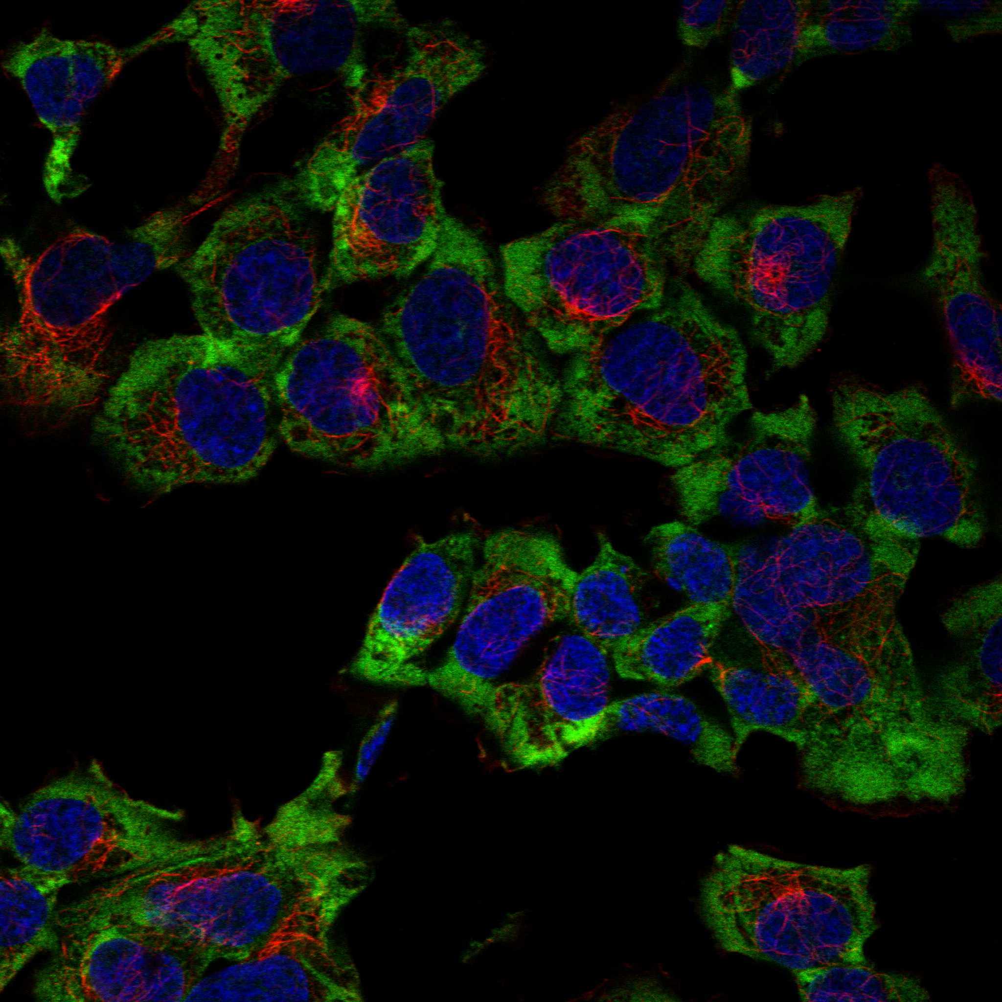
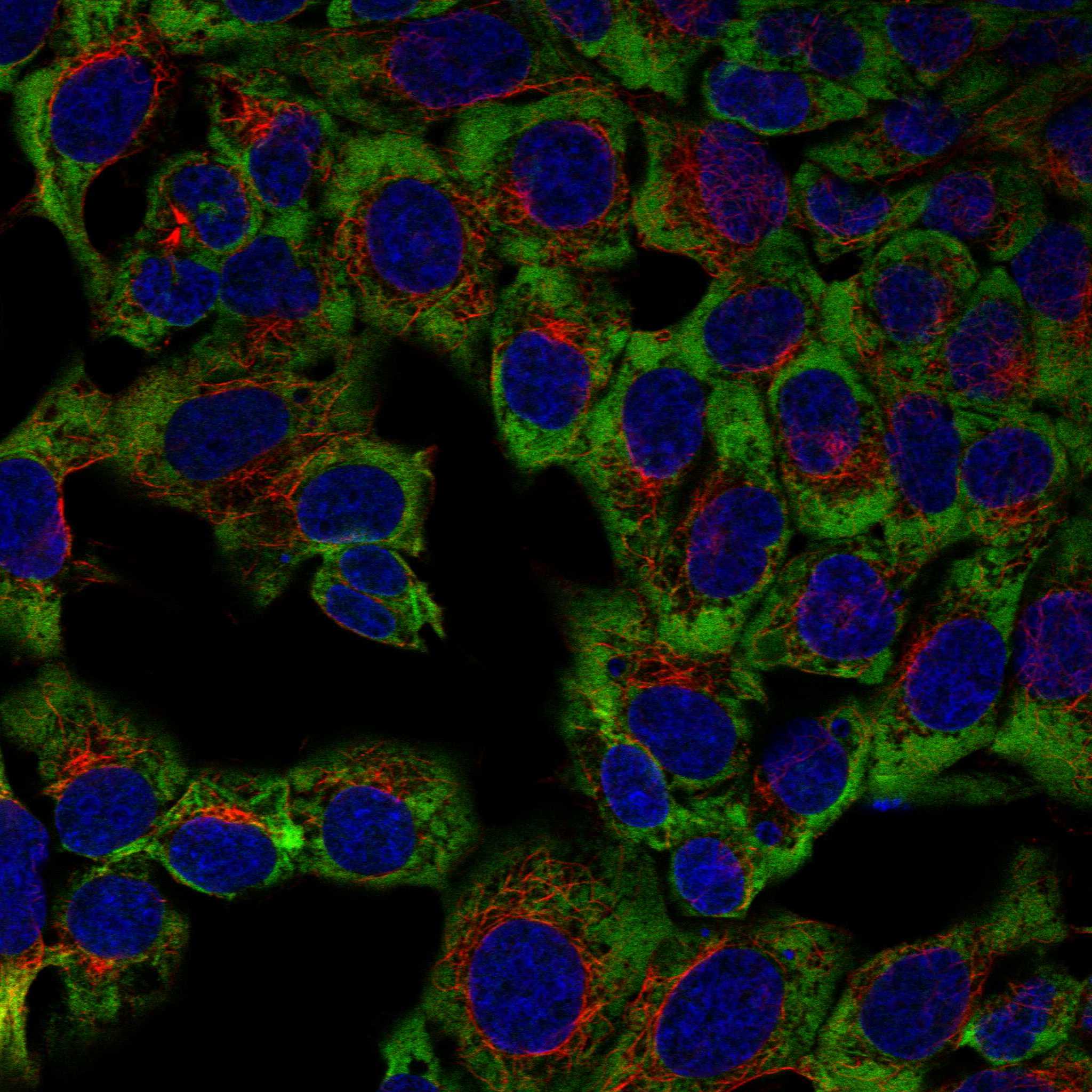
ICC-IF cell lines (HPA subcellular resource): HPA ICC-IF images show AR in HAP1, Rh30, U2OS, with annotated localisation: Cytosol (supported) (HPA subcellular).
Technical controls: Include a no-primary, secondary-only control and a host-species-matched isotype control, then confirm specificity with AR knockout material or a validated peptide block (standard IHC practice). Block endogenous peroxidase for chromogenic IHC and assess gland lumina for nonspecific signal (standard IHC practice).
⚠️Feasibility: Keep fixation consistent across test and control sections; prolonged fixation can reduce epitope accessibility (standard IHC practice). Optimize heat-induced antigen retrieval for the FFPE antibody, while frozen-section IF may avoid retrieval but requires separate validation (standard IHC/IF practice). Score cellular staining separately from luminal deposits in epididymis, and interpret nuclear and cytoplasmic signal in light of AR's ligand-dependent localization (UniProt P10275 subcellular location; HPA: cytosol supported).

HPA tissue IHC evidence for AR

Comprehensive Human Protein Atlas IHC scoring per tissue (reliability: Enhanced — High consistency between antibody staining and RNA expression data.). Rows are taken directly from the HPA tissue chart — click any row's HPA link to view the source.

Positive expression · recommended positive controls

TissueCell typeLevelEvidenceSource
Epididymis Glandular cells High Protein (IHC) HPA →
Seminal vesicle Glandular cells High Protein (IHC) HPA →
Breast Glandular cells Medium Protein (IHC) HPA →
Endometrium Cells in endometrial stroma Medium Protein (IHC) HPA →
Fallopian tube Glandular cells Medium Protein (IHC) HPA →

Undetected expression · recommended negative controls

TissueCell typeLevelEvidenceSource
Adipose tissue Adipocytes Not detected Protein (IHC) HPA →
Adrenal gland Glandular cells Not detected Protein (IHC) HPA →
Appendix Glandular cells Not detected Protein (IHC) HPA →
Bone marrow Hematopoietic cells Not detected Protein (IHC) HPA →
Bronchus Respiratory epithelial cells Not detected Protein (IHC) HPA →
Section 3

Advanced AR IHC Tips

Troubleshoot AR staining in paraffin sections by checking retrieval, nuclear localisation, antibody epitope coverage and cell specific controls before scoring.

How should I retrieve AR when nuclear staining is weak?
Use Tris-EDTA pH 9.0 HIER at 95–98 °C for 20 min (page retrieval setting). Allow sections to cool in retrieval buffer, then compare a known positive glandular control with the test section using the same detection conditions (standard IHC practice; HPA: high in epididymal glandular cells). If nuclei remain weak, compare a shorter heating period or a citrate buffer at pH 6.0 on adjacent sections while checking tissue integrity (standard IHC practice). Score nuclear and cytoplasmic signal separately because AR can move from cytoplasm to nucleus after ligand binding (UniProt P10275 localisation).
How can fixation explain weak or uneven AR staining?
Record fixative, fixation duration and section thickness before changing the antibody concentration, because fixation and processing can alter epitope accessibility (standard IHC practice). Compare affected sections with a similarly processed positive control containing glandular cells, such as epididymis or seminal vesicle (HPA: high in epididymal and seminal vesicle glandular cells). For formalin fixed paraffin sections, repeat the specified Tris-EDTA pH 9.0 retrieval at 95–98 °C for 20 min before judging a block negative (page retrieval setting). If staining varies between the tissue edge and centre, inspect preservation and compare another block or section level before assigning an AR score (standard IHC practice).
Should AR appear in nuclei, cytoplasm, or both?
Score nuclear and cytoplasmic staining separately in chromogenic sections, since AR is predominantly cytoplasmic without ligand and can enter the nucleus after ligand binding (UniProt P10275 localisation). Nuclear staining in reproductive organ cells is a useful IHC reference pattern; glandular cells of epididymis and seminal vesicle show high expression (HPA: tissue IHC). Cytoplasmic signal alone needs careful comparison with controls because a separate cell imaging dataset reports cytosolic localisation (HPA: subcellular). Check that brown signal lies within intact cell boundaries and that the counterstain permits reliable identification of nuclei; exclude precipitate and damaged tissue from compartment scoring (standard IHC practice).
Could antibody epitope coverage explain an unexpected AR pattern?
Check the catalog antibody’s stated immunogen and epitope against the AR sequence before interpreting a negative section, because the record lists 4 isoforms (UniProt P10275 isoforms). AR has no transmembrane segment, and its ligand binding domain spans residues 669–900, so assess whether the documented epitope falls in a region represented by the isoform of interest (UniProt P10275 topology and domains). Documented modified residues include positions 83, 96, 225, 258 and 269 (UniProt P10275 modified residues). If epitope coverage remains uncertain, compare a second IHC validated antibody with a documented, distinct epitope on adjacent sections and retain the same cellular scoring rules (standard IHC practice).
How should I compare AR immunofluorescence with chromogenic IHC?
Treat IF as a separate validation experiment and compare its nuclear and cytoplasmic AR patterns with the chromogenic section from the same specimen where feasible (standard IF practice; UniProt P10275 localisation). Multiplex AR with a validated marker for the expected glandular cell population, since epididymal and seminal vesicle glandular cells show high AR staining in tissue IHC (HPA: tissue IHC). Select fluorophores after inspecting unstained tissue autofluorescence, and include single stain controls to assess channel bleed through (standard IF practice). Because AR has no transmembrane segment, use a controlled permeabilisation step to reach intracellular epitopes, then check that nuclear counterstaining supports the claimed compartment assignment (UniProt P10275 topology; standard IF practice).
What should I change when AR chromogenic staining has high background?
Inspect a no primary control to separate nonspecific detection signal from staining attributable to the AR antibody (standard IHC practice). Block endogenous peroxidase before enzyme based chromogenic detection, and check whether diffuse brown signal persists in regions lacking intact cells (standard IHC practice). Titrate the catalog antibody and detection reagent on adjacent sections while keeping Tris-EDTA pH 9.0, 95–98 °C, 20 min retrieval constant (page retrieval setting; standard IHC practice). Compare background with a positive glandular control and a reference cell population reported as undetected, such as adipocytes, while evaluating tissue preservation and morphology (HPA: high in epididymal glandular cells; HPA: AR not detected in adipocytes; standard IHC practice).
How should I quantify AR across paraffin sections? ⚠ ANSWER MARKED FOR VERIFICATION
Define the cell population and nuclear scoring threshold before reviewing treatment groups, then report the percentage of positive nuclei and staining intensity (standard IHC practice). An H-score can sum intensity weighted percentages across 0–3 intensity categories, giving a 0–300 scale when every cell is assigned a category (standard IHC practice). Normalise positive counts to all evaluable nuclei in the same annotated cell population, and report density per mm² only when area is the relevant denominator (standard IHC practice). Keep cytoplasmic scores separate because AR localisation can change with ligand binding, and exclude damaged or necrotic regions consistently (UniProt P10275 localisation; standard IHC practice).
How can I distinguish true AR staining from artefact?
A credible positive should occupy an identifiable cell and show a compartment pattern consistent with AR biology; ligand binding can shift AR from cytoplasm into the nucleus (UniProt P10275 localisation). Compare glandular cells in epididymis or seminal vesicle with the test population, while recognising that HPA reports no detection in adipocytes (HPA: tissue IHC). Treat staining confined to cut edges, folds or necrotic areas cautiously, and inspect a no primary control for endogenous enzyme signal or detection artefact (standard IHC practice). Recheck apparently positive cells in an unexpected compartment or cell population against morphology, counterstain and an independently processed section before reporting their AR status (standard IHC practice).
Boster reagents

Best AR / Androgen receptor IHC Antibodies

Catalog anti-AR antibodies have human paraffin-section IHC images and a T47D-cell IF image; selected products list Human, Mouse, and Rat reactivity (catalog image alts, reactivity).

Real IHC data Immunohistochemical analysis of paraffin-embedded human prostate carcinoma, using Androgen Receptor Antibody.
Anti-Androgen Receptor AR Rabbit Monoclonal Antibody
Cat # M00542-1
Real IHC data Immunohistochemical analysis of paraffin-embedded human prostate, using Androgen Receptor Antibody.
Anti-Androgen Receptor AR Rabbit Monoclonal Antibody
Cat # M00542-2
Real IHC data Immunohistochemical analysis of paraffin-embedded human prostate carcinoma, using Androgen Receptor(AR-V7 specific) Antibody.
Anti-Androgen Receptor (AR-V7 specific) Rabbit Monoclonal Antibody
Cat # M00542-3
Real IF data IF analysis of Androgen Receptor/AR using anti-Androgen Receptor/AR antibody (A00542). Androgen Receptor/AR was detected in immunocytochemical section of T47D cells. Enzyme antigen retrieval was performed using IHC enzyme antigen retrieval reagent (AR0022) for 15 mins. The cells were blocked with 10% goat serum. And then incubated with 5μg/mL rabbit anti-Androgen Receptor/AR Antibody (A00542) overnight at 4°C. DyLight®488 Conjugated Goat Anti-Rabbit IgG (BA1127) was used as secondary antibody at 1:100 dilution and incubated for 30 minutes at 37°C. The section was counterstained with DAPI. Visualize using a fluorescence microscope and filter sets appropriate for the label used.
Anti-Androgen Receptor/AR Antibody ®
Cat # A00542

M00542-1 shows paraffin-embedded human prostate carcinoma IHC; M00542-2 shows paraffin-embedded human prostate IHC; M00542-3 shows paraffin-embedded human prostate carcinoma IHC with an AR-V7-specific antibody (catalog image alts). A00542 shows IF in T47D cells and lists IF/ICC applications, while the three M00542 products list IHC applications (catalog image alts, applications).

Which to pick: For FFPE IHC, choose the rabbit monoclonal M00542-1 or M00542-2; choose M00542-3 when the catalog’s AR-V7-specific antibody is needed (catalog titles, applications, IHC image alts). For IF/ICC, A00542 has a T47D IF image and a 5 μg/mL ICC/IF recommendation; M00542-1 and M00542-2 also list IF/ICC (catalog image alts, dilution, applications). For Mouse or Rat work, M00542-2 lists IHC and IF/ICC with those species, while A00542 lists IF/ICC with those species (catalog applications, reactivity).

Each figure is that product's own IHC / IF validation image from its datasheet.

References

  1. UniProt Consortium. UniProt entry P10275 (ANDR_HUMAN, Androgen receptor).
  2. Human Protein Atlas. AR tissue IHC expression (reliability: Enhanced).
  3. Human Protein Atlas. AR subcellular location (ICC-IF): Localized to the cytosol..
  4. Human Protein Atlas. AR antibody validation summary (3 antibodies).
  5. Gender- and Grade-Dependent Activation of Androgen Receptor Signaling in Adult-Type Diffuse Gliomas: Epigenetic Insights from a Retrospective Cohort Study. Biomedicines 2025 — PMC12561669.
  6. Chromosome X aneusomy and androgen receptor gene copy number aberrations in apocrine carcinoma of the breast. Virchows Archiv : an international journal of pathology 2021 — PMC8364532.
  7. Immunohistochemical Characterization of the Androgen Receptor in Breast Cancer and Its Relationship with Breast Cancer Subtypes. Medical sciences (Basel, Switzerland) 2026 — PMC13515587.
  8. PIK3CA mutations in androgen receptor-positive triple negative breast cancer confer sensitivity to the combination of PI3K and androgen receptor inhibitors. Breast cancer research : BCR 2014 — PMC4187324.
  9. PubMed PMID:3216866 — UniProt-cited evidence.
  10. PubMed PMID:3174628 — UniProt-cited evidence.
  11. PubMed PMID:2911578 — UniProt-cited evidence.