ARF1 / ADP-ribosylation factor 1 · IHC design guide

Design Immunohistochemistry for ARF1

Plan chromogenic ARF1 IHC-P using colon glandular cells as a high-staining reference (HPA tissue IHC). Assess cytoplasmic staining while accounting for uncertain tissue-IHC reliability (HPA tissue IHC).

Evidence assembled Oct 2026 · For research use; verify linked source records and product datasheet before use
Immunohistochemistry protocol sheet for ARF1 (IHC for ARF1): expected localisation Cytoplasmic tissue staining (HPA tissue IHC), antibody M01279-1, validated IHC image, and IHC protocol steps
Printable ARF1 IHC protocol sheet — expected localisation Cytoplasmic tissue staining (HPA tissue IHC), antibody M01279-1, controls and protocol steps. Open the full ARF1 IHC guide →

ARF1 Immunohistochemistry Experimental Design Guide

Expected localisation, validated protocols, controls and antibodies — the at-a-glance facts below, then the full design guide.

Must know before staining
Expected localisation Cytoplasmic tissue staining (HPA tissue IHC)
Staining pattern General cytoplasmic staining in glandular and neuronal cells (HPA tissue IHC)
Antigen retrieval EDTA pH 8.0 HIER, heat-mediated (datasheet M01279-1)
Positive control ⓘ Adrenal gland+4 more · see all
Negative control ⓘ Ovary
Important caveats
Reasons your staining may differ from the expected pattern.
Fixation Matched tissue-IHC evidence does not establish the fixation claim. Validate the specimen-specific method before use. (selected-SKU IHC image M01279-1)
Caveat Staining reliability is uncertain (HPA tissue IHC)
Regulation No expression regulator annotated (UniProt)
Isoform / epitope No annotated isoforms; mature chain spans residues 2–181 (UniProt)
Section 1

Recommended ARF1 IHC & IF Protocols

Start with the catalog antibody’s IHC-P protocol (datasheet: M01279-1), then compare the four published ARF1 IHC protocols below (PMC5129974; PMC7778598; PMC4941279; PMC5295416).

Recommended immunohistochemistry (IHC-P) protocol parameters
SampleParaffin-embedded human colon tissue; fixative not specified (datasheet M01279-1)
FixationImage fixative and duration unreported (datasheet M01279-1); verify before use.
Sectioning4–5 µm sections on charged slides (standard)
DeparaffinisationXylene, graded ethanol series to water (standard)
Antigen retrievalHeat retrieval: EDTA pH 8.0 (datasheet M01279-1); 20 min, 95–100 °C (standard)
Peroxidase block3% H2O2, 10 min, room temperature (standard)
Blocking10% goat serum (datasheet M01279-1)
Primary antibodyRabbit monoclonal (clone GFA-1) anti-ARF1, 1:50 (datasheet M01279-1)
Primary incubationOvernight at 4 °C (datasheet M01279-1)
DetectionHRP-conjugated secondary, DAB chromogen (datasheet M01279-1)
CounterstainHematoxylin, blue, dehydrate and mount (standard)
Expected resultARF1-positive staining in glandular cells of adrenal gland (HPA tissue IHC: High). HPA tissue profile: General cytoplasmic expression. No signal in the no-primary control.
💡Decision noteTry heat-mediated EDTA retrieval at pH 8.0 first (datasheet: M01279-1). The breast tumor protocol reports proprietary heat retrieval reagents without their composition (PMC4941279).
Section 2

What Is the Expected ARF1 Staining Pattern?

In paraffin section IHC, expect general cytoplasmic ARF1 staining in glandular, respiratory epithelial and neuronal cells reported as positive by HPA (HPA: tissue IHC). Golgi membrane association is biologically compatible with cytoplasmic staining, but a discrete Golgi pattern is not required (UniProt P84077: location; HPA: general cytoplasmic expression). ARF1 has no transmembrane segment (UniProt P84077: topology). Interpret intensity cautiously because HPA rates tissue IHC reliability Uncertain (HPA: tissue IHC reliability).

What am I looking at on my slide?
Cytoplasmic stain is strong in colon or duodenal glandular cells and in bronchial respiratory epithelium (HPA: High in these cells).This fits the reported positive pattern; compare the named cells within each section rather than treating every cell as equally positive (HPA: tissue IHC). Cytoplasmic staining is consistent with HPA's tissue profile and UniProt's Golgi association (HPA: general cytoplasmic expression; UniProt P84077: location).
Staining appears exclusively nuclear, with little cytoplasmic signal in a known positive cell population (HPA: general cytoplasmic expression).Treat this as a suspect compartment pattern: nuclear enrichment is not the reported tissue pattern or UniProt location (HPA: tissue IHC; UniProt P84077: location). Check antibody specificity and detection controls before scoring it as ARF1 (standard IHC practice).
Ovarian stromal cells stain strongly, or stain dominates cells other than the named positive population (HPA: ovarian stroma Not detected; HPA: tissue IHC positives).Investigate cross-reactivity or endogenous chromogenic activity with appropriate controls (standard IHC practice). The HPA negative observation applies to ovarian stromal cells, not the entire ovary, and its reliability rating limits certainty (HPA: tissue IHC).
A broad chromogenic haze covers stroma and cells without a clear cytoplasmic boundary (HPA: general cytoplasmic expression).Diffuse background cannot establish a cell specific ARF1 result (standard IHC practice). Compare a no-primary control, blocking and detection conditions, then reassess named positive cells (standard IHC practice; HPA: tissue IHC positives).
Colon glandular cells lack detectable stain while the control section also lacks a clear positive population (HPA: colon glandular cells High).An absent signal in a reported positive population calls for an assay check before biological interpretation (standard IHC practice). HPA's Uncertain reliability also means one negative section should not be used to claim universal absence (HPA: tissue IHC reliability).
💡Expected ARF1 appearanceA convincing positive is predominantly cytoplasmic chromogenic stain in reported High populations such as colon glandular cells, with a clear cell associated pattern; isolated nuclear stain or uniform tissue haze is suspect (HPA: tissue IHC; UniProt P84077: location; standard IHC practice).
How each factor affects the staining
Compartment and membrane association (UniProt P84077: location and topology).ARF1 associates with Golgi membranes and has no transmembrane segment; HPA describes tissue staining broadly as cytoplasmic (UniProt P84077: location and topology; HPA: tissue IHC). Do not require a sharply resolved Golgi signal in chromogenic sections (standard IHC interpretation).
Cell population and intensity (HPA: tissue IHC).HPA reports High staining in adrenal, colon, duodenal and epididymal glandular cells, bronchial respiratory epithelium, and caudate and cortical neurons; endometrial glandular cells are Medium (HPA: tissue IHC). Ovarian stromal cells are Not detected (HPA: tissue IHC).
Evidence strength and antibody validation (HPA: tissue IHC; HPA: antibody CAB007742).The tissue profile is Uncertain because antibody staining and RNA show medium consistency; CAB007742 is also rated Uncertain for IHC (HPA: tissue IHC reliability; HPA: antibody CAB007742). Use orthogonal controls before making a strong negative or unexpected localization claim (standard IHC practice).
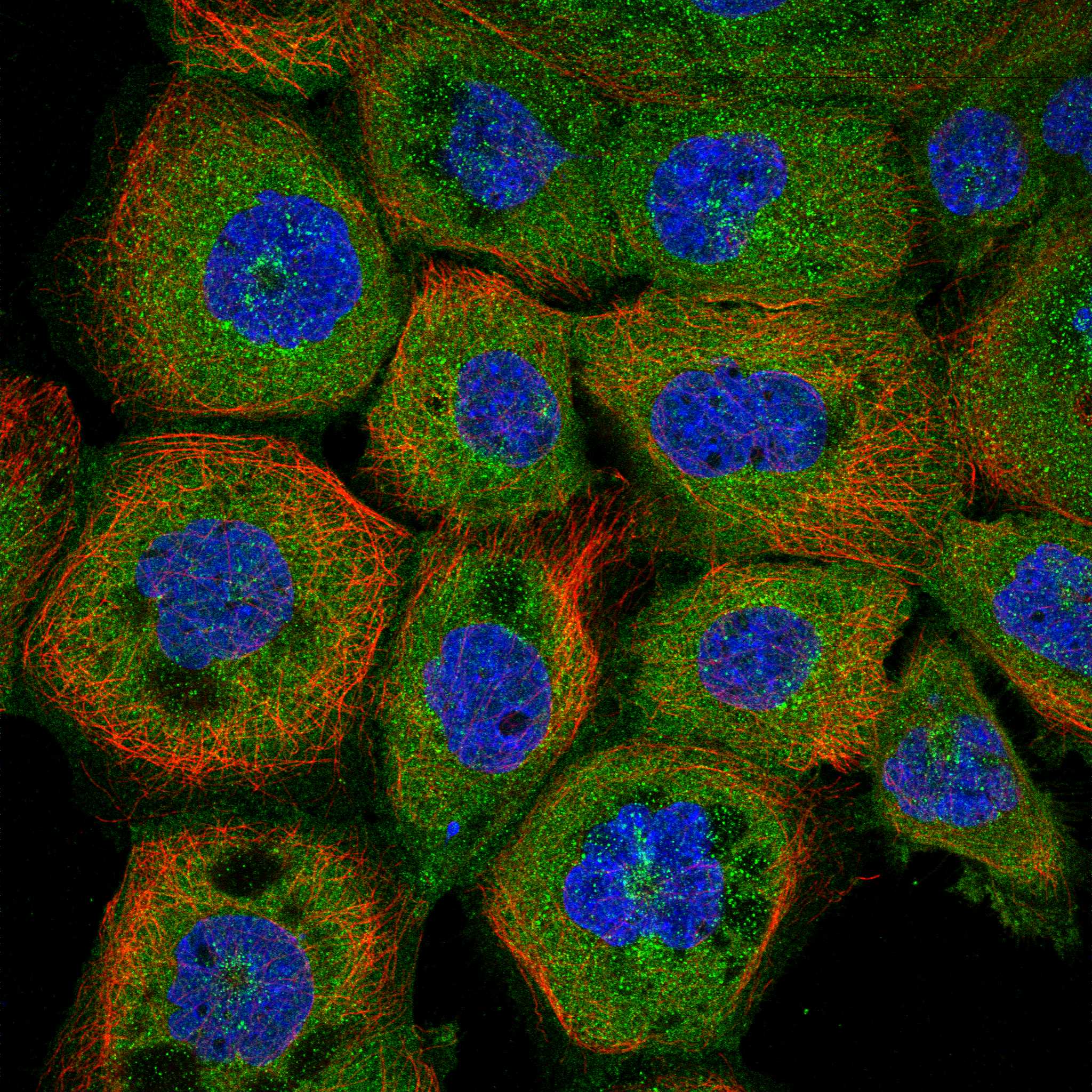
IF/ICC: What pattern is reported? (HPA: subcellular ICC-IF).HPA reports mainly cytosolic ARF1 with an additional plasma membrane location; both locations are approved in ICC-IF, and CAB007742 is Supported for ICC (HPA: subcellular ICC-IF; HPA: antibody CAB007742). These cell imaging observations do not establish the appearance of a paraffin section (HPA: assay scope).
Protein architecture (UniProt P84077: topology and processing).No transmembrane segment, signal peptide, propeptide or annotated isoform is listed (UniProt P84077: topology and processing). These annotations do not identify the antibody epitope or establish a preferred antigen retrieval condition (UniProt P84077: annotation scope).
Why is my staining missing, weak or wrong?
SituationLikely causeNext action
No stain in colon glandular cells (HPA: colon glandular cells High).Detection failure or an unsuitable assay condition is possible; HPA does not establish a target specific fixation effect (standard IHC practice; HPA: evidence scope).Check a reported positive control, antibody dilution, detection reagents and the assay's retrieval settings (standard IHC practice). Record any retrieval change as an empirical optimization, not an ARF1 specific requirement (standard IHC practice).
Nuclei dominate the signal (HPA: general cytoplasmic expression).A compartment mismatch raises concern about nonspecific signal or interpretation (HPA: tissue IHC; UniProt P84077: location).Compare no-primary and known positive controls, then score cytoplasm separately from nuclei (standard IHC practice; HPA: tissue IHC positives).
Strong stain appears in ovarian stromal cells (HPA: ovarian stroma Not detected).Cross-reactivity or endogenous detection activity is possible; the HPA result is Uncertain (standard IHC practice; HPA: tissue IHC reliability).Check no-primary and detection controls, and confirm the cell identity before calling an unexpected ARF1 positive (standard IHC practice; HPA: ovarian stroma Not detected).
Diffuse brown background obscures cell borders (standard IHC observation).Nonspecific antibody binding or endogenous chromogenic activity can obscure the reported cytoplasmic pattern (standard IHC practice; HPA: general cytoplasmic expression).Review blocking, primary antibody dilution, washes and detection controls; judge the result in the named positive cells (standard IHC practice; HPA: tissue IHC positives).
Only a faint perinuclear focus is visible in a reported High cell population (HPA: tissue IHC positives).Golgi association can make the focus biologically plausible, but HPA reports general cytoplasmic staining and rates tissue reliability Uncertain (UniProt P84077: location; HPA: tissue IHC).Compare the complete cytoplasmic pattern with a positive control and repeat scoring across cells before calling the section negative (standard IHC practice; HPA: tissue IHC positives).
A section differs from an ICC-IF image (HPA: subcellular ICC-IF; HPA: tissue IHC).HPA reports mainly cytosol plus plasma membrane in ICC-IF, while its tissue IHC profile is general cytoplasmic (HPA: subcellular ICC-IF; HPA: tissue IHC).Interpret the paraffin section against HPA tissue cell populations and its Uncertain IHC rating; use ICC-IF localization as context (HPA: tissue IHC; HPA: subcellular ICC-IF).

Sample controls for ARF1 IHC & IF

🧪Run colon first; its glandular cells should stain (HPA: High in colon glandular cells; selected-SKU IHC caption: ARF1 detected in human colon). Use ovarian stroma cells as the negative tissue compartment (HPA: Not detected in ovarian stroma cells); on the colon slide, treat any unstained nonglandular cells as a local background reference, since the supplied HPA row establishes staining only for glandular cells (HPA: High in colon glandular cells).
Positive control tissue: Adrenal gland (Glandular cells, HPA High)
Negative control tissue: Ovary (HPA Not detected)
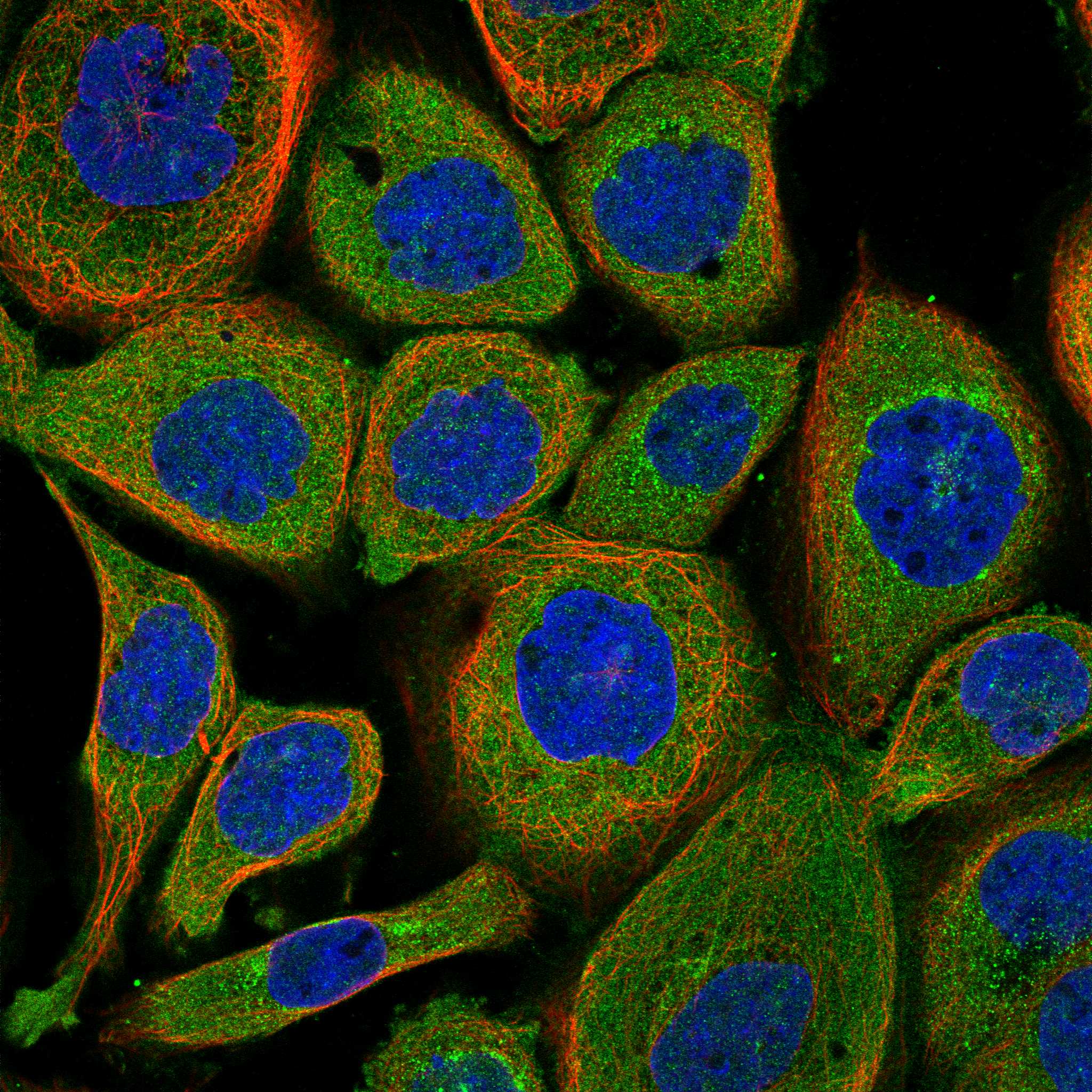
ICC-IF cell lines (HPA subcellular resource): HPA ICC-IF images show ARF1 in A-431, U-251MG, U2OS, with annotated localisation: Cytosol (approved) (HPA subcellular).
Technical controls: Include a no-primary (secondary-only) control, an isotype control matched to the primary antibody’s rabbit host and clonality, and a knockout sample or validated peptide-block control if available (selected-SKU IHC caption: rabbit primary; standard IHC practice). For colon sections developed with HRP/DAB, quench endogenous peroxidase and check the no-primary slide for residual staining (selected-SKU IHC caption: colon, HRP/DAB; standard IHC practice).
⚠️Feasibility: A target-specific fixation window or fixation effect is unreported, and the selected-SKU paraffin-section caption does not state a fixative (selected-SKU IHC caption: fixative not stated). The reported IHC condition uses heat-mediated EDTA retrieval at pH 8.0, a 1:50 rabbit primary overnight at 4°C, and HRP/DAB detection (selected-SKU IHC caption). The supplied evidence does not establish whether frozen sections or IF are easier; ICC-IF images are available for A-431, U-251MG and U2OS, while the colon HRP/DAB workflow calls for an endogenous peroxidase check (HPA: ICC-IF image cell lines; selected-SKU IHC caption: colon, HRP/DAB; standard IHC practice).

HPA tissue IHC evidence for ARF1

Comprehensive Human Protein Atlas IHC scoring per tissue (reliability: Uncertain — Medium consistency between antibody staining and RNA expression data.). Rows are taken directly from the HPA tissue chart — click any row's HPA link to view the source.

Positive expression · recommended positive controls

TissueCell typeLevelEvidenceSource
Adrenal gland Glandular cells High Protein (IHC) HPA →
Bronchus Respiratory epithelial cells High Protein (IHC) HPA →
Caudate Neuronal cells High Protein (IHC) HPA →
Cerebral cortex Neuronal cells High Protein (IHC) HPA →
Colon Glandular cells High Protein (IHC) HPA →

Undetected expression · recommended negative controls

TissueCell typeLevelEvidenceSource
Ovary Ovarian stroma cells Not detected Protein (IHC) HPA →
Section 3

Advanced ARF1 IHC Tips

Use the catalog antibody’s paraffin-section colon example as the IHC starting point, then assess staining against ARF1 localisation and appropriate controls.

How should I optimize antigen retrieval when ARF1 staining is weak?
Start with heat-mediated retrieval in EDTA pH 8.0 for paraffin sections (datasheet M01279-1). The documented colon IHC example used this retrieval before 10% goat-serum blocking and antibody incubation at 1:50 overnight at 4°C (datasheet M01279-1). If staining remains weak, vary heating duration on adjacent sections while keeping the antibody dilution and detection conditions fixed, and check whether tissue morphology deteriorates (standard IHC practice). Compare the resulting cytoplasmic staining within the same cell type; retrieval that raises diffuse background or damages morphology does not establish stronger ARF1 detection (HPA: general cytoplasmic expression; standard IHC practice).
Can fixation explain absent or uneven ARF1 staining in paraffin sections?
Target-specific fixation sensitivity is unknown: the selected paraffin-section colon caption does not state a fixative, and no fixation comparison is supplied (datasheet M01279-1). Record the fixative and fixation duration for each specimen, then compare sections processed through the same retrieval, antibody incubation and chromogenic detection workflow (standard IHC practice). Inspect poorly stained regions for tissue damage or uneven processing before changing antibody concentration, because either can complicate interpretation of an IHC result (standard IHC practice). Neither the reported tissue staining pattern nor ARF1’s membrane association demonstrates how this antibody responds to a particular fixative (HPA: tissue IHC; UniProt P84077 localisation; datasheet M01279-1).
What intracellular staining pattern should I expect for ARF1 in IHC?
Evaluate ARF1 staining primarily in the cytoplasm, while allowing for a more concentrated perinuclear pattern consistent with its Golgi association (HPA: general cytoplasmic expression; UniProt P84077 localisation). ARF1 has no transmembrane segment; its myristoylated amino terminus mediates membrane association, with more stable anchoring after GTP loading (UniProt P84077 topology and localisation). A separate cell-imaging dataset reports mainly cytosolic localisation with additional plasma-membrane localisation, so an exclusively sharp membrane rim is not required in chromogenic sections (HPA: subcellular localisation). Compare compartments within intact cells and across similarly processed sections before treating a different pattern as biological redistribution (standard IHC practice).
How should unknown epitope location affect ARF1 IHC interpretation?
The supplied ARF1 record lists 0 isoforms and a chain spanning residues 2–181, but it does not map this antibody’s epitope (UniProt P84077 processing and isoforms; datasheet M01279-1). Its amino terminus is modified and participates in membrane anchoring, so epitope accessibility cannot be inferred from sequence position without an antibody epitope assignment (UniProt P84077 modified residues and localisation). Do not interpret stronger staining after retrieval as evidence that a particular terminal epitope has been exposed (standard IHC practice). Use a validated independent antibody or a suitable loss-of-target control, if available, to test whether the same cellular pattern depends on ARF1 (standard IHC practice).
How can IF help assess an ambiguous chromogenic ARF1 pattern?
Use IF as a separate localisation check, pairing ARF1 with a marker for the expected cell type, such as a glandular epithelial marker when examining colon glands (HPA: high in colon glandular cells; standard IF practice). Choose fluorophores after inspecting unstained tissue for autofluorescence; a spectrally separated far-red channel can help when shorter wavelengths carry substantial background (standard IF practice). Because ARF1 can occupy cytosol and the cytoplasmic face of membranes, test permeabilisation suitable for intracellular access, while recognising that this antibody’s epitope side is unreported (HPA: subcellular localisation; UniProt P84077 topology; datasheet M01279-1). Interpret IF compartment detail alongside the paraffin-section IHC result, without assuming identical preparation effects (standard IHC/IF practice).
How do I reduce diffuse DAB background without losing ARF1 signal?
Begin with a no-primary control and inspect whether colour persists in the tissue or concentrates at section edges (standard IHC practice). Include a peroxidase-blocking step before HRP detection, and control DAB development consistently; these are general chromogenic workflow measures, not ARF1-specific findings (standard IHC practice). The documented example used 10% goat serum, 1:50 primary antibody overnight at 4°C, and a peroxidase-conjugated secondary for 30 minutes at 37°C (datasheet M01279-1). If background remains high, adjust blocking or antibody concentration one variable at a time, checking that expected cellular staining is retained (HPA: general cytoplasmic expression; standard IHC practice).
How should I score ARF1 staining across paraffin sections? ⚠ ANSWER MARKED FOR VERIFICATION
Define the scored cell population and cytoplasmic compartment before reviewing treatment groups, because the reported tissue IHC profile is broadly cytoplasmic (HPA: tissue IHC; standard IHC practice). For a chosen cell type, report the percentage of positive cells and an H-score from 0–300 based on staining intensity; keep thresholds constant across slides (standard IHC practice). Normalise positive-cell counts to the number of evaluable cells, or report stained-cell density per mm² of evaluable tissue when cell counts are impractical (standard IHC practice). Exclude folds, damaged margins and necrotic areas, and analyse comparable anatomical regions with matched staining batches (standard IHC practice).
Which findings support true ARF1 staining rather than artefact?
A reproducible cytoplasmic signal in intact glandular cells of colon is compatible with the reported high tissue staining and general cytoplasmic profile (HPA: colon glandular cells high; HPA: tissue IHC profile). Check whether staining also appears in a no-primary control or follows cut edges, folds, necrosis or residual endogenous peroxidase activity; these patterns support a technical explanation (standard IHC practice). Strong staining confined to an unexpected compartment or cell population needs independent validation before assigning it to ARF1 (UniProt P84077 localisation; standard IHC practice). Treat HPA tissue comparisons as context rather than proof, because their ARF1 antibody staining reliability is rated uncertain (HPA: reliability Uncertain).
Boster reagents

Best ARF1 / ADP-ribosylation factor 1 IHC Antibodies

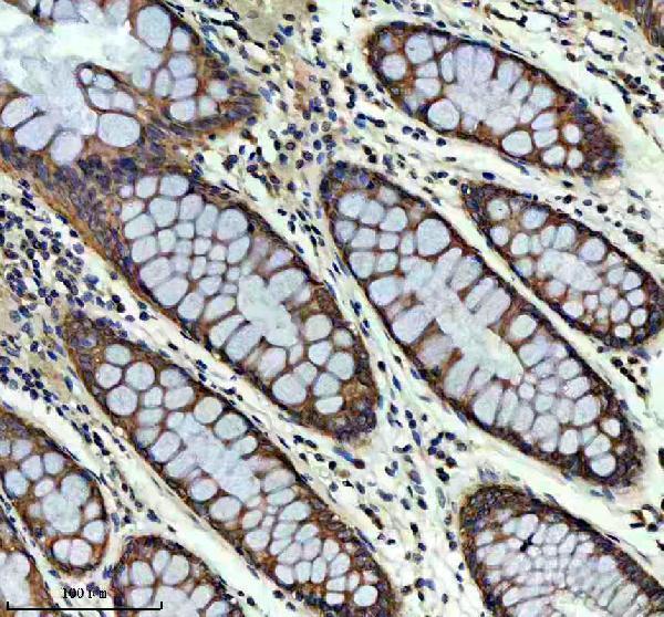
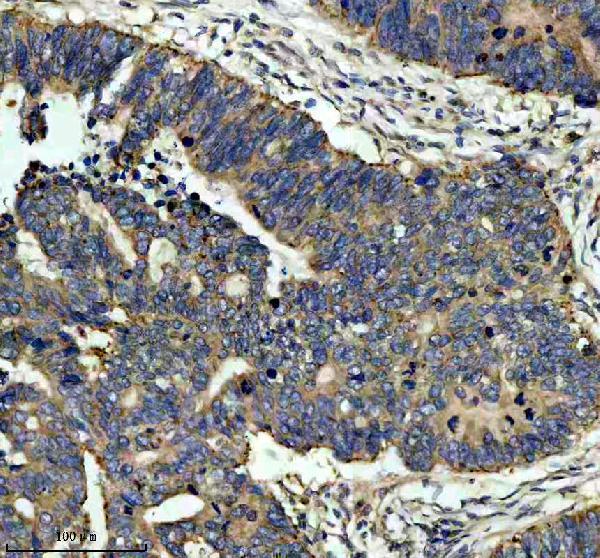
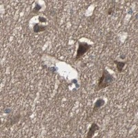
M01279-1 has IHC images from human paraffin sections of colon, colon cancer, and thyroid cancer (catalog IHC captions). Human, mouse, and rat reactivity is listed (catalog reactivity).

Real IHC data IHC analysis of ARF1 using anti-ARF1 antibody (M01279-1). ARF1 was detected in a paraffin-embedded section of human colon tissue. Heat mediated antigen retrieval was performed in EDTA buffer (pH 8.0, epitope retrieval solution). The tissue section was blocked with 10% goat serum. The tissue section was then incubated with 1:50 rabbit anti-ARF1 Antibody (M01279-1) overnight at 4°C. Peroxidase Conjugated Goat Anti-rabbit IgG was used as secondary antibody and incubated for 30 minutes at 37°C. The tissue section was developed using HRP Conjugated Rabbit IgG Super Vision Assay Kit (Catalog # SV0002) with DAB as the chromogen.
Anti-ARF1 Rabbit Monoclonal Antibody
Cat # M01279-1

M01279-1 is a rabbit monoclonal antibody listed for IHC (catalog applications and antibody details). Its IHC captions show staining in human paraffin sections of colon, colon cancer, and thyroid cancer at 1:50 (catalog IHC captions).

Which to pick: For tissue IHC, choose M01279-1 based on its human paraffin-section images; the fixative is unreported (catalog IHC captions). No IF/ICC application or IF image is supplied for M01279-1, so this payload does not establish an IF/ICC choice (catalog applications and image data). For mouse or rat tissue, M01279-1 lists reactivity with both species, but its supplied IHC images show human tissue only (catalog reactivity and IHC captions).

Each figure is that product's own IHC / IF validation image from its datasheet.

References

  1. UniProt Consortium. UniProt entry P84077 (ARF1_HUMAN, ADP-ribosylation factor 1).
  2. Human Protein Atlas. ARF1 tissue IHC expression (reliability: Uncertain).
  3. Human Protein Atlas. ARF1 subcellular location (ICC-IF): Mainly localized to the cytosol. In addition localized to the plasma membrane..
  4. Human Protein Atlas. ARF1 antibody validation summary (1 antibodies).
  5. ARF1 promotes prostate tumorigenesis via targeting oncogenic MAPK signaling. Oncotarget 2016 — PMC5129974.
  6. Anti-allergic drug azelastine suppresses colon tumorigenesis by directly targeting ARF1 to inhibit IQGAP1-ERK-Drp1-mediated mitochondrial fission. Theranostics 2021 — PMC7778598.
  7. ADP-ribosylation factor 1 expression regulates epithelial-mesenchymal transition and predicts poor clinical outcome in triple-negative breast cancer. Oncotarget 2016 — PMC4941279.
  8. Suppression of breast cancer metastasis through the inactivation of ADP-ribosylation factor 1. Oncotarget 2016 — PMC5295416.
  9. PubMed PMID:2474826 — UniProt-cited evidence.
  10. PubMed PMID:1899243 — UniProt-cited evidence.
  11. PubMed PMID:1577740 — UniProt-cited evidence.