This website uses cookies to ensure you get the best experience on our website.


- Table of Contents
Plan ARG1 staining in paraffin sections using hepatocytes and bone marrow hematopoietic cells as positive references (HPA tissue IHC). This guide covers fixation, staining interpretation, and controls for the expected cytosolic signal (UniProt).

Expected localisation, validated protocols, controls and antibodies — the at-a-glance facts below, then the full design guide.
| Expected localisation | Cytosolic; neutrophil azurophil granules (UniProt) | |
| Staining pattern | High hepatocyte and marrow-cell staining; cytosolic expected (HPA tissue IHC; UniProt) | |
| Antigen retrieval | EDTA pH 8.0 HIER, heat-mediated (datasheet A01106) | |
| Positive control | Bone marrow+3 more · see all | |
| Negative control | Adipose tissue+4 more · see all |
| Fixation | Keep fixation consistent across paraffin sections. (standard IHC practice; not target-specific) | |
| Caveat | Bone marrow staining varies across cell subsets (HPA tissue IHC) | |
| Regulation | Expressed in granuloma macrophages (UniProt) | |
| Isoform / epitope | Three isoforms; epitope differences are unspecified (UniProt) |
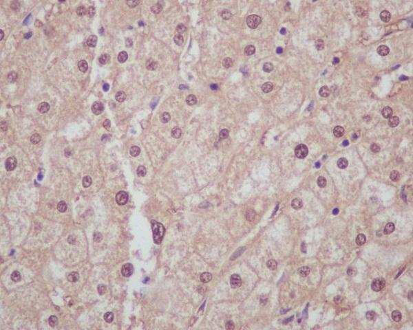
The catalog antibody has an IHC-P protocol (datasheet: A01106). Two reports describe ARG1 staining of paraffin sections (PMC11648120; PMC10371158).
| Sample | Paraffin-embedded human liver cancer tissue; fixative not specified (datasheet A01106) |
| Fixation | Image fixative and duration unreported (datasheet A01106); verify before use. |
| Sectioning | 4–5 µm sections on charged slides (standard) |
| Deparaffinisation | Xylene, graded ethanol series to water (standard) |
| Antigen retrieval | Heat retrieval: EDTA pH 8.0 (datasheet A01106); 20 min, 95–100 °C (standard) |
| Peroxidase block | 3% H2O2, 10 min, room temperature (standard) |
| Blocking | 10% goat serum (datasheet A01106) |
| Primary antibody | Rabbit anti-ARG1, 2-5 μg/ml (datasheet A01106) |
| Primary incubation | Overnight at 4 °C (datasheet A01106) |
| Detection | Streptavidin-biotin complex (SABC), DAB chromogen (datasheet A01106) |
| Counterstain | Hematoxylin, blue, dehydrate and mount (standard) |
| Expected result | ARG1-positive staining in hematopoietic cells of bone marrow (HPA tissue IHC: High). HPA tissue profile: Selective expression in liver and subsets of bone marrow cells. No signal in the no-primary control. |
ARG1 is cytosolic, with additional localization to neutrophil azurophil granules and no transmembrane segment (UniProt P05089: subcellular location and topology). In paraffin-section IHC, expect strong staining in hepatocytes and subsets of bone-marrow hematopoietic cells; spleen red-pulp cells and preleptotene spermatocytes show medium staining (HPA: tissue IHC). HPA rates the tissue pattern Enhanced, citing consistency between antibody staining and RNA expression (HPA: reliability).
| Strong cytoplasmic staining in hepatocytes or a subset of bone-marrow hematopoietic cells. | This matches the principal positive tissue pattern (HPA: High in hepatocytes and bone-marrow hematopoietic cells). Score the stained cell population and its cytoplasmic distribution together; a high-intensity field alone does not show that the expected cells carry the signal (general IHC interpretation). |
| Staining is chiefly nuclear or outlines cell membranes, with little cytoplasmic signal. | That distribution conflicts with the reported cytosolic location and absence of a transmembrane segment (UniProt P05089: location and topology). Treat it as suspect, then compare the slide with a known-positive tissue and appropriate detection controls before interpreting the compartment as ARG1 (general IHC practice). |
| Prominent staining appears in cells reported as negative, such as adipocytes or respiratory epithelial cells. | HPA reports ARG1 as not detected in those cell types (HPA: adipose tissue and bronchus IHC). Consider antibody cross-reactivity or detection-related signal, including endogenous activity with chromogenic detection (general IHC practice). Check cell identity and controls before treating unexpected staining as biological expression. |
| Weak, widespread color obscures both expected positive cells and surrounding tissue. | A diffuse field is difficult to reconcile with selective liver and bone-marrow expression (HPA: tissue IHC profile). It may reflect nonspecific antibody binding, inadequate blocking or washing, or detection background (general IHC practice). Judge the pattern against negative controls and the expected positive cell population. |
| No staining appears in hepatocytes on a technically readable section. | HPA reports high hepatocyte staining, making liver a useful positive reference (HPA: Liver, hepatocytes High). First check that the reference section, primary antibody, detection reagents, and run controls performed as intended (general IHC practice). The supplied sources do not identify ARG1-specific fixation sensitivity or an antigen-retrieval requirement. |
| Tissue and cell selection | Liver hepatocytes and subsets of bone-marrow hematopoietic cells are high; spleen red-pulp cells and preleptotene spermatocytes are medium (HPA: tissue IHC). Skin keratinocytes are low, so weak staining there is less decisive than a liver reference (HPA: Skin, keratinocytes Low). |
| Compartment and immune-cell context | Cytosol is the main reported location; neutrophils can also contain ARG1 in azurophil granules (UniProt P05089: subcellular location). UniProt also reports ARG1 in macrophages in mycobacterial granulomas and ILC2s during lung disease, so cell context matters when reviewing immune infiltrates (UniProt P05089: tissue specificity). |
| Antibody evidence and molecular variation | Five listed antibodies have Enhanced IHC validation, a category supported by independent antibodies or orthogonal data (HPA: antibody validation). UniProt lists three isoforms, but these payloads give no epitope mapping or isoform-specific IHC performance; do not infer which isoforms a chosen antibody detects (UniProt P05089: isoforms). |
| IF/ICC Q: Is a fluorescence pattern established here? | A: No ICC-IF images or HPA subcellular location are available (HPA: subcellular record). Cytosolic ARG1 is a location expectation from UniProt, not an HPA-validated ICC-IF staining pattern (UniProt P05089: location; HPA: subcellular record). |
| Situation | Likely cause | Next action |
|---|---|---|
| The liver positive reference has no visible hepatocyte signal. | A failed staining run or an unsuitable antibody setting is possible (general IHC practice); HPA reports hepatocytes as High (HPA: Liver IHC). | Review run controls and reagent performance, then optimize the IHC-validated antibody within its documented IHC conditions (general IHC practice). No ARG1-specific retrieval setting or dilution is supplied here. |
| Only nuclei or cell borders stain strongly. | The dominant compartment does not match reported cytosolic ARG1 or its lack of a transmembrane segment (UniProt P05089: location and topology). | Compare with hepatocyte staining and detection controls; assess whether counterstain, pigment, or nonspecific detection is being mistaken for target signal (general IHC practice). |
| Unexpected epithelial or adipocyte staining dominates the slide. | Those cell types are reported as not detected in the cited HPA tissues (HPA: bronchus respiratory epithelium; adipose adipocytes). Cross-reactivity or detection background is possible (general IHC practice). | Confirm the cell type and compare a negative tissue, a known-positive liver section, and appropriate detection controls before assigning ARG1 positivity (general IHC practice). |
| Brown color appears across much of a blood-rich section. | Chromogenic background, including endogenous enzyme activity, can complicate interpretation (general IHC practice); HPA describes a selective ARG1 tissue pattern (HPA: tissue IHC profile). | Examine the appropriate detection control and verify that the laboratory's endogenous-activity blocking step worked; score only recognizable cells with the expected distribution (general IHC practice). |
| Spleen or testis staining looks weaker than liver. | HPA reports medium staining in spleen red-pulp cells and preleptotene spermatocytes, versus high staining in hepatocytes (HPA: tissue IHC). | Compare the specified cell populations and compartments rather than demanding equal intensity across tissues (HPA: tissue IHC; general IHC interpretation). |
| An IF/ICC result is being used to confirm the IHC compartment. | HPA supplies no ICC-IF images or subcellular localization call for ARG1 (HPA: subcellular record). | Describe the IF/ICC observation as experimental, and use the cytosolic location as a UniProt-based expectation; do not label the fluorescence pattern HPA-validated (UniProt P05089: location; HPA: subcellular record). |
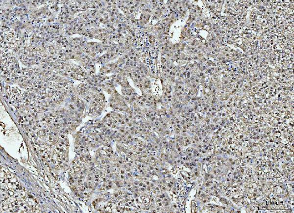
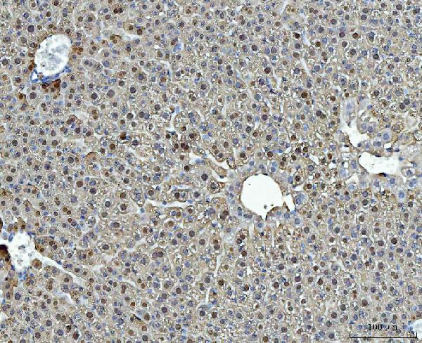
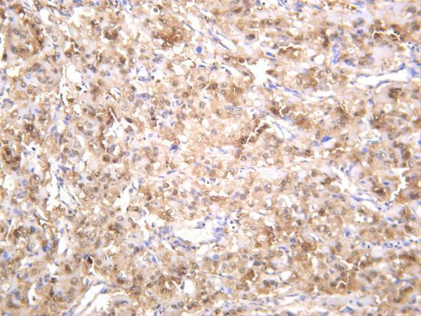
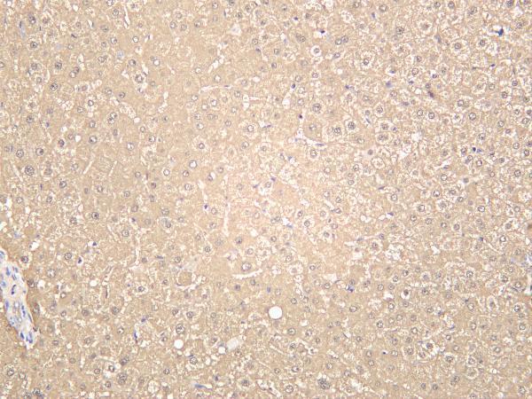


Comprehensive Human Protein Atlas IHC scoring per tissue (reliability: Enhanced — High consistency between antibody staining and RNA expression data.). Rows are taken directly from the HPA tissue chart — click any row's HPA link to view the source.
| Tissue | Cell type | Level | Evidence | Source |
|---|---|---|---|---|
| Bone marrow | Hematopoietic cells | High | Protein (IHC) | HPA → |
| Liver | Hepatocytes | High | Protein (IHC) | HPA → |
| Spleen | Cells in red pulp | Medium | Protein (IHC) | HPA → |
| Testis | Preleptotene spermatocytes | Medium | Protein (IHC) | HPA → |
| Tissue | Cell type | Level | Evidence | Source |
|---|---|---|---|---|
| Adipose tissue | Adipocytes | Not detected | Protein (IHC) | HPA → |
| Adrenal gland | Glandular cells | Not detected | Protein (IHC) | HPA → |
| Appendix | Glandular cells | Not detected | Protein (IHC) | HPA → |
| Breast | Adipocytes | Not detected | Protein (IHC) | HPA → |
| Bronchus | Respiratory epithelial cells | Not detected | Protein (IHC) | HPA → |
Troubleshoot ARG1 staining in paraffin sections by checking retrieval, cell identity and cytoplasmic localisation before comparing staining scores.
The catalog lists anti-ARG1 antibodies with IHC figures from human liver or liver cancer tissue, plus mouse and rat liver tissue (IHC captions); two antibodies also list IF among their applications (catalog: applications).
A01106 has paraffin-section IHC figures from human liver cancer and mouse and rat liver (IHC captions); its listed reactivity also includes monkey (catalog: reactivity). M01106-5 has IHC figures from human hepatocellular carcinoma and human, mouse and rat liver (IHC captions), while M01106-2 has a paraffin-section IHC figure from human liver (IHC caption).
Which to pick: For tissue IHC on paraffin sections, choose A01106 for human, mouse or rat samples with directly pictured examples (A01106 IHC captions), or M01106-2 for human liver with a pictured paraffin-section example (M01106-2 IHC caption). For IF/ICC, M01106-2 lists both applications and is a rabbit monoclonal (catalog: M01106-2 applications and clonality); its supplied figure shows IHC only (M01106-2 IHC caption). For cross-species IHC and IF, M01106-5 is a rabbit monoclonal listed for human, mouse and rat (catalog: M01106-5 applications, clonality and reactivity); its IHC captions do not report the fixative or section processing.