This website uses cookies to ensure you get the best experience on our website.


- Table of Contents
Plan ARHGAP35 IHC using the cytoplasmic tissue pattern and high staining in breast glandular and cerebellar Purkinje cells as benchmarks (HPA tissue IHC). For paraffin sections, start the IHC-validated antibody at 1:100–1:300 (datasheet: A03592) and score staining by cell type.

Expected localisation, validated protocols, controls and antibodies — the at-a-glance facts below, then the full design guide.
| Expected localisation | Cytoplasmic staining in most tissues (HPA tissue IHC) | |
| Staining pattern | Cytoplasmic signal across most tissue cell types (HPA tissue IHC) | |
| Antigen retrieval | Tris-EDTA pH 9.0 HIER, 95–98 °C, 20 min (rule: nuclear antigen) | |
| Positive control | Breast+4 more · see all | |
| Negative control | Liver |
| Fixation | Matched tissue-IHC evidence does not establish the fixation claim. Validate the specimen-specific method before use. (selected-SKU IHC image A03592) | |
| Caveat | Signaling may shift protein to membrane ruffles (UniProt) | |
| Regulation | Expression regulation unreported (UniProt) | |
| Isoform / epitope | No annotated isoforms; one intact chain (UniProt) |
The catalog antibody’s IHC-P protocol (datasheet) is paired with a published human gastric cancer tissue microarray procedure (PMC9342288).
| Sample | Paraffin-embedded human brain tissue; fixative not specified (datasheet A03592) |
| Fixation | Image fixative and duration unreported (datasheet A03592); verify before use. |
| Sectioning | 4–5 µm sections on charged slides (standard) |
| Deparaffinisation | Xylene, graded ethanol series to water (standard) |
| Antigen retrieval | Heat-induced epitope retrieval in Tris-EDTA buffer, pH 9.0, 20 min at 95–98 °C (standard rule: nuclear antigen) |
| Peroxidase block | 3% H2O2, 10 min, room temperature (standard) |
| Blocking | 10% normal serum of the secondary host, 30 min, room temperature (standard) |
| Primary antibody | Rabbit anti-ARHGAP35, 1:100-1:300 (datasheet A03592) |
| Primary incubation | Overnight at 4 °C (standard) |
| Detection | HRP-polymer secondary, DAB chromogen 5–10 min (standard) |
| Counterstain | Hematoxylin, blue, dehydrate and mount (standard) |
| Expected result | ARHGAP35-positive staining in glandular cells of breast (HPA tissue IHC: High). HPA tissue profile: Cytoplasmic expression in most tissues. No signal in the no-primary control. |
In paraffin-section IHC, expect predominantly cytoplasmic ARHGAP35 staining in many tissues, with strong staining reported in breast glandular cells, cerebellar Purkinje cells and cortical neurons (HPA: tissue IHC). ARHGAP35 has no transmembrane segment; UniProt also lists nuclear, cytoskeletal, basal-body and context-dependent membrane localization (UniProt Q9NRY4: localization and topology). The HPA tissue profile is Approved, with medium staining–RNA consistency and external verification pending (HPA: reliability).
| Clear cytoplasmic staining in breast or endometrial glandular cells, Purkinje cells, or cortical neurons. | This matches reported high staining in those cell populations and the predominantly cytoplasmic tissue profile (HPA: tissue IHC). Score the identified cells and compartment, rather than judging a section only by its overall color (general IHC practice). |
| Staining is concentrated in extracellular spaces or as granular deposits over the section, without a clear cellular pattern. | That distribution does not match the reported cytoplasmic tissue pattern and warrants an artifact check (HPA: tissue IHC; general IHC practice). Nuclear staining alone needs more careful interpretation: UniProt lists the nucleus, although HPA describes tissue IHC as mainly cytoplasmic (UniProt Q9NRY4: localization; HPA: tissue IHC). |
| Strong chromogenic staining appears in liver cholangiocytes while the chosen positive cells show little staining. | HPA reports ARHGAP35 as not detected in cholangiocytes, so verify cell identity and investigate antibody cross-reactivity or endogenous detection activity (HPA: liver IHC; general IHC practice). This comparison raises a concern; it does not, by itself, identify which mechanism produced the color. |
| Diffuse color covers cells and tissue spaces, obscuring cell boundaries. | The slide cannot support reliable compartment or cell-type scoring when background masks those features (general IHC practice). Compare a control lacking primary antibody and review blocking, washing and detection steps to locate nonspecific color (general IHC practice). |
| No signal is seen in a selected HPA high-staining tissue, including its reported positive cell population. | First confirm that the expected cells are present and identifiable; then assess staining-run controls, antibody conditions, antigen retrieval and detection (HPA: tissue IHC; general IHC practice). One negative section cannot establish biological absence, especially with HPA external verification still pending (HPA: reliability). |
| Which compartment should guide IHC scoring? | Use the predominantly cytoplasmic HPA tissue pattern as the primary comparison (HPA: tissue IHC). UniProt also lists nucleus, cytoskeleton and basal body, plus membrane-ruffle localization after specified signaling, so an unusual intracellular pattern merits validation rather than automatic rejection (UniProt Q9NRY4: localization). |
| What distinctive ciliary result is documented? | HPA reports high staining at cilia tips in fallopian-tube and nasopharyngeal ciliated cells (HPA: tissue IHC). Evaluate that feature only where identifiable ciliated cells are present; absence of visible tips elsewhere is not evidence of target loss (general IHC practice). |
| How should tissue choice affect the comparison? | HPA reports low tissue RNA specificity and cytoplasmic protein expression in most tissues, but lists both high-staining cell populations and low-staining populations (HPA: tissue IHC). A low-staining population is a weaker sensitivity check than one reported high; cholangiocytes provide a reported not-detected comparison (HPA: tissue IHC). |
| How strong is the IHC validation? | Three listed antibodies have Approved IHC status: HPA055184, HPA056470 and CAB037311 (HPA: antibody validation). The tissue profile has medium staining–RNA consistency and awaits external verification (HPA: reliability); Approved should therefore be read with that qualification. |
| IF/ICC Q: Should nuclear bodies be expected in IHC? | HPA ICC-IF mainly localizes ARHGAP35 to nuclear bodies, with additional microtubule and aggresome localization (HPA: subcellular ICC-IF). That result is a useful modality-specific comparison; it does not replace the predominantly cytoplasmic paraffin-tissue IHC expectation (HPA: tissue IHC). |
| Situation | Likely cause | Next action |
|---|---|---|
| A reported high-staining population is blank. | The expected cells may be absent from the section, or the staining run may have failed (HPA: tissue IHC; general IHC practice). | Confirm histology and run controls, then review antigen retrieval, primary-antibody conditions and detection using the selected antibody's IHC-P instructions (general IHC practice). |
| Cytoplasmic signal is weak only in a low-staining population. | HPA lists some populations as low, including ovarian follicle cells and skeletal-muscle myocytes (HPA: tissue IHC). | Compare an appropriate HPA high-staining population in the same run before interpreting weak color as a technical failure (HPA: tissue IHC; general IHC practice). |
| Cholangiocytes stain strongly. | This conflicts with HPA's not-detected cholangiocyte result; cross-reactivity or endogenous detection activity is possible (HPA: liver IHC; general IHC practice). | Check morphology and a control lacking primary antibody; review endogenous-enzyme blocking if the chromogenic detection system requires it (general IHC practice). |
| Color is diffuse or pools beyond cell boundaries. | Nonspecific background or chromogen deposit can obscure the expected cytoplasmic pattern (HPA: tissue IHC; general IHC practice). | Inspect controls and reagent deposits, then review blocking, washing and detection conditions before assigning compartment scores (general IHC practice). |
| Nuclear staining dominates the tissue section. | UniProt permits nuclear localization, while HPA describes tissue IHC as predominantly cytoplasmic; the discordance needs context (UniProt Q9NRY4: localization; HPA: tissue IHC). | Compare expected positive cells and controls, and seek independent antibody or orthogonal evidence before calling the nuclear pattern ARHGAP35-specific (general IHC practice). |
| A cilia-tip signal is missing. | The field may lack identifiable ciliated cells, or cilia may be poorly represented in the section (general IHC practice). | Confirm ciliated-cell morphology in fallopian-tube or nasopharyngeal tissue before assessing the HPA high cilia-tip pattern (HPA: tissue IHC; general IHC practice). |
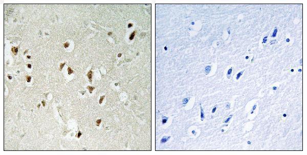
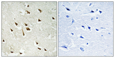
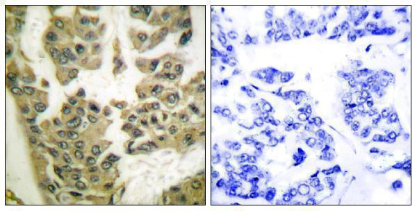


Comprehensive Human Protein Atlas IHC scoring per tissue (reliability: Approved — Medium consistency between antibody staining and RNA expression data. Pending external verification.). Rows are taken directly from the HPA tissue chart — click any row's HPA link to view the source.
| Tissue | Cell type | Level | Evidence | Source |
|---|---|---|---|---|
| Breast | Glandular cells | High | Protein (IHC) | HPA → |
| Cerebellum | Purkinje cells | High | Protein (IHC) | HPA → |
| Cerebral cortex | Neuronal cells | High | Protein (IHC) | HPA → |
| Endometrium | Glandular cells | High | Protein (IHC) | HPA → |
| Epididymis | Glandular cells | High | Protein (IHC) | HPA → |
| Tissue | Cell type | Level | Evidence | Source |
|---|---|---|---|---|
| Liver | Cholangiocytes | Not detected | Protein (IHC) | HPA → |
Troubleshoot ARHGAP35 staining in paraffin sections by checking retrieval, compartment, cell identity and controls before comparing signal across samples.
Two antibodies have human paraffin-section IHC images (catalog image captions); both list Human, Mouse, and Rat reactivity and IF applications (catalog application and reactivity lists).
A03592 has a human paraffin-embedded brain IHC image with a peptide-blocked control and lists IF (A03592 image caption; catalog application list). P30433 has human paraffin-embedded brain and breast carcinoma IHC images with peptide-blocked controls and lists IF and ICC (P30433 image captions; catalog application list).
Which to pick: For tissue IHC, choose A03592 for ARHGAP35 or P30433 when Tyr1105 phosphorylation is the target; each has its own human paraffin-section IHC image, and neither caption reports the fixative (catalog titles; respective IHC image captions). For IF/ICC, P30433 lists both applications and is described as polyclonal, while A03592 lists IF and has no reported clonality; neither has an IF image in the payload (catalog application lists; P30433 dilution record; A03592 catalog record; catalog IF image lists). Both list Human, Mouse, and Rat reactivity for cross-species work, while their supplied IHC images show human tissue (catalog reactivity lists; respective IHC image captions).