ARHGDIA / Rho GDP-dissociation inhibitor 1 · IHC design guide

Design Immunohistochemistry for ARHGDIA

Plan chromogenic IHC of paraffin sections around the cytoplasmic tissue pattern reported for ARHGDIA (HPA tissue IHC). Start with the IHC-validated antibody at 1:100–1:300 (datasheet: A03135-1), and compare staining across cells and controls.

Evidence assembled Oct 2026 · For research use; verify linked source records and product datasheet before use
Immunohistochemistry protocol sheet for ARHGDIA (IHC for ARHGDIA): expected localisation Cytoplasmic across tissues (HPA tissue IHC), antibody A03135-1, validated IHC image, and IHC protocol steps
Printable ARHGDIA IHC protocol sheet — expected localisation Cytoplasmic across tissues (HPA tissue IHC), antibody A03135-1, controls and protocol steps. Open the full ARHGDIA IHC guide →

ARHGDIA Immunohistochemistry Experimental Design Guide

Expected localisation, validated protocols, controls and antibodies — the at-a-glance facts below, then the full design guide.

Must know before staining
Expected localisation Cytoplasmic across tissues (HPA tissue IHC)
Staining pattern Widespread cytoplasmic staining in tissue cells (HPA tissue IHC)
Antigen retrieval Citrate pH 6.0 HIER, 95–98 °C, 20 min (rule: cytoplasmic / membrane antigen)
Positive control ⓘ Appendix+4 more · see all
Negative control ⓘ None in HPA (detected in all 45 tissues); use no-primary + isotype controls
Important caveats
Reasons your staining may differ from the expected pattern.
Fixation Keep fixation consistent across paraffin sections. (standard IHC practice; not target-specific)
Caveat Staining and RNA show medium consistency (HPA tissue IHC)
Regulation Expression regulation is unreported (UniProt)
Isoform / epitope 2 isoforms; epitope coverage is unreported (UniProt)
Section 1

Recommended ARHGDIA IHC & IF Protocols

The catalog antibody’s IHC-P protocol is accompanied by three published ARHGDIA IHC methods (PMC4991396; PMC5250662; PMC3445469).

Recommended immunohistochemistry (IHC-P) protocol parameters
SampleParaffin-embedded human cervix carcinoma tissue; fixative not specified (datasheet A03135-1)
FixationImage fixative and duration unreported (datasheet A03135-1); verify before use.
Sectioning4–5 µm sections on charged slides (standard)
DeparaffinisationXylene, graded ethanol series to water (standard)
Antigen retrievalHeat-induced epitope retrieval in citrate buffer, pH 6.0, 20 min at 95–98 °C (standard rule: cytoplasmic / membrane antigen)
Peroxidase block3% H2O2, 10 min, room temperature (standard)
Blocking10% normal serum of the secondary host, 30 min, room temperature (standard)
Primary antibodyRabbit anti-ARHGDIA, 1:100 - 1:300 (datasheet A03135-1)
Primary incubationOvernight at 4 °C (standard)
DetectionHRP-polymer secondary, DAB chromogen 5–10 min (standard)
CounterstainHematoxylin, blue, dehydrate and mount (standard)
Expected resultARHGDIA-positive staining in glandular cells of appendix (HPA tissue IHC: High). HPA tissue profile: Ubiquitous cytoplasmic expression. No signal in the no-primary control.
💡Decision noteStart with citrate pH 6.0 HIER at 95–98 °C for 20 min (page retrieval rule); ARHGDIA is cytoplasmic (UniProt P52565).
Section 2

What Is the Expected ARHGDIA Staining Pattern?

ARHGDIA is expected mainly in the cytoplasm, with a cytosolic distribution consistent with its lack of a transmembrane segment (UniProt P52565; HPA: supported cytosol). Tissue IHC shows broad cytoplasmic staining, including high staining in several glandular cell populations and lung macrophages (HPA: ubiquitous cytoplasmic expression; High in listed cells). HPA rates the tissue staining Approved, with medium consistency between staining and RNA data (HPA: reliability summary).

What am I looking at on my slide?
Cytoplasmic staining in appendix, cervix, gallbladder or thyroid glandular cells.This matches the reported compartment and high staining in those cell populations (UniProt P52565: cytoplasm; HPA: High in glandular cells). Interpret the stained cells on the section rather than treating every cell in the tissue as equally positive.
Strong nuclear-only staining, with little or no cytoplasmic signal.This conflicts with the reported cytoplasmic and supported cytosolic locations (UniProt P52565; HPA: subcellular). Check the staining controls and antibody conditions before scoring it as ARHGDIA; nuclear staining alone does not establish target localisation.
Strong staining in a cell population reported as Low by HPA.For example, strong signal in skeletal myocytes or bronchial respiratory epithelial cells differs from HPA's Low observations (HPA: Low in those cells). Assess morphology and controls for cross-reactivity or endogenous detection activity; Low is not an absence claim.
Uniform haze across cells, stroma and clear areas.A pattern that obscures cell boundaries cannot establish the expected cytoplasmic distribution (UniProt P52565; HPA: cytoplasmic expression). In chromogenic IHC, background can arise from detection activity or inadequate blocking or washing (general IHC practice).
No cytoplasmic signal in a selected high-staining reference tissue.An absent signal in appendix glandular cells or lung macrophages conflicts with the reported High observations (HPA: High in those cells). First confirm the intended cells are present, then assess antibody and detection controls; one negative section cannot establish absent ARHGDIA.
💡Expected ARHGDIA appearanceCall a section positive when the expected cells show predominantly cytoplasmic chromogen, potentially strong in HPA High populations; isolated nuclear signal or uniform noncellular colour is suspect (UniProt P52565: cytoplasm; HPA: High populations; general IHC interpretation).
How each factor affects the staining
Compartment and topologyARHGDIA is cytoplasmic and has no transmembrane segment (UniProt P52565); HPA supports cytosol by ICC-IF (HPA: subcellular). Use a cytoplasmic pattern as the localisation check, without requiring a membrane rim.
Cell population and tissue choiceHPA reports High staining in lung macrophages, pancreatic exocrine glandular cells and testicular Leydig cells, but Low staining in cardiomyocytes and skeletal myocytes (HPA: tissue IHC). Compare like cell populations when judging intensity.
Strength of tissue evidenceHPA calls the tissue IHC profile Approved and describes medium consistency with RNA data (HPA: reliability summary). The listed antibodies are IHC Approved; the payload does not designate either as IHC Enhanced (HPA: antibody validation).
Isoforms and modificationsUniProt lists two isoforms and several modified residues, including phosphorylation and acetylation (UniProt P52565). No supplied epitope map links these features to the catalog antibody, so their effects on IHC staining cannot be predicted.
Processing and secretionUniProt lists no signal peptide, propeptide or transmembrane segment, and describes a 2–204 chain (UniProt P52565). These annotations support looking for intracellular staining; they do not define a tissue-specific staining threshold.
IF/ICC cross-check?HPA reports supported cytosol localisation and ICC-IF images in A-431, U-251MG and U2OS (HPA: subcellular). IF/ICC can check compartment agreement; those images do not establish an IHC-P staining condition.
Why is my staining missing, weak or wrong?
SituationLikely causeNext action
Known high-staining cells remain blank.The selected cell population may be absent, or the IHC detection run may have failed (HPA: High cell populations; general IHC practice).Confirm the cells on a counterstained section; inspect a concurrent positive control, reagent application and chromogen development before interpreting the target section (general IHC practice).
Signal appears only in nuclei.The compartment conflicts with cytoplasm and supported cytosol (UniProt P52565; HPA: subcellular); the observation alone does not identify the cause.Review morphology and a no-primary control, then reassess the antibody staining conditions before calling the nuclear pattern specific (general IHC practice).
Brown colour is widespread beyond cell boundaries.Endogenous detection activity or nonspecific background may obscure cellular localisation (general chromogenic IHC practice).Compare a no-primary control; check the detection system's blocking and washing steps, then rescore only interpretable cellular staining (general IHC practice).
A Low-rated population stains as strongly as the selected positive population.This differs from HPA's relative observations; nonspecific antibody binding or endogenous detection activity is possible (HPA: Low and High cell populations; general IHC practice).Identify each cell type on the counterstain and compare controls. Treat HPA's Low rating as a reference observation, not proof that every positive cell is artefactual (HPA: tissue IHC).
Only the tissue edge stains strongly.Uneven reagent coverage or edge-related background can distort a chromogenic section (general IHC practice).Check section coverage and control slides; judge the expected cytoplasmic pattern in intact interior cells before assigning an intensity score (general IHC practice).
IHC and an IF/ICC image appear to disagree.HPA's supported IF/ICC result concerns cytosolic localisation in imaged cell lines, whereas its tissue IHC profile describes cell populations in sections (HPA: subcellular; tissue IHC).Compare the compartment within clearly identified cells first. Investigate controls and specimen identity before treating a difference in intensity across preparations as a localisation conflict (general IHC/IF practice).

Sample controls for ARHGDIA IHC & IF

🧪Run cervix first and expect staining in glandular cells (HPA: High in cervix glandular cells). HPA detects ARHGDIA in all 45 scored tissues, so use no-primary and isotype controls for negative comparisons; any less-stained neighboring cells on the positive slide are background references, not validated antigen-negative cells (HPA: no negative tissue rows).
Positive control tissue: Appendix (Glandular cells, HPA High)
Negative control tissue: None in HPA: ARHGDIA is detected in all 45 scored tissues. Use a no-primary (secondary-only) and an isotype control instead.
ICC-IF cell lines (HPA subcellular resource): HPA ICC-IF images show ARHGDIA in A-431, U-251MG, U2OS, with annotated localisation: Cytosol (supported) (HPA subcellular).
Technical controls: Include a no-primary, secondary-only slide and an isotype control matched to the primary antibody’s host species and clonality; confirm biological specificity with a knockout sample or peptide competition, which is shown for the catalog antibody in cervix carcinoma (selected SKU A03135-1 tissue-IHC caption). For chromogenic cervix sections, quench endogenous peroxidase and compare glandular staining against the negative controls (standard IHC practice; HPA: High in cervix glandular cells).
⚠️Feasibility: A target-specific fixation window or fixation effect is unreported, and the selected A03135-1 paraffin-section caption does not state the fixative (selected SKU tissue-IHC caption). Retrieval dependency is unreported; optimize antigen retrieval empirically for paraffin sections (standard IHC practice). The supplied evidence does not establish whether frozen sections or IF are easier; in cervix, assess gland-associated chromogen background against the no-primary slide (HPA: High in cervix glandular cells; standard IHC practice).

HPA tissue IHC evidence for ARHGDIA

Comprehensive Human Protein Atlas IHC scoring per tissue (reliability: Approved — Medium consistency between antibody staining and RNA expression data.). Rows are taken directly from the HPA tissue chart — click any row's HPA link to view the source.

Positive expression · recommended positive controls

TissueCell typeLevelEvidenceSource
Appendix Glandular cells High Protein (IHC) HPA →
Cervix Glandular cells High Protein (IHC) HPA →
Gallbladder Glandular cells High Protein (IHC) HPA →
Lung Macrophages High Protein (IHC) HPA →
Pancreas Exocrine glandular cells High Protein (IHC) HPA →

Undetected expression · recommended negative controls

TissueCell typeLevelEvidenceSource
None in HPA: ARHGDIA is detected in all 45 scored tissues. Use a no-primary (secondary-only) and an isotype control instead.
Section 3

Advanced ARHGDIA IHC Tips

Troubleshoot ARHGDIA staining by checking cytoplasmic localisation, section quality, controls, and the cell populations included in each score.

How should I adjust retrieval when cytoplasmic ARHGDIA staining is weak?
Use citrate pH 6.0 heat-induced epitope retrieval at 95–98 °C for 20 min (page retrieval setting). For weak staining, first check that sections stayed covered by buffer and that the bath reached its set temperature; compare a known positive section processed alongside the sample (general IHC practice; HPA: high in cervix glandular cells). If staining remains weak, test another retrieval buffer or pH on matched sections as a fallback, holding antibody and detection conditions constant (general IHC practice). Score cytoplasmic signal and tissue damage together: ARHGDIA is cytoplasmic, so stronger diffuse counterstain or torn sections do not establish improved detection (UniProt P52565 localisation; general IHC practice).
Could fixation explain weak ARHGDIA staining in my paraffin sections?
Target-specific sensitivity of ARHGDIA staining to fixation is unknown from the supplied evidence; the selected paraffin-section caption does not report its fixative (catalog caption: A03135-1). Record the fixative, fixation duration, processing history, and section age for each block before comparing staining (general IHC practice). On sections from comparable blocks, keep citrate pH 6.0 retrieval at 95–98 °C for 20 min constant while checking whether staining varies with processing history (page retrieval setting; general IHC practice). Include a concurrently stained positive control and assess morphology, since differences in staining alone cannot identify fixation as the cause (HPA: high in cervix glandular cells; general IHC practice).
What staining pattern should I expect, and how should I assess membranous signal?
Expect predominantly cytoplasmic staining in tissue sections: UniProt places ARHGDIA in the cytoplasm, and HPA reports ubiquitous cytoplasmic expression (UniProt P52565 localisation; HPA: tissue profile). Use a 2-compartment review of cytoplasm and nucleus in intact cells, noting any sharp membrane or nuclear predominance separately (general IHC practice). ARHGDIA lacks a transmembrane segment, although it interacts with Rho proteins involved in membrane recycling; that biology alone does not validate a membrane-only stain (UniProt P52565 topology and function). Compare suspect areas with a positive glandular-cell control and a reagent control before assigning an unusual pattern to ARHGDIA (HPA: high in cervix glandular cells; general IHC practice).
Can epitope accessibility or isoforms explain discordant ARHGDIA staining?
ARHGDIA has 2 listed isoforms and several annotated modified residues, including phosphoserines at positions 34 and 47 (UniProt P52565 isoforms and modified residues). The supplied evidence does not map the catalog antibody’s epitope or establish its isoform selectivity, so staining differences cannot be assigned to either mechanism (catalog caption: A03135-1; UniProt P52565 isoforms). If results disagree across specimens, compare adjacent sections using the same citrate pH 6.0 retrieval and detection run, then check whether the difference persists with an independently validated antibody of known epitope (page retrieval setting; general IHC practice). Peptide blocking in the selected image supports binding to that peptide under the pictured conditions but does not identify which isoform is stained (catalog caption: A03135-1).
How can IF help assess an ambiguous chromogenic ARHGDIA pattern?
Use IF as a complementary localisation check, pairing ARHGDIA with a marker of the expected cell type, such as a macrophage marker when examining lung macrophages (HPA: high in lung macrophages; general IF practice). Choose spectrally separated fluorophores and assign the weaker signal to a channel with low tissue autofluorescence; inspect unstained tissue in each channel before interpreting overlap (general IF practice). Because ARHGDIA is cytosolic and has no transmembrane segment, optimise permeabilisation to admit antibody to the cytosol while preserving morphology (HPA: cytosol supported; UniProt P52565 topology; general IF practice). Keep the 2 channels and their single-stain controls separate during acquisition, then compare cellular distribution with adjacent chromogenic sections (general IF/IHC practice).
How do I distinguish ARHGDIA signal from diffuse chromogenic background?
Check whether brown deposit follows intact cytoplasm and expected cell populations rather than pooling at edges, folds, or necrotic areas (HPA: ubiquitous cytoplasmic expression; general IHC practice). Run a no-primary control through the same secondary, peroxidase block, and DAB steps to reveal detection-related signal (general chromogenic IHC practice). Compare this control with a positive section containing glandular cells or macrophages; HPA reports high staining in several glandular populations and lung macrophages (HPA: tissue IHC). If background persists, optimise blocking, washing, and the catalog antibody concentration on matched sections, recording 1 variable changed per comparison (general IHC practice). Peptide blocking is shown for the selected paraffin-section image, but it does not replace tissue and reagent controls (catalog caption: A03135-1; general IHC practice).
What should I score when ARHGDIA staining varies across cell populations? ⚠ ANSWER MARKED FOR VERIFICATION
Define the cell population and region before scoring, since HPA reports high ARHGDIA staining in some glandular cells and lung macrophages but low staining in several other populations (HPA: tissue IHC). For a cell-based readout, report percent positive cells and an H-score using 0–3 intensity grades; score cytoplasm consistently across sections (general IHC practice; UniProt P52565 localisation). Normalise each result to the number of evaluable cells in that defined population, and report excluded necrotic or folded areas (general IHC practice). If counting positive cells per mm², also report cellular density or the eligible tissue area so differences in tissue composition are visible (general IHC practice).
When is an apparent ARHGDIA-positive cell a convincing true positive?
A convincing result shows reproducible cytoplasmic signal in intact cells and appropriate control staining; cytosolic localisation is supported independently of tissue staining (HPA: cytosol supported; UniProt P52565 localisation; general IHC practice). For context, HPA reports high staining in cervix glandular cells and lung macrophages, while its tissue IHC reliability is Approved with medium RNA–protein consistency (HPA: tissue IHC). Inspect 4 common artefact contexts separately: section edges, folds, necrosis, and endogenous peroxidase activity revealed by the no-primary control (general chromogenic IHC practice). Predominantly nuclear or sharply membrane-restricted deposit warrants control review before interpretation because ARHGDIA is cytoplasmic and lacks a transmembrane segment (UniProt P52565 localisation and topology; general IHC practice).
Boster reagents

Best ARHGDIA / Rho GDP-dissociation inhibitor 1 IHC Antibodies

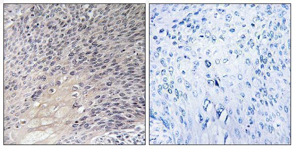
A03135-1 has an IHC image from paraffin-embedded human cervix carcinoma (catalog IHC image caption) and lists IHC, IF and ICC applications for human, mouse and rat (catalog applications/reactivity).

Real IHC data Immunohistochemistry analysis of paraffin-embedded human cervix carcinoma tissue, using ARHGDIA Antibody. The picture on the right is blocked with the synthesized peptide.
Anti-ARHGDIA/Rhogdi Antibody
Cat # A03135-1

A03135-1 is the sole rendered card; its IHC image shows paraffin-embedded human cervix carcinoma with a peptide-blocked comparison (catalog IHC image caption). IF and ICC are listed applications, and human, mouse and rat are listed reactivities; no IF image is supplied (catalog applications/reactivity; catalog IF image alts).

Which to pick: Choose A03135-1 for paraffin-section IHC because its own caption documents that sample preparation; the fixative is unreported (catalog IHC image caption). A03135-1 is also listed for IF/ICC, although it has no IF image (catalog applications; catalog IF image alts). For cross-species work, both A03135-1 and the monoclonal M03135-2 list human, mouse and rat reactivity, but only A03135-1 has an IHC image in the payload (catalog reactivity; catalog clone; catalog IHC image alts).

Each figure is that product's own IHC / IF validation image from its datasheet.

References

  1. UniProt Consortium. UniProt entry P52565 (GDIR1_HUMAN, Rho GDP-dissociation inhibitor 1).
  2. Human Protein Atlas. ARHGDIA tissue IHC expression (reliability: Approved).
  3. Human Protein Atlas. ARHGDIA subcellular location (ICC-IF): Localized to the cytosol..
  4. Human Protein Atlas. ARHGDIA antibody validation summary (2 antibodies).
  5. Interplay between PCBP2 and miRNA modulates ARHGDIA expression and function in glioma migration and invasion. Oncotarget 2016 — PMC4991396.
  6. Downregulation of ARHGDIA contributes to human glioma progression through activation of Rho GTPase signaling pathway. Tumour biology : the journal of the International Society for Oncodevelopmental Biology and Medicine 2016 — PMC5250662.
  7. Proteomic analysis of adrenocorticotropic hormone treatment of an infantile spasm model induced by N-methyl-D-aspartic acid and prenatal stress. PloS one 2012 — PMC3445469.
  8. PubMed PMID:8262133 — UniProt-cited evidence.
  9. PubMed PMID:14702039 — UniProt-cited evidence.
  10. PubMed PMID:16625196 — UniProt-cited evidence.