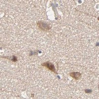
ARHGEF7 / Rho guanine nucleotide exchange factor 7 · IHC design guide

Design Immunohistochemistry for ARHGEF7

Plan chromogenic IHC-P for ARHGEF7 with antibody titration and tissue controls. Expect broad cytoplasmic staining in tissue sections (HPA tissue IHC); UniProt also places the protein at cell adhesions (UniProt).

Evidence assembled Oct 2026 · For research use; verify linked source records and product datasheet before use
Immunohistochemistry protocol sheet for ARHGEF7 (IHC for ARHGEF7): expected localisation Tissue cytoplasm (HPA tissue IHC); cell adhesions molecularly (UniProt), antibody A02764-2, validated IHC image, and IHC protocol steps
Printable ARHGEF7 IHC protocol sheet — expected localisation Tissue cytoplasm (HPA tissue IHC); cell adhesions molecularly (UniProt), antibody A02764-2, controls and protocol steps. Open the full ARHGEF7 IHC guide →

ARHGEF7 Immunohistochemistry Experimental Design Guide

Expected localisation, validated protocols, controls and antibodies — the at-a-glance facts below, then the full design guide.

Must know before staining
Expected localisation Tissue cytoplasm (HPA tissue IHC); cell adhesions molecularly (UniProt)
Staining pattern Broad cytoplasmic staining across cell types (HPA tissue IHC)
Antigen retrieval EDTA pH 8.0 HIER, heat-mediated (datasheet A02764-2)
Positive control ⓘ Adipose tissue+4 more · see all
Negative control ⓘ Salivary gland+1 more · see all
Important caveats
Reasons your staining may differ from the expected pattern.
Fixation Keep tissue fixation consistent across samples. (standard IHC practice; not target-specific)
Caveat Some cell types lack detectable staining (HPA tissue IHC)
Regulation No expression regulator annotated (UniProt)
Isoform / epitope 6 isoforms; variant epitope coverage is unknown (UniProt)
Section 1

Recommended ARHGEF7 IHC & IF Protocols

Compare the catalog antibody’s IHC-P protocol (datasheet A02764-2) with the published colorectal adenocarcinoma IHC protocol (PMC6192735).

Recommended immunohistochemistry (IHC-P) protocol parameters
SampleParaffin-embedded human liver cancer tissue; fixative not specified (datasheet A02764-2)
FixationImage fixative and duration unreported (datasheet A02764-2); verify before use.
Sectioning4–5 µm sections on charged slides (standard)
DeparaffinisationXylene, graded ethanol series to water (standard)
Antigen retrievalHeat retrieval: EDTA pH 8.0 (datasheet A02764-2); 20 min, 95–100 °C (standard)
Peroxidase block3% H2O2, 10 min, room temperature (standard)
Blocking10% goat serum (datasheet A02764-2)
Primary antibodyRabbit anti-ARHGEF7, 2-5 μg/ml (datasheet A02764-2)
Primary incubationOvernight at 4 °C (datasheet A02764-2)
DetectionHRP-conjugated secondary, DAB chromogen (datasheet A02764-2)
CounterstainHematoxylin, blue, dehydrate and mount (standard)
Expected resultARHGEF7-positive staining in adipocytes of adipose tissue (HPA tissue IHC: Medium). HPA tissue profile: Ubiquitous cytoplasmic expression. No signal in the no-primary control.
💡Decision noteStart with heat-mediated EDTA pH 8.0 retrieval (datasheet A02764-2); the published IHC protocol also used EDTA pH 8.0 (PMC6192735).
Section 2

What Is the Expected ARHGEF7 Staining Pattern?

Expect predominantly cytoplasmic ARHGEF7 staining in several cell types, including adipocytes, glandular cells, glia and neurons (HPA tissue IHC: ubiquitous cytoplasmic expression; listed cells Medium). Signal may also occur at the cell cortex and adhesions (UniProt Q14155 localisation). ARHGEF7 has no transmembrane segment (UniProt Q14155 topology). HPA rates its tissue IHC profile Approved, with medium consistency between antibody staining and RNA expression (HPA tissue IHC).

What am I looking at on my slide?
Cytoplasmic staining in adipocytes or breast, adrenal, appendix or duodenal glandular cells.These are plausible positive cells; HPA reports Medium staining in each (HPA tissue IHC). Judge their signal against neighbouring background and tissue morphology (general IHC practice).
Signal is predominantly nuclear, with little or no cytoplasmic staining.This departs from the reported cytoplasmic profile and UniProt localisation (HPA tissue IHC; UniProt Q14155). Check counterstain overlap and detection background before assigning it to ARHGEF7 (general IHC practice).
Strong staining in salivary glandular cells or smooth muscle cells.Both are listed as Not detected (HPA tissue IHC). Consider cross-reactivity or endogenous detection activity; compare with a no-primary control and review morphology (general IHC practice).
Diffuse colour covers cells and surrounding tissue without clear cell boundaries.This obscures the expected cytoplasmic pattern (HPA tissue IHC). Review blocking, washing, primary concentration and detection exposure with a no-primary control (general IHC practice).
No signal in an intact expected-positive region, such as cerebral cortex neurons.HPA reports Medium neuronal staining there (HPA tissue IHC). First check the positive control, tissue preservation and IHC-P workflow before treating the absence as biological (general IHC practice).
💡Expected ARHGEF7 appearanceCall a result positive when identifiable expected cells show predominantly cytoplasmic signal of roughly Medium intensity (HPA tissue IHC); dominant nuclear signal or strong staining in HPA Not detected cell types warrants investigation (HPA tissue IHC; UniProt Q14155 localisation).
How each factor affects the staining
Antibody evidenceHPA004744 is listed as IHC Approved, while the tissue profile has medium staining–RNA consistency (HPA antibodies; HPA tissue IHC). Interpret a surprising result with controls rather than treating approval as proof of specificity in every section (general IHC practice).
Subcellular distributionARHGEF7 is reported at the cortex, ruffles, lamellipodia and cell adhesions; only a small proportion is at focal adhesions (UniProt Q14155 localisation). A punctate peripheral accent can fit, but focal adhesion staining need not dominate an IHC section.
Tissue and cell contextHPA describes ubiquitous cytoplasmic expression and low RNA tissue specificity, yet individual cells range from Medium to Not detected (HPA tissue IHC). Score the named cell population, not an entire organ as uniformly positive.
Isoforms and epitope coverageUniProt lists 6 isoforms (UniProt Q14155). The supplied record gives no antibody epitope, so it cannot establish which isoforms HPA004744 detects; seek epitope documentation before attributing a staining difference to isoform choice.
Processing and topologyUniProt records one chain spanning residues 1–803, no signal peptide or propeptide, and no transmembrane segment (UniProt Q14155). The record provides no basis to expect a cleaved, secreted or membrane-spanning staining pattern.
Antigen retrievalThe supplied HPA and UniProt records give no ARHGEF7-specific retrieval condition or fixation sensitivity. Use the catalog antibody's IHC-P protocol for a starting condition, then assess morphology and controls (general IHC practice).
IF/ICC Q: Is an HPA cell-image pattern available?A: No main ICC-IF location or cell-line images are supplied (HPA subcellular). UniProt reports cortex and adhesion localisation, but the supplied HPA record cannot validate an IF/ICC image pattern (UniProt Q14155; HPA subcellular).
Why is my staining missing, weak or wrong?
SituationLikely causeNext action
The expected-positive control is blank.An IHC-P processing, reagent or detection failure is possible (general IHC practice); HPA reports Medium staining in the named positive cells (HPA tissue IHC).Check the catalog antibody's IHC-P instructions, reagent activity and control section before interpreting test tissue as negative (general IHC practice).
Signal is weak in expected-positive cells.The detection setting may be insufficient, or the sampled cells may differ from the HPA-listed population (general IHC practice; HPA tissue IHC).Confirm cell identity, then optimise primary concentration and development time using positive and no-primary controls (general IHC practice).
Nearly every structure has brown colour.Diffuse background may arise from insufficient blocking, washing or excessive detection development (general IHC practice).Inspect the no-primary control; adjust blocking, washes and development while preserving visible expected cytoplasm (general IHC practice; HPA tissue IHC).
Nuclei appear to carry the strongest signal.Counterstain or nonspecific detection may be mistaken for target signal; HPA's reported pattern is cytoplasmic (general IHC practice; HPA tissue IHC).Compare chromogen and counterstain appearances and review the no-primary control before scoring nuclear positivity (general IHC practice).
Salivary glandular or smooth muscle cells stain strongly.Cross-reactivity or endogenous detection activity is possible because these cells are Not detected in the supplied tissue profile (HPA tissue IHC; general IHC practice).Compare a no-primary control and a listed positive tissue; review cell identity and detection blocking (general IHC practice; HPA tissue IHC).
Peripheral dots are absent despite clear cytoplasmic staining.Only a small proportion of ARHGEF7 is reported at focal adhesions, so such detail may be subtle (UniProt Q14155 localisation).Assess the cytoplasmic cell pattern first; reserve a claim of focal adhesion localisation for evidence that resolves those structures (HPA tissue IHC; UniProt Q14155 localisation).

Sample controls for ARHGEF7 IHC & IF

🧪Run adipose tissue first and look for staining in adipocytes (HPA: Medium in adipocytes). Run salivary gland as the negative tissue (HPA: Not detected in glandular cells); on the adipose slide, non-adipocyte stromal cells should show background-level staining as provisional internal comparators, since their ARHGEF7 status is not established by the supplied HPA row.
Positive control tissue: Adipose tissue (Adipocytes, HPA Medium)
Negative control tissue: Salivary gland (HPA Not detected)
ICC-IF cell lines (HPA subcellular resource): HPA carries no ICC-IF cell line for ARHGEF7; derive a cell-line control from the positive tissue's cell type (Adipocytes) and confirm it by RNA or western blot first.
Technical controls: Include no-primary (secondary-only) and concentration-matched rabbit IgG isotype controls matched to the primary antibody’s clonality, plus an ARHGEF7 knockout specimen if available (selected tissue-IHC caption: rabbit primary and anti-rabbit secondary). Block endogenous peroxidase before HRP/DAB detection, and distinguish staining from the clear lipid spaces in adipocytes (selected tissue-IHC caption: HRP/DAB; standard IHC practice).
⚠️Feasibility: A target-specific fixation window or fixation effect is unreported, and the selected A02764-2 tissue-IHC caption does not state a fixative. Its paraffin-section workflow uses heat retrieval in EDTA at pH 8.0, but does not establish whether retrieval is required; the supplied evidence does not show that frozen sections or IF/ICC are easier (selected tissue-IHC caption). In adipose tissue, lipid spaces can complicate interpretation of staining around adipocytes (standard IHC practice).

HPA tissue IHC evidence for ARHGEF7

Comprehensive Human Protein Atlas IHC scoring per tissue (reliability: Approved — Medium consistency between antibody staining and RNA expression data.). Rows are taken directly from the HPA tissue chart — click any row's HPA link to view the source.

Positive expression · recommended positive controls

TissueCell typeLevelEvidenceSource
Adipose tissue Adipocytes Medium Protein (IHC) HPA →
Adrenal gland Glandular cells Medium Protein (IHC) HPA →
Appendix Glandular cells Medium Protein (IHC) HPA →
Breast Glandular cells Medium Protein (IHC) HPA →
Caudate Glial cells Medium Protein (IHC) HPA →

Undetected expression · recommended negative controls

TissueCell typeLevelEvidenceSource
Salivary gland Glandular cells Not detected Protein (IHC) HPA →
Smooth muscle Smooth muscle cells Not detected Protein (IHC) HPA →
Section 3

Advanced ARHGEF7 IHC Tips

Troubleshooting ARHGEF7 staining in paraffin sections starts with the catalog antibody’s documented retrieval conditions and a compartment-aware reading of the signal.

What retrieval should I use when ARHGEF7 staining is weak in paraffin sections?
Start with heat-mediated retrieval in EDTA buffer at pH 8.0 (datasheet A02764-2). The documented paraffin-section stain then used 10% goat serum and 2 μg/ml primary antibody overnight at 4°C (datasheet A02764-2). If signal remains weak, compare retrieval duration on adjacent sections while holding antibody concentration, detection and tissue handling constant (standard IHC practice). Include a section processed without primary antibody to distinguish weak specific staining from detection background (standard IHC practice). Record the retrieval conditions used for every comparison, because the caption reports the buffer and pH but gives no heating time (datasheet A02764-2).
Could fixation explain inconsistent ARHGEF7 staining across paraffin blocks?
Target-specific sensitivity to fixation is unknown; the selected paraffin-section caption does not state its fixative (datasheet A02764-2). Compare blocks with documented fixation histories and process adjacent sections together using EDTA at pH 8.0 (datasheet A02764-2; standard IHC practice). Keep section thickness, retrieval, 2 μg/ml primary concentration and overnight 4°C incubation consistent while assessing the difference (datasheet A02764-2; standard IHC practice). Examine morphology and staining in an internal reference cell population before attributing a weak result to fixation (standard IHC practice). Neither tissue expression patterns nor ARHGEF7’s intracellular location establishes a fixation effect for this antibody (HPA tissue IHC; UniProt Q14155 subcellular location).
Should I expect a membrane rim or diffuse cytoplasmic ARHGEF7 staining?
Read the stain primarily as cytoplasmic, with possible enrichment near cell adhesions or projections rather than a uniform membrane rim (HPA: ubiquitous cytoplasmic expression; UniProt Q14155 subcellular location). ARHGEF7 is reported at the cell cortex, ruffles and lamellipodia, while only a small proportion is detected at focal adhesions (UniProt Q14155 subcellular location). It has no transmembrane segment, so a crisp circumferential membrane pattern alone needs scrutiny (UniProt Q14155 topology; standard IHC interpretation). Compare well-preserved cells away from section edges and use the same retrieval and DAB development across sections (standard IHC practice). Report cytoplasmic intensity separately from any junctional accentuation rather than treating both patterns as equivalent (standard IHC scoring practice).
How should I interpret variable staining if the antibody epitope is unspecified?
ARHGEF7 has 6 annotated isoforms, and the supplied caption does not identify this antibody’s epitope (UniProt Q14155 isoforms; datasheet A02764-2). Its CH, SH3, DH and PH domains occupy distinct regions, so isoform coverage cannot be inferred without the antibody’s mapped immunogen (UniProt Q14155 domains; standard antibody validation practice). Multiple phosphorylation sites are annotated, including serines at positions 153 and 518, but these annotations do not establish altered recognition in sections (UniProt Q14155 modified residues). If staining differs between specimens, check section quality and processing first, then seek epitope and isoform coverage information before assigning a biological cause (standard IHC practice). Document the antibody identifier, retrieval conditions and scoring compartment for comparison (datasheet A02764-2; standard IHC practice).
How can I check a suspected ARHGEF7 pattern by multiplex IF?
Use multiplex IF as a separate check of cell identity and intracellular pattern; the supplied subcellular record lists no ICC/IF images for comparison (HPA subcellular). Pair ARHGEF7 with a marker for the expected cell type, such as a neuronal marker when examining neuronal cells, and assess signal within marker-positive cells (HPA: cerebral cortex neuronal cells, medium; standard IF practice). Choose fluorophores after checking tissue autofluorescence in an unstained section, and include single-label controls to assess channel bleed-through (standard IF practice). ARHGEF7 lacks a transmembrane segment, so permeabilisation should permit antibody access to its intracellular epitope, whose exact position is unspecified here (UniProt Q14155 topology; datasheet A02764-2). Do not transfer the paraffin IHC incubation directly to IF without validation (datasheet A02764-2; standard IF practice).
How do I reduce diffuse brown staining without losing ARHGEF7 signal?
First examine a section without primary antibody to identify background from the detection system (standard IHC practice). The documented paraffin workflow used 10% goat serum, 2 μg/ml primary overnight at 4°C, an anti-rabbit peroxidase secondary for 30 minutes at 37°C, and DAB development (datasheet A02764-2). A peroxidase block and careful DAB timing are general chromogenic IHC measures, not evidence specific to ARHGEF7 (standard IHC practice). If the control is clean but tissue staining remains diffuse, titrate the primary around the documented concentration and compare washed, adjacent sections (datasheet A02764-2; standard IHC practice). Preserve interpretable cytoplasmic signal while checking for stronger staining at tissue edges or damaged areas (HPA: ubiquitous cytoplasmic expression; standard IHC practice).
What should I score when comparing ARHGEF7 staining between specimens? ⚠ ANSWER MARKED FOR VERIFICATION
Define the cell population and cytoplasmic compartment before scoring, since the tissue profile describes ubiquitous cytoplasmic expression (HPA tissue IHC; standard IHC scoring practice). Record the percentage of positive cells and intensity, or use a cytoplasmic H-score from 0–300 with fixed intensity thresholds (standard IHC scoring practice). For spatial questions, count positive cells per mm² of viable tissue and report the sampled area (standard IHC scoring practice). Normalise comparisons to the same cell type, viable tissue area, section processing and image acquisition settings (standard IHC scoring practice). Keep any focal adhesion-like accentuation separate from bulk cytoplasmic scoring, because only a small ARHGEF7 fraction is reported at focal adhesions (UniProt Q14155 subcellular location).
How can I distinguish genuine ARHGEF7 staining from artefact?
Favor reproducible cytoplasmic staining in intact cells, with possible cortical or adhesion-associated accentuation (HPA: ubiquitous cytoplasmic expression; UniProt Q14155 subcellular location). Check cell identity: medium staining is reported in cerebral cortex neuronal cells and adipocytes, whereas salivary gland glandular cells and smooth muscle cells were not detected in the listed tissue results (HPA tissue IHC). Treat isolated nuclear staining, tissue-edge halos and necrotic deposits cautiously and compare them with morphology and the no-primary control (UniProt Q14155 subcellular location; standard IHC interpretation). Evaluate brown signal remaining without primary antibody for endogenous enzyme or detection-system contribution (standard IHC practice). The tissue profile is approved with medium consistency against RNA, so use controls and replicate sections before drawing a biological conclusion (HPA tissue IHC reliability; standard IHC practice).
Boster reagents

Best ARHGEF7 / Rho guanine nucleotide exchange factor 7 IHC Antibodies

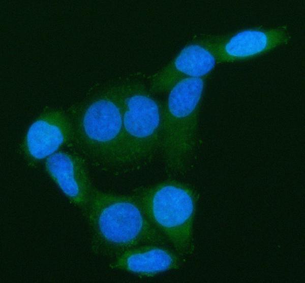
Two anti-ARHGEF7 antibodies have human paraffin-section IHC images (catalog IHC captions); one also has IF images from HeLa cells and human paraffin sections (A02764-2 IF captions).

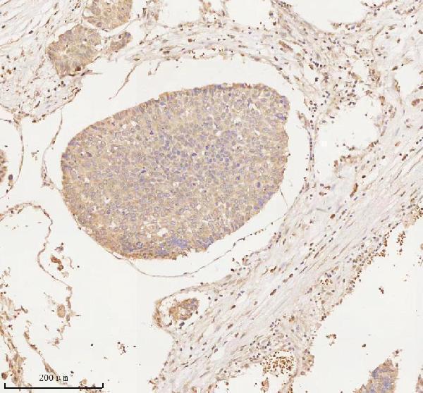
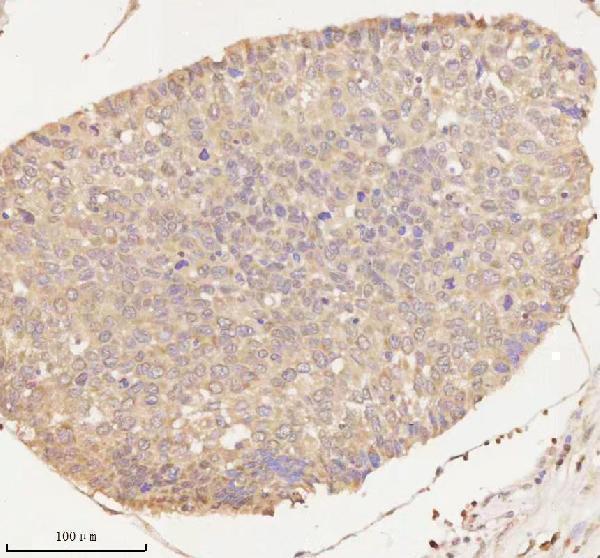
Real IHC data IHC analysis of ARHGEF7 using anti-ARHGEF7 antibody (A02764-2). ARHGEF7 was detected in a paraffin-embedded section of human liver cancer tissue. Heat mediated antigen retrieval was performed in EDTA buffer (pH 8.0, epitope retrieval solution). The tissue section was blocked with 10% goat serum. The tissue section was then incubated with 2 μg/ml rabbit anti-ARHGEF7 Antibody (A02764-2) overnight at 4°C. Peroxidase Conjugated Goat Anti-rabbit IgG was used as secondary antibody and incubated for 30 minutes at 37°C. The tissue section was developed using HRP Conjugated Rabbit IgG Super Vision Assay Kit (Catalog # SV0002) with DAB as the chromogen.
Anti-ARHGEF7 Antibody ®
Cat # A02764-2
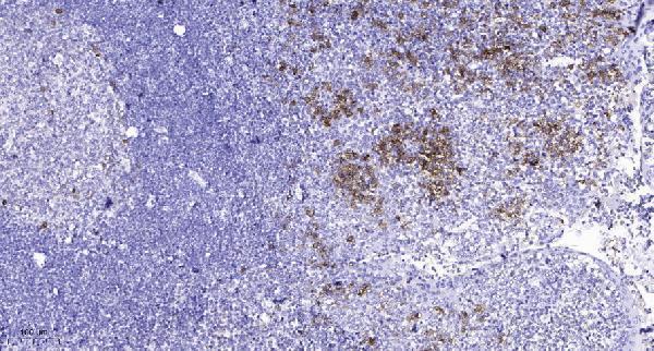
Real IHC data Immunohistochemical analysis of paraffin-embedded human tonsil. 1, Antibody was diluted at 1:200(4° overnight). 2, Tris-EDTA,pH9.0 was used for antigen retrieval. 3,Secondary antibody was diluted at 1:200(room temperature, 45min).
Anti-ARHGEF7/Beta Pix Antibody
Cat # A02764

A02764 will render with an IHC image of paraffin-embedded human tonsil and lists IHC and IF applications (A02764 catalog and IHC caption). A02764-2 will render with an IHC image of paraffin-embedded human liver cancer tissue; its catalog also includes IF images from HeLa cells and human paraffin sections (A02764-2 catalog, IHC and IF captions).

Which to pick: For tissue IHC, choose A02764 for the documented tonsil example using Tris-EDTA at pH 9.0, or A02764-2 for the documented liver cancer example using EDTA at pH 8.0; both captions describe paraffin sections and leave the fixative unreported (respective IHC captions). For IF/ICC, A02764-2 has image examples, while A02764 lists IF/ICC without an IF image in the payload (respective catalogs). Both list Human, Mouse and Rat reactivity, but their supplied IHC images show human tissue; A02764 is identified as polyclonal, while A02764-2 has no clonality stated (respective catalogs).

Each figure is that product's own IHC / IF validation image from its datasheet.

References

  1. UniProt Consortium. UniProt entry Q14155 (ARHG7_HUMAN, Rho guanine nucleotide exchange factor 7).
  2. Human Protein Atlas. ARHGEF7 tissue IHC expression (reliability: Approved).
  3. Human Protein Atlas. ARHGEF7 subcellular location (ICC-IF): Highest expression in JURKAT: 75.7 nTPM.
  4. Human Protein Atlas. ARHGEF7 antibody validation summary (1 antibodies).
  5. ARHGEF7 promotes metastasis of colorectal adenocarcinoma by regulating the motility of cancer cells. International journal of oncology 2018 — PMC6192735.
  6. Arhgef7 is essential for granule cell precursor proliferation and migration during cerebellum development. iScience 2026 — PMC12828525.
  7. PubMed PMID:8590280 — UniProt-cited evidence.
  8. PubMed PMID:9726964 — UniProt-cited evidence.
  9. PubMed PMID:17974005 — UniProt-cited evidence.