ARID1B / AT-rich interactive domain-containing protein 1B · IHC design guide

Design Immunohistochemistry for ARID1B

Use nuclear staining as the ARID1B readout in paraffin sections, with hematopoietic and neuronal cells as high-staining references (HPA tissue IHC). Start the catalog antibody at 1:100–1:300 and score each cell type against a negative control (datasheet A02556-1; standard IHC practice).

Evidence assembled Oct 2026 · For research use; verify linked source records and product datasheet before use
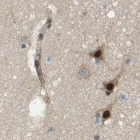
Immunohistochemistry protocol sheet for ARID1B (IHC for ARID1B): expected localisation Mainly nuclear in tissue (HPA tissue IHC), antibody A02556-1, validated IHC image, and IHC protocol steps
Printable ARID1B IHC protocol sheet — expected localisation Mainly nuclear in tissue (HPA tissue IHC), antibody A02556-1, controls and protocol steps. Open the full ARID1B IHC guide →

ARID1B Immunohistochemistry Experimental Design Guide

Expected localisation, validated protocols, controls and antibodies — the at-a-glance facts below, then the full design guide.

Must know before staining
Expected localisation Mainly nuclear in tissue (HPA tissue IHC)
Staining pattern Nuclear staining across tissue cell types (HPA tissue IHC)
Antigen retrieval Tris-EDTA pH 8.0 HIER, heat-mediated (datasheet A02556-1)
Positive control ⓘ Bone marrow+4 more · see all
Negative control ⓘ None in HPA (detected in all 44 tissues); use no-primary + isotype controls
Important caveats
Reasons your staining may differ from the expected pattern.
Fixation Keep fixation consistent across sections (standard IHC practice; not target-specific)
Caveat Low-staining cell populations may be missed (HPA tissue IHC)
Regulation BAF subunits switch during neural development (UniProt)
Isoform / epitope 4 isoforms; check epitope coverage (UniProt)
Section 1

Recommended ARID1B IHC & IF Protocols

The catalog antibody’s IHC-P protocol (datasheet: A02556-1) is accompanied by four published ARID1B IHC protocols (PMC6032401; PMC6396229; PMC8034473; PMC5687163).

Recommended immunohistochemistry (IHC-P) protocol parameters
SampleParaffin-embedded Human breast cancer tissue; fixative not specified (datasheet A02556-1)
FixationImage fixative and duration unreported (datasheet A02556-1); verify before use.
Sectioning4–5 µm sections on charged slides (standard)
DeparaffinisationXylene, graded ethanol series to water (standard)
Antigen retrievalHeat retrieval: Tris-EDTA pH 8.0 (datasheet A02556-1); 20 min, 95–100 °C (standard)
Peroxidase block3% H2O2, 10 min, room temperature (standard)
Blocking10% normal serum of the secondary host, 30 min, room temperature (standard)
Primary antibodyRabbit anti-ARID1B, 1:100-1:300 (datasheet A02556-1)
Primary incubationOvernight at 4 °C (standard)
DetectionHRP-polymer secondary, DAB chromogen 5–10 min (standard)
CounterstainHematoxylin, blue, dehydrate and mount (standard)
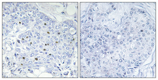
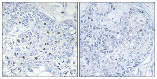
Expected resultARID1B-positive staining in hematopoietic cells of bone marrow (HPA tissue IHC: High). HPA tissue profile: Mainly nuclear expression in all tissues. No signal in the no-primary control.
💡Decision noteStart with heat-mediated Tris-EDTA retrieval at pH 8.0 (datasheet: A02556-1); use sodium citrate when reproducing the specified published protocols (PMC6032401; PMC6396229).
Section 2

What Is the Expected ARID1B Staining Pattern?

ARID1B should stain mainly nuclei across tissues (HPA: tissue IHC profile; UniProt Q8NFD5: nucleus). High staining is reported in cortical neurons, cerebellar molecular-layer cells, and several hematopoietic, glandular, and squamous epithelial populations (HPA: tissue IHC). It has no transmembrane segment (UniProt Q8NFD5: topology). HPA rates its tissue IHC evidence Approved, with medium consistency between staining and RNA expression (HPA: tissue IHC reliability).

What am I looking at on my slide?
Distinct nuclear staining in cortical neuronal cells or bone marrow hematopoietic cells.This fits two reported high-staining cell populations (HPA: cerebral cortex, High in neuronal cells; bone marrow, High in hematopoietic cells). Assess nuclei within the named population rather than judging the whole tissue by its overall color (general IHC practice).
Predominantly cytoplasmic or membrane staining with little nuclear signal.This conflicts with the mainly nuclear tissue pattern (HPA: tissue IHC profile; UniProt Q8NFD5: nucleus). Review controls and detection before assigning it to ARID1B. Additional membrane and cytosol locations in ICC-IF are uncertain (HPA: subcellular location).
Strong staining in an unexpected cell population while the expected nuclei are faint.Compare the cells with HPA’s reported populations: for example, prostate glandular cells have Low staining, whereas oral mucosal squamous epithelial cells have High staining (HPA: tissue IHC). An unexpected pattern may reflect cross-reactivity or endogenous detection activity; it alone does not identify the cause (general IHC practice).
Diffuse color over tissue and spaces between cells, without clear nuclear boundaries.Treat this as background until controls show otherwise (general IHC practice). ARID1B’s reported IHC pattern is mainly nuclear (HPA: tissue IHC profile); broad haze cannot be scored as nuclear positivity simply because the tissue has reported expression.
No nuclear signal in a reported high-staining population.A negative result in, for example, esophageal squamous epithelial cells conflicts with the reported High staining (HPA: esophagus). Check the assay with a known-positive section before interpreting a study sample as negative (general IHC practice). HPA provides no negative tissue list (HPA: tissue IHC).
💡Expected ARID1B appearanceCall a section positive when identifiable cells show predominantly nuclear chromogenic staining, including strong signal where HPA reports High staining; diffuse haze or dominant nonnuclear color is suspect (HPA: tissue IHC profile and cell-level staining; general IHC practice).
How each factor affects the staining
Cell population and tissue contextARID1B is broadly distributed, but reported levels vary by cell population: High in cerebellar molecular-layer cells and Low in liver cholangiocytes, for example (HPA: tissue IHC). Choose and score the specific cells of interest; Low does not mean absent.
Antibody validationHPA016511 has Approved IHC and Supported ICC status; HPA075291 has Supported ICC status with no IHC status listed (HPA: antibodies). ICC support alone does not establish an IHC result. The tissue IHC profile has medium staining–RNA consistency (HPA: reliability).
Isoforms and epitope coverageUniProt lists four ARID1B isoforms (UniProt Q8NFD5: isoforms). The supplied records do not locate an antibody epitope relative to them, so the staining pattern cannot establish which isoforms were detected.
Detection chemistryEndogenous enzyme activity can imitate chromogenic staining when an enzyme-based detection system is used; inspect the relevant detection control before interpreting color as target signal (general IHC practice). This is an assay consideration, not a reported ARID1B property.
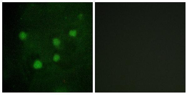
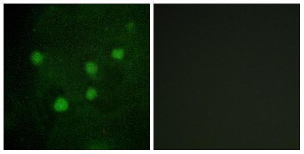
IF/ICC Q: where should fluorescence appear?Mainly in the nucleoplasm (HPA: ICC-IF, supported). Plasma membrane and cytosol are additional uncertain locations (HPA: ICC-IF). Use the separate IF/ICC guide for that application; these ICC-IF observations do not change the mainly nuclear tissue IHC expectation (HPA: tissue IHC).
Why is my staining missing, weak or wrong?
SituationLikely causeNext action
Expected nuclei are blank in a reported High population.The assay may have failed, or the examined section may not contain the named population (general IHC practice); HPA reports High staining only for specified cells, such as epididymal glandular cells (HPA: epididymis).Confirm cell identity and run a reported high-staining tissue alongside the sample. Review antigen retrieval, primary antibody incubation, and detection with the antibody’s IHC instructions (general IHC practice).
Brown signal dominates cytoplasm or cell borders.The pattern conflicts with mainly nuclear tissue IHC (HPA: tissue IHC profile). ICC-IF membrane and cytosol assignments are uncertain and do not validate a dominant chromogenic pattern (HPA: subcellular location).Compare with a reported high-staining tissue and a detection control; inspect whether nuclear signal is present before scoring the section (general IHC practice).
Staining appears in unexpected cells, including populations reported Low.Unexpected color could arise from cross-reactivity or endogenous detection activity (general IHC practice). Low is a reported level, not proof that every cell must be blank (HPA: tissue IHC levels).Check the staining distribution against the named HPA cell population, then compare a control omitting primary antibody and an independent antibody where available (general IHC practice).
Diffuse background hides nuclear boundaries.Nonspecific binding or excess detection signal can obscure the compartment being scored (general IHC practice); the expected tissue pattern is mainly nuclear (HPA: tissue IHC profile).Inspect the no-primary control, review blocking and washing, and adjust antibody or detection conditions within the assay instructions (general IHC practice).
Chromogenic color persists without primary antibody.The primary antibody cannot account for that control signal; endogenous detection activity or reagent background is possible (general IHC practice).Check the detection chemistry and its appropriate blocking or control steps, then repeat the section before calling ARID1B positive (general IHC practice).
An ICC-IF image appears broader than the tissue IHC pattern.ICC-IF supports nucleoplasmic localization but lists cytosol and plasma membrane as uncertain; tissue IHC is mainly nuclear (HPA: subcellular location; tissue IHC profile).Interpret each application against its own validated pattern and controls. For IHC, score identifiable tissue-cell nuclei; consult the separate IF/ICC guide for fluorescence setup (general IHC practice; HPA: tissue IHC profile).

Sample controls for ARID1B IHC & IF

🧪Run bone marrow first: hematopoietic cells should show nuclear staining (HPA: High in hematopoietic cells; UniProt Q8NFD5: nucleus). HPA detects ARID1B in all 44 scored tissues, so there is no supported negative tissue or confirmed internal negative cell population; cells without specific nuclear staining would show only the counterstained nucleus, while no-primary and isotype controls carry the negative comparison (HPA: no negative tissue rows; standard IHC practice).
Positive control tissue: Bone marrow (Hematopoietic cells, HPA High)
Negative control tissue: None in HPA: ARID1B is detected in all 44 scored tissues. Use a no-primary (secondary-only) and an isotype control instead.
ICC-IF cell lines (HPA subcellular resource): HPA ICC-IF images show ARID1B in A-431, U-251MG, U2OS, RT-4, KOLF2.1J, with annotated localisation: Nucleoplasm (supported) (HPA subcellular).
Technical controls: Include a no-primary, secondary-only control and an isotype control matched to the primary antibody’s host species and antibody class; use ARID1B knockout material or an immunogen-peptide block as a biological specificity control (standard IHC practice; A02556-1 caption: peptide-block control). Block endogenous peroxidase when using chromogenic detection in bone marrow (standard IHC practice).
⚠️Feasibility: A target-specific fixation window or fixation effect is unreported in the supplied evidence, and the exact A02556-1 paraffin-section caption does not state a fixative (A02556-1 caption). That caption uses high-pressure, high-temperature Tris-EDTA retrieval at pH 8.0, but does not establish a required retrieval condition for bone marrow (A02556-1 caption: human breast cancer). The supplied evidence does not establish whether frozen sections or IF are easier; bone marrow hematopoietic cells can produce endogenous-peroxidase background in chromogenic IHC (standard IHC practice).

HPA tissue IHC evidence for ARID1B

Comprehensive Human Protein Atlas IHC scoring per tissue (reliability: Approved — Medium consistency between antibody staining and RNA expression data.). Rows are taken directly from the HPA tissue chart — click any row's HPA link to view the source.

Positive expression · recommended positive controls

TissueCell typeLevelEvidenceSource
Bone marrow Hematopoietic cells High Protein (IHC) HPA →
Cerebellum Cells in molecular layer High Protein (IHC) HPA →
Cerebral cortex Neuronal cells High Protein (IHC) HPA →
Epididymis Glandular cells High Protein (IHC) HPA →
Esophagus Squamous epithelial cells High Protein (IHC) HPA →

Undetected expression · recommended negative controls

TissueCell typeLevelEvidenceSource
None in HPA: ARID1B is detected in all 44 scored tissues. Use a no-primary (secondary-only) and an isotype control instead.
Section 3

Advanced ARID1B IHC Tips

Troubleshoot ARID1B chromogenic IHC by checking nuclear localisation, the tested retrieval conditions, and matched section controls (UniProt Q8NFD5; datasheet A02556-1).

Which retrieval conditions should I try first for weak ARID1B staining?
Start with heat-mediated Tris-EDTA at pH 8.0 for ARID1B retrieval in paraffin sections (datasheet A02556-1). The selected antibody image used high pressure and temperature with this buffer, followed by a 1:100 primary dilution overnight at 4 °C (caption A02556-1). If staining remains weak, compare retrieval duration or heating intensity on adjacent sections while holding antibody dilution and detection constant (standard IHC practice). Check that any gain appears mainly in nuclei, the expected compartment, rather than across the whole section (UniProt Q8NFD5 localisation; HPA tissue IHC). Record the tested heating conditions because the caption gives no retrieval duration (caption A02556-1).
Could fixation explain inconsistent nuclear staining between paraffin blocks?
Target-specific fixation sensitivity for ARID1B is unknown from the supplied evidence, and the selected paraffin-section caption does not state its fixative (caption A02556-1). Record the fixative, fixation duration, processing history, and section age for each block before comparing staining (standard IHC practice). Stain adjacent sections together using Tris-EDTA at pH 8.0 and a consistent primary incubation; the selected image used 1:100 overnight at 4 °C (datasheet A02556-1; caption A02556-1). Include a known staining control in each run to separate a block effect from a run effect (standard IHC practice). Interpret a block difference cautiously until those variables are matched (standard IHC practice).
Where should I expect ARID1B signal in a tissue section?
Score convincing ARID1B staining primarily in nuclei: UniProt lists the nucleus, and HPA reports mainly nuclear expression across tissues (UniProt Q8NFD5 localisation; HPA tissue IHC). HPA subcellular data place the principal signal in the nucleoplasm, while plasma membrane and cytosol assignments are uncertain (HPA subcellular). Compare chromogen with the nuclear counterstain at high magnification so staining on adjacent cell borders does not appear nuclear (standard IHC practice). ARID1B has no transmembrane segment, so prominent membrane-only staining warrants a specificity check (UniProt Q8NFD5 topology). Review a no-primary control and the selected antibody’s peptide-preabsorbed comparison when investigating unexpected compartments (standard IHC practice; caption A02556-1).
Could isoforms or epitope masking change my ARID1B IHC result?
ARID1B has 4 listed isoforms, and its ARID domain spans residues 1136–1227 (UniProt Q8NFD5 isoforms and domains). The supplied caption gives no antibody epitope, so it cannot establish which isoforms the selected antibody detects (caption A02556-1). Check the immunogen sequence against the isoforms before treating a negative nuclear result as protein loss (standard antibody validation practice; UniProt Q8NFD5 isoforms). UniProt lists modified residues, including dimethylarginine at 487 and phosphoserine at 585, but their effect on this antibody’s staining is unreported (UniProt Q8NFD5 modified residues; caption A02556-1). Compare independently mapped epitopes if isoform coverage matters to the study (standard antibody validation practice).
How can I use IF to investigate an ambiguous chromogenic pattern?
On a matched section, pair ARID1B with a marker for the cell population being assessed and a nuclear counterstain, then compare nuclear overlap cell by cell (standard IF practice; UniProt Q8NFD5 localisation). Choose fluorophores after checking the tissue’s autofluorescence and the available filter channels, and include single-stain controls for multiplexing (standard IF practice). ARID1B has no transmembrane segment and is chiefly nucleoplasmic, so assess nuclear access when selecting permeabilisation conditions; the antibody epitope is unreported (UniProt Q8NFD5 topology; HPA subcellular; caption A02556-1). Check whether apparent cytosolic or membrane fluorescence persists in controls before comparing it with chromogenic IHC (HPA subcellular; standard IF practice). Keep IF fixation and antibody conditions separately documented because the supplied tissue caption describes paraffin IHC (caption A02556-1).
How do I reduce diffuse brown staining without losing nuclear signal?
First inspect a no-primary section for detection-system background and compare it with the stained section under the same illumination (standard IHC practice). Use a peroxidase block before chromogenic detection, then reassess blocking, primary concentration, wash stringency, and DAB development one variable at a time (standard IHC practice). The selected image used 1:100 antibody overnight at 4 °C, which provides a documented starting condition rather than a universal optimum (caption A02556-1). Check whether any adjustment preserves discrete nuclear staining, the expected ARID1B pattern (UniProt Q8NFD5 localisation; HPA tissue IHC). The caption includes a peptide-preabsorbed negative comparison, which can help assess staining attributable to that antibody under the tested conditions (caption A02556-1).
What should I measure when comparing ARID1B across samples? ⚠ ANSWER MARKED FOR VERIFICATION
Define the cell population and nuclear positivity threshold before scoring, using the same rules across sections (standard IHC quantification practice; HPA tissue IHC). Report the percentage of positive nuclei and, when staining intensity is reproducible, a nuclear H-score; give the number of evaluable cells and fields (standard IHC quantification practice). Normalise positive counts to all evaluable nuclei in that cell population, or report positive cells per mm² when tissue area is the relevant denominator (standard IHC quantification practice). Exclude necrotic, folded, and poorly counterstained regions using predefined criteria (standard IHC quantification practice). Interpret small differences cautiously because HPA describes only medium consistency between antibody staining and RNA expression (HPA tissue IHC).
When should I doubt an apparent ARID1B-positive IHC result?
A convincing result should show interpretable nuclear staining in intact cells, consistent with ARID1B’s nuclear localisation and HPA’s mainly nuclear tissue pattern (UniProt Q8NFD5 localisation; HPA tissue IHC). Treat dominant membrane-only or diffuse extracellular colour as suspect because HPA’s membrane assignment is uncertain and UniProt lists no transmembrane segment (HPA subcellular; UniProt Q8NFD5 topology). Compare the apparent positive cells with the intended cell population rather than pooling every cell type in the field (standard IHC interpretation practice). Inspect section edges and necrotic areas for artefact, and use a no-primary control to assess endogenous enzyme or detection background (standard IHC practice). The selected antibody’s peptide-preabsorbed comparison provides an additional specificity check for its tested paraffin-section conditions (caption A02556-1).
Boster reagents

Best ARID1B / AT-rich interactive domain-containing protein 1B IHC Antibodies

Two anti-ARID1B antibodies have paraffin-section IHC images from human breast cancer and IF images from HUVEC cells (catalog captions); both list human and rat reactivity (catalog reactivity).

Real IHC data Immunohistochemical analysis of paraffin-embedded Human breast cancer. Antibody was diluted at 1:100 (4° overnight). High-pressure and temperature Tris-EDTA, pH8.0 was used for antigen retrieval. Negetive contrl (right) obtaned from antibody was pre-absorbed by immunogen peptide.
Anti-BAF250b ARID1B Antibody
Cat # A02556-1
Real IHC data Immunohistochemical analysis of paraffin-embedded Human breast cancer. Antibody was diluted at 1:100(4° overnight). High-pressure and temperature Tris-EDTA,pH8.0 was used for antigen retrieval. Negetive contrl (right) obtaned from antibody was pre-absorbed by immunogen peptide.
Anti-BAF250B ARID1B Antibody
Cat # A30528

A02556-1 lists IHC and IF (catalog applications), with images from paraffin-embedded human breast cancer and HUVEC cells, respectively (A02556-1 captions). A30528 lists IHC, IF and ICC (catalog applications), with images from paraffin-embedded human breast cancer and HUVEC cells for IHC and IF, respectively (A30528 captions).

Which to pick: For tissue IHC, either SKU has its own paraffin-embedded human breast cancer image (respective IHC captions); the fixative is unreported in both captions. For IF, both have HUVEC images (respective IF captions); choose A30528 if ICC is required because it also lists ICC, while A02556-1 lists IF (catalog applications). Both are rabbit polyclonal antibodies (catalog host and dilution_raw) and list human and rat reactivity (catalog reactivity), but their supplied IHC images show human tissue only (respective IHC captions).

Each figure is that product's own IHC / IF validation image from its datasheet.

References

  1. UniProt Consortium. UniProt entry Q8NFD5 (ARI1B_HUMAN, AT-rich interactive domain-containing protein 1B).
  2. Human Protein Atlas. ARID1B tissue IHC expression (reliability: Approved).
  3. Human Protein Atlas. ARID1B subcellular location (ICC-IF): Mainly localized to the nucleoplasm. In addition localized to the plasma membrane and cytosol..
  4. Human Protein Atlas. ARID1B antibody validation summary (2 antibodies).
  5. ARID1B as a Potential Therapeutic Target for ARID1A-Mutant Ovarian Clear Cell Carcinoma. International journal of molecular sciences 2018 — PMC6032401.
  6. Upregulated expression of AT-rich interactive domain-containing protein 1B predicts poor prognosis in patients with triple-negative breast cancer. Oncology letters 2019 — PMC6396229.
  7. ARID1B/SUB1-activated lncRNA HOXA-AS2 drives the malignant behaviour of hepatoblastoma through regulation of HOXA3. Journal of cellular and molecular medicine 2021 — PMC8034473.
  8. Expression of ARID1B Is Associated With Poor Outcomes and Predicts the Benefit from Adjuvant Chemotherapy in Bladder Urothelial Carcinoma. Journal of Cancer 2017 — PMC5687163.
  9. PubMed PMID:14574404 — UniProt-cited evidence.
  10. PubMed PMID:9804814 — UniProt-cited evidence.
  11. PubMed PMID:8896557 — UniProt-cited evidence.