This website uses cookies to ensure you get the best experience on our website.


- Table of Contents
Plan chromogenic ARL6IP1 IHC-P with the catalog antibody starting at 2 μg/mL (datasheet: IHC-P). Expect cytoplasmic tissue staining (HPA tissue IHC), with ER membrane localisation as molecular context (UniProt); interpret intensity cautiously because tissue staining reliability is uncertain (HPA tissue IHC).

Expected localisation, validated protocols, controls and antibodies — the at-a-glance facts below, then the full design guide.
| Expected localisation | Cytoplasmic tissue staining (HPA tissue IHC); ER membrane (UniProt) | |
| Staining pattern | Duodenal glandular cells: cytoplasmic staining (HPA tissue IHC) | |
| Antigen retrieval | Citrate pH 6.0 HIER, 95–98 °C, 20 min (rule: cytoplasmic / membrane antigen) | |
| Positive control | Duodenum+4 more · see all | |
| Negative control | Adipose tissue+4 more · see all |
| Fixation | Keep fixation consistent across sections (standard IHC practice; not target-specific) | |
| Caveat | Staining may detect proteins from more than one gene (HPA tissue IHC) | |
| Regulation | Highest in early myeloid progenitors (UniProt) | |
| Isoform / epitope | 3 isoforms; epitope coverage is unreported (UniProt) |
The catalog antibody’s IHC-P protocol is accompanied by one published ARL6IP1 IHC protocol using human oral squamous cell carcinoma sections (PMC13383874).
| Sample | human bladder tissue; fixation not specified (datasheet A08487) |
| Fixation | Image fixative and duration unreported (datasheet A08487); verify before use. |
| Sectioning | 4–5 µm sections on charged slides (standard) |
| Deparaffinisation | Xylene, graded ethanol series to water (standard) |
| Antigen retrieval | Heat-induced epitope retrieval in citrate buffer, pH 6.0, 20 min at 95–98 °C (standard rule: cytoplasmic / membrane antigen) |
| Peroxidase block | 3% H2O2, 10 min, room temperature (standard) |
| Blocking | 10% normal serum of the secondary host, 30 min, room temperature (standard) |
| Primary antibody | Rabbit anti-ARL6IP1, 2 μg/mL (datasheet A08487) |
| Primary incubation | Overnight at 4 °C (standard) |
| Detection | HRP-polymer secondary, DAB chromogen 5–10 min (standard) |
| Counterstain | Hematoxylin, blue, dehydrate and mount (standard) |
| Expected result | ARL6IP1-positive staining in glandular cells of duodenum (HPA tissue IHC: High). HPA tissue profile: Cytoplasmic expression in several tissues. No signal in the no-primary control. |
ARL6IP1 is a membrane protein concentrated in ER tubules and sheet edges; expect predominantly cytoplasmic staining rather than a nuclear pattern (UniProt Q15041 localization and topology). In paraffin sections, HPA reports high staining in duodenal and small-intestinal glandular cells, among others, but rates tissue IHC reliability Uncertain because staining and RNA data have medium consistency and the antibody may target proteins from more than one gene (HPA tissue IHC).
| Granular or reticular cytoplasmic stain in intestinal glandular cells, with little nuclear signal. | This fits the reported cytoplasmic tissue pattern and ER localization (HPA tissue IHC; UniProt Q15041 localization). High staining in duodenum and small intestine makes these useful comparison tissues, but a matching image alone does not establish antibody specificity because HPA rates the tissue evidence Uncertain (HPA tissue IHC). |
| Strong, sharply nuclear staining dominates the section. | Nuclear dominance is discordant with the reported ER membrane location and cytoplasmic tissue pattern (UniProt Q15041 localization; HPA tissue IHC). Treat it as a possible staining artifact or off-target signal; review morphology, detection controls and independent antibody evidence before assigning it to ARL6IP1. |
| The strongest signal appears in an unexpected cell population while the expected glandular cells remain weak. | For duodenum or small intestine, this reverses the HPA high-glandular-cell pattern (HPA tissue IHC). Possible explanations include cross-reactivity or endogenous detection activity. Because HPA cautions that the antibody may recognize products of more than one gene, confirm cell identity and compare suitable detection controls (HPA tissue IHC). |
| Color is widespread across cells, stroma and empty areas without a clear cellular pattern. | This does not resolve the reported cytoplasmic distribution (HPA tissue IHC). In routine chromogenic IHC, widespread haze can arise from nonspecific antibody binding, inadequate blocking, residual detection activity or excessive development. Inspect a primary-antibody-omission control and adjust one workflow variable at a time. |
| A duodenal or small-intestinal section shows no glandular-cell signal. | HPA records high staining in those glandular cells, so a blank section warrants a technical check (HPA tissue IHC). Verify tissue identity, section integrity, retrieval and detection performance using appropriate controls. Do not infer biological absence from one negative stain, especially given HPA's Uncertain tissue reliability (HPA tissue IHC). |
| Membrane topology and access | ARL6IP1 has three transmembrane segments, with annotated cytoplasmic and lumenal regions (UniProt Q15041 topology). Epitope access could depend on antibody binding site and section processing; the supplied records do not identify the epitope or establish a target-specific antigen-retrieval condition. |
| Tissue choice and conflicting evidence | HPA reports high staining in cardiomyocytes, whereas UniProt says ARL6IP1 was not detected in heart; HPA also reports no staining in bone-marrow hematopoietic cells despite UniProt expression in hematopoietic lineages (HPA tissue IHC; UniProt Q15041 tissue specificity). Avoid treating either tissue as a definitive positive or negative control. |
| Antibody evidence | HPA lists tissue IHC as Uncertain and antibody HPA045307 as IHC Uncertain; its tissue summary cautions that antibodies may target proteins from more than one gene (HPA tissue IHC; HPA antibody validation). Interpret even correctly located signal provisionally and seek independent confirmation for consequential conclusions. |
| Isoforms and processing | UniProt lists three isoforms, one chain spanning residues 1–203, no annotated signal peptide or propeptide, and no glycosylation sites (UniProt Q15041 isoforms and processing). These annotations do not show which isoforms the IHC antibody recognizes or support an expected shedding pattern. |
| IF/ICC: what localization should appear? | An ER pattern is the supported subcellular result; HPA shows ICC-IF images from A-431, U-251MG and U2OS and rates HPA045307 ICC Supported (HPA subcellular; HPA antibody validation). HPA cautions that this localization evidence involves antibodies targeting proteins from multiple genes (HPA subcellular). |
| Situation | Likely cause | Next action |
|---|---|---|
| High signal in nuclei rather than cytoplasm. | The location conflicts with ER localization and reported cytoplasmic tissue staining (UniProt Q15041 localization; HPA tissue IHC). | Check the primary-antibody-omission control, counterstain and cell boundaries; repeat with an independent antibody if available. Record the pattern as unresolved until specificity is supported. |
| No glandular staining in duodenum or small intestine. | These cells have high HPA tissue staining, but that evidence is rated Uncertain (HPA tissue IHC). A technical failure is possible. | Confirm section identity and morphology, then check the run's positive and detection controls. Review retrieval and dilution as general IHC variables without assuming a known ARL6IP1-specific setting. |
| Diffuse chromogen obscures cell boundaries. | Nonspecific binding, residual endogenous detection activity or prolonged development can create broad background in chromogenic IHC. | Compare a primary-antibody-omission section; review blocking, washes and development time. Score localization only after cellular boundaries are interpretable. |
| Cardiomyocytes stain strongly but another source suggests no heart expression. | HPA reports high cardiomyocyte staining, while UniProt reports no detection in heart (HPA tissue IHC; UniProt Q15041 tissue specificity). | Report the disagreement with the result. Assess antibody specificity independently before using heart as a reference tissue. |
| Bone-marrow hematopoietic cells are unstained. | HPA records no detected tissue staining, while UniProt reports expression across hematopoietic lineages (HPA tissue IHC; UniProt Q15041 tissue specificity). | Do not use a negative marrow section alone to rule out ARL6IP1. Check technical controls and seek independent evidence if marrow expression matters. |
| Two antibodies yield different cytoplasmic patterns. | HPA's tissue assessment is Uncertain and cautions about recognition of proteins from more than one gene (HPA tissue IHC). | Document each antibody's validation status and compare staining in the same cell populations and controls. Withhold a definitive localization or expression call until the discrepancy is resolved. |
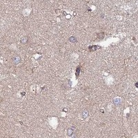


Comprehensive Human Protein Atlas IHC scoring per tissue (reliability: Uncertain — Medium consistency between antibody staining and RNA expression data. Caution, targets protein from more than one gene. Pending external verification.). Rows are taken directly from the HPA tissue chart — click any row's HPA link to view the source.
| Tissue | Cell type | Level | Evidence | Source |
|---|---|---|---|---|
| Duodenum | Glandular cells | High | Protein (IHC) | HPA → |
| Heart muscle | Cardiomyocytes | High | Protein (IHC) | HPA → |
| Ovary | Follicle cells | High | Protein (IHC) | HPA → |
| Small intestine | Glandular cells | High | Protein (IHC) | HPA → |
| Smooth muscle | Smooth muscle cells | High | Protein (IHC) | HPA → |
| Tissue | Cell type | Level | Evidence | Source |
|---|---|---|---|---|
| Adipose tissue | Adipocytes | Not detected | Protein (IHC) | HPA → |
| Bone marrow | Hematopoietic cells | Not detected | Protein (IHC) | HPA → |
| Fallopian tube | Glandular cells | Not detected | Protein (IHC) | HPA → |
| Lymph node | Germinal center cells | Not detected | Protein (IHC) | HPA → |
| Oral mucosa | Squamous epithelial cells | Not detected | Protein (IHC) | HPA → |
These troubleshooting steps center on chromogenic IHC for ARL6IP1 in paraffin sections; IF/ICC is addressed separately.
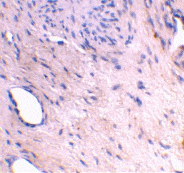
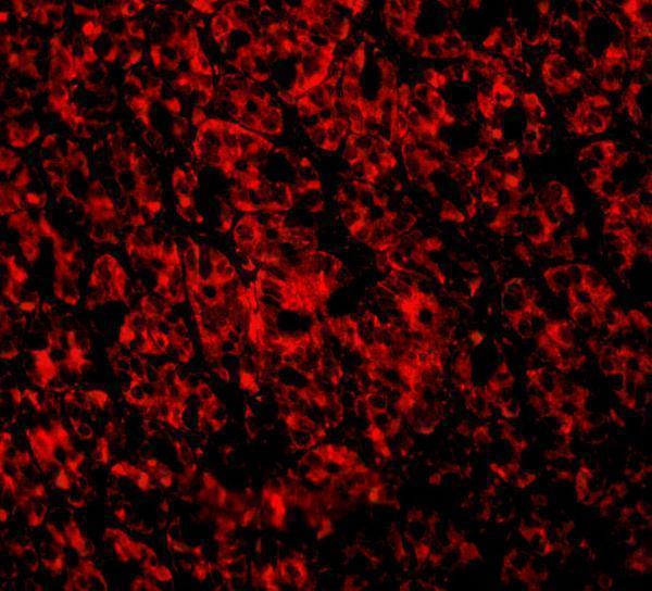
A08487 has IHC-P and IF applications for human and mouse samples (catalog applications/reactivity), with IHC shown in human bladder tissue and IF shown in mouse intestine cells (image captions).
A08487 will render with an IHC image of human bladder tissue stained at 2 μg/mL (IHC image caption). Its IF image shows mouse intestine cells stained at 2 μg/mL (IF image caption).
Which to pick: For tissue IHC, choose A08487: IHC-P is listed (catalog applications), and its IHC image shows human bladder tissue; the caption does not report the fixative or section processing (IHC image caption). For IF/ICC, A08487 lists IF and shows staining in mouse intestine cells (catalog applications; IF image caption). For human and mouse work, A08487 lists both species and has a rabbit host; its clonality is unreported (catalog reactivity/host/clone).