ARSB / Arylsulfatase B · IHC design guide

Design Immunohistochemistry for ARSB

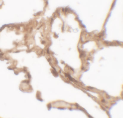
Plan chromogenic ARSB IHC in paraffin sections using kidney tubules, cardiomyocytes or cervical glands as high-staining references (HPA tissue IHC). Assess granular cytoplasmic staining and use the catalog antibody’s human lung IHC image at 2.5 µg/mL as a dilution reference (HPA tissue IHC; datasheet: IHC image).

Evidence assembled Oct 2026 · For research use; verify linked source records and product datasheet before use
Immunohistochemistry protocol sheet for ARSB (IHC for ARSB): expected localisation Granular cytoplasm in tissue (HPA tissue IHC), antibody A01609, validated IHC image, and IHC protocol steps
Printable ARSB IHC protocol sheet — expected localisation Granular cytoplasm in tissue (HPA tissue IHC), antibody A01609, controls and protocol steps. Open the full ARSB IHC guide →

ARSB Immunohistochemistry Experimental Design Guide

Expected localisation, validated protocols, controls and antibodies — the at-a-glance facts below, then the full design guide.

Must know before staining
Expected localisation Granular cytoplasm in tissue (HPA tissue IHC)
Staining pattern Granular cytoplasmic staining across many cell types (HPA tissue IHC)
Antigen retrieval Citrate pH 6.0 HIER, 95–98 °C, 20 min (rule: cytoplasmic / membrane antigen)
Positive control ⓘ Cervix+4 more · see all
Negative control ⓘ Ovary
Important caveats
Reasons your staining may differ from the expected pattern.
Fixation Keep fixation consistent across paraffin sections. (standard IHC practice; not target-specific)
Caveat Ovarian stroma is undetected despite broad tissue staining (HPA tissue IHC)
Regulation No regulator of ARSB abundance annotated (UniProt)
Isoform / epitope 2 isoforms; no transmembrane epitope orientation (UniProt)
Section 1

Recommended ARSB IHC & IF Protocols

The catalog antibody’s IHC-P protocol is accompanied by three published ARSB IHC protocols covering prostate tissue and frozen rat kidney sections (PMC4125552; PMC3763935; PMC3866634).

Recommended immunohistochemistry (IHC-P) protocol parameters
SampleTissue sections; selected-image fixative not specified (standard IHC workflow)
FixationImage fixative and duration unreported (datasheet A01609); verify before use.
Sectioning4–5 µm sections on charged slides (standard)
DeparaffinisationXylene, graded ethanol series to water (standard)
Antigen retrievalHeat-induced epitope retrieval in citrate buffer, pH 6.0, 20 min at 95–98 °C (standard rule: cytoplasmic / membrane antigen)
Peroxidase block3% H2O2, 10 min, room temperature (standard)
Blocking10% normal serum of the secondary host, 30 min, room temperature (standard)
Primary antibodyRabbit anti-ARSB, 1:100–1:500 starting range (standard)
Primary incubationOvernight at 4 °C (standard)
DetectionHRP-polymer secondary, DAB chromogen 5–10 min (standard)
CounterstainHematoxylin, blue, dehydrate and mount (standard)
Expected resultARSB-positive staining in glandular cells of cervix (HPA tissue IHC: High). HPA tissue profile: Ubiquitous cytoplasmic expression often with a granular pattern. No signal in the no-primary control.
💡Decision noteStart with citrate pH 6.0 retrieval at 95–98 °C for 20 min (page retrieval rule), then compare staining with the published conditions.
Section 2

What Is the Expected ARSB Staining Pattern?

ARSB is annotated in lysosomes and at the cell surface, with no transmembrane segment (UniProt P15848). In paraffin-section IHC, expect widespread, often granular cytoplasmic staining (HPA tissue IHC). Kidney tubular cells, cardiomyocytes and cervical glandular cells are high-staining examples (HPA tissue IHC). HPA rates the tissue pattern Approved, with medium agreement between staining and RNA data; external verification remains pending (HPA tissue IHC).

What am I looking at on my slide?
Granular cytoplasmic staining in kidney tubular cells, cardiomyocytes or cervical glandular cells.This matches the reported cytoplasmic pattern in cells rated High (HPA tissue IHC). Compare cell identity and distribution before scoring intensity; the HPA assessment is Approved but awaits external verification (HPA tissue IHC).
Predominantly nuclear staining, with little granular cytoplasmic signal.A nuclear-dominant result conflicts with the reported tissue pattern (HPA tissue IHC) and the lysosomal annotation (UniProt P15848). Treat it as a possible artefact; examine controls and morphology before calling it ARSB.
Strong staining of ovarian stromal cells.Those cells are reported as Not detected (HPA tissue IHC). Investigate cross-reactivity or endogenous chromogenic activity (general IHC practice); one unexpected specimen alone cannot establish which explanation applies.
Diffuse brown haze obscures cell boundaries or granules.The pattern is difficult to interpret against HPA’s often granular cytoplasmic profile (HPA tissue IHC). Background from the detection system or inadequate blocking is possible (general IHC practice); use controls to localize the source.
No staining in kidney tubular cells on the test section.Kidney tubular cells are a High reference (HPA tissue IHC), so a blank result warrants a run-level check. Confirm tissue integrity and counterstained morphology, then assess retrieval, antibody and detection controls (general IHC practice).
💡Expected ARSB appearanceCall a positive result when identifiable kidney tubular cells, cardiomyocytes or cervical glandular cells show convincing granular cytoplasmic chromogen (HPA tissue IHC); isolated nuclear staining or diffuse haze is not the reported tissue pattern (HPA tissue IHC).
How each factor affects the staining
Reference cell type and intensityUse the named cells when comparing sections: kidney tubules, cardiomyocytes and cervical glandular cells are High, while ovarian stroma is Not detected (HPA tissue IHC). A whole-tissue label can conceal differences between its cell types.
Compartment annotationLysosome and cell surface are both annotated, and ARSB has no transmembrane segment (UniProt P15848). In tissue IHC, HPA describes a ubiquitous, often granular cytoplasmic pattern (HPA tissue IHC); a membrane rim alone does not reproduce that description.
Antibody evidenceHPA037770 and HPA037771 each have Approved IHC status (HPA antibodies). The overall tissue profile has medium agreement with RNA data and awaits external verification (HPA tissue IHC), so assess unexpected patterns with controls.
Q: Should IF/ICC look identical to tissue IHC?A: HPA reports mainly Golgi localization in ICC-IF, with that main location Approved (HPA subcellular). Tissue IHC is described as often granular and cytoplasmic (HPA tissue IHC). Interpret each assay against its own reference pattern.
Why is my staining missing, weak or wrong?
SituationLikely causeNext action
Kidney tubular cells are blank.A High reference lacks its expected signal (HPA tissue IHC); the failed step is unknown.Check section morphology, a run control, retrieval conditions and detection reagents in sequence (general IHC practice). Do not infer ARSB absence from this slide alone.
Only nuclei stain strongly.Nuclear dominance disagrees with granular cytoplasmic tissue staining (HPA tissue IHC); nonspecific signal is possible.Review cell boundaries and control sections, then reassess antibody concentration and detection background (general IHC practice).
Ovarian stromal cells stain strongly.The reported level is Not detected (HPA tissue IHC); cross-reactivity or endogenous activity remains possible (general IHC practice).Compare with a control omitting the primary antibody and with a named high-staining cell population (general IHC practice; HPA tissue IHC).
Brown signal appears without primary antibody.The primary-independent signal points to endogenous activity or a detection-system source (general IHC practice).Check the appropriate enzyme-blocking step and detection-only control before interpreting cellular ARSB staining (general IHC practice).
Background masks granular cytoplasmic detail.Diffuse deposition prevents comparison with the reported tissue pattern (HPA tissue IHC); blocking or chromogen development may contribute (general IHC practice).Review blocking, antibody concentration, wash steps and development time against the run controls (general IHC practice).
Every cell type appears equally dark.Saturated chromogen can obscure intensity differences (general IHC practice); HPA reports High, Medium, Low and Not detected cell groups (HPA tissue IHC).Reduce development or antibody concentration as indicated by controls, then reassess identifiable cell types (general IHC practice).

Sample controls for ARSB IHC & IF

🧪Run cervix first: glandular cells should stain strongly (HPA: High in cervical glandular cells). Use ovarian stroma as the negative tissue (HPA: Not detected in ovarian stroma cells); on the cervix slide, treat cells without signal above local background as internal negative comparators, since HPA does not identify a negative cervical cell type (HPA: cervix row).
Positive control tissue: Cervix (Glandular cells, HPA High)
Negative control tissue: Ovary (HPA Not detected)
ICC-IF cell lines (HPA subcellular resource): HPA ICC-IF images show ARSB in A-431, U-251MG, U2OS, with annotated localisation: Golgi apparatus (approved) (HPA subcellular).
Technical controls: Include a no-primary, secondary-only control; an isotype control matched to the primary antibody’s host species, immunoglobulin class, and clonality; and ARSB knockout tissue or a validated peptide-block control (standard IHC practice). Block endogenous peroxidase for chromogenic detection, and check endogenous biotin if using avidin–biotin detection; compare background in cervical glands across controls (standard IHC practice).
⚠️Feasibility: No target-specific fixation window or retrieval requirement is reported in the supplied evidence, and the selected A01609 lung IHC caption leaves the fixative unreported (selected A01609 tissue-IHC caption). Start with the catalog antibody’s IHC-P protocol and optimize antigen retrieval empirically; the evidence does not establish whether frozen sections or IF/ICC are easier for ARSB (supplied target/application evidence). No cervix-specific artefact is reported, so assess glandular background against the controls (HPA: High in cervical glandular cells; supplied target/application evidence).

HPA tissue IHC evidence for ARSB

Comprehensive Human Protein Atlas IHC scoring per tissue (reliability: Approved — Medium consistency between antibody staining and RNA expression data. Pending external verification.). Rows are taken directly from the HPA tissue chart — click any row's HPA link to view the source.

Positive expression · recommended positive controls

TissueCell typeLevelEvidenceSource
Cervix Glandular cells High Protein (IHC) HPA →
Heart muscle Cardiomyocytes High Protein (IHC) HPA →
Kidney Cells in tubules High Protein (IHC) HPA →
Adrenal gland Glandular cells Medium Protein (IHC) HPA →
Appendix Glandular cells Medium Protein (IHC) HPA →

Undetected expression · recommended negative controls

TissueCell typeLevelEvidenceSource
Ovary Ovarian stroma cells Not detected Protein (IHC) HPA →
Section 3

Advanced ARSB IHC Tips

Troubleshoot ARSB staining in paraffin section IHC using the page retrieval setting, the catalog antibody’s tissue image, and compartment specific controls.

How should I adjust retrieval when ARSB staining is weak or diffuse?
Start with citrate pH 6.0 heat induced retrieval at 95–98 °C for 20 min (page retrieval setting). If signal is weak, check that sections reached the stated temperature throughout the run, then compare a slightly longer retrieval time on adjacent sections while holding antibody concentration and detection constant (standard IHC practice). Score granular cytoplasmic staining separately from diffuse background, because the reported tissue pattern is often granular and cytoplasmic (HPA: tissue IHC profile). Include a known positive section in each comparison, such as kidney tubules, so a change in retrieval is not mistaken for a change in tissue expression (HPA: High in kidney tubular cells; standard IHC practice).
Could fixation explain inconsistent ARSB staining between paraffin blocks?
ARSB specific fixation sensitivity is unknown from the supplied evidence; record each block’s fixative, fixation duration, and processing history before comparing staining (supplied evidence: no target specific fixation study; standard IHC practice). The human lung image reports staining at 2.5 μg/mL but does not state its fixative, so it cannot establish a fixation condition for other blocks (A01609 tissue IHC caption). Run blocks through the same citrate pH 6.0 retrieval and detection workflow, with a positive control section in each batch (page retrieval setting; standard IHC practice). If signal varies, compare staining within similarly processed tissue and assess morphology before attributing the difference to ARSB expression (standard IHC practice).
Which compartments should count as plausible ARSB staining in tissue?
Assess granular cytoplasmic staining first, since that is the reported tissue IHC pattern for ARSB (HPA: tissue IHC profile). Lysosomal and cell surface localisation are annotated for the protein, while an independent cell imaging summary calls the Golgi apparatus its main location (UniProt P15848 subcellular location; HPA: subcellular summary). ARSB has a signal peptide at residues 1–36 and no annotated transmembrane segment, so a sharp membrane outline alone needs corroboration rather than automatic acceptance (UniProt P15848 processing and topology). Compare the pattern across positive cells and controls; predominantly nuclear or uniformly extracellular chromogen should prompt a review of background and tissue morphology (standard IHC practice).
Can processing or glycosylation change which ARSB epitope is detected?
Confirm the catalog antibody’s immunogen or mapped epitope before interpreting isoform specific staining; the supplied record lists 2 isoforms but gives no antibody epitope (UniProt P15848 isoforms; supplied evidence: epitope unspecified). The annotated signal peptide spans residues 1–36, and the listed protein chain spans 37–533, so an epitope in the removed signal sequence would require separate validation (UniProt P15848 processing). Six glycosylation sites are annotated at residues 188, 279, 291, 366, 426, and 458; the payload does not show that any one site alters this antibody’s IHC binding (UniProt P15848 glycosylation; supplied evidence: no epitope study). Compare adjacent sections under identical retrieval and detection conditions before assigning a staining difference to an isoform or modification (standard IHC practice).
How can IF help assess the ARSB pattern seen by chromogenic IHC?
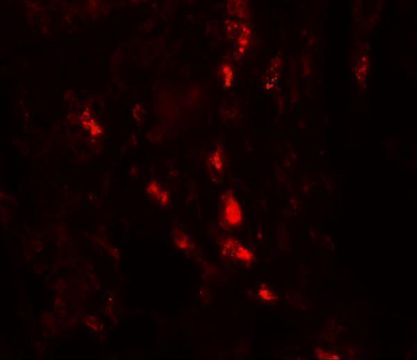
Use IF as a separate localisation check, since this page’s curated antibody protocol is for IHC in paraffin sections (application scope). In kidney, multiplex ARSB with a validated tubular epithelial marker to identify the cell population reported as strongly positive, and use single stain controls to check channel bleed through (HPA: High in kidney tubular cells; standard IF practice). Choose a red or far red ARSB fluorophore after checking tissue autofluorescence in an unstained section (standard IF practice). The antibody epitope’s membrane side is unspecified; compare mild permeabilisation with no permeabilisation before interpreting intracellular versus surface signal (supplied evidence: epitope unspecified; UniProt P15848 lysosomal and cell surface locations; standard IF practice).
What should I check when ARSB DAB staining looks widespread?
Compare the section with a no primary control and review the peroxidase block, because endogenous enzyme activity can deposit chromogen independently of antibody binding (standard chromogenic IHC practice). Titrate the catalog antibody around the pictured 2.5 μg/mL condition on serial sections, keeping retrieval, incubation, and DAB development matched (A01609 tissue IHC caption; standard IHC practice). Examine tissue edges, folds, damaged areas, and deposits separately from intact cells, where uneven reagent exposure can complicate interpretation (standard IHC practice). A broad distribution alone is not grounds to reject signal: the reported ARSB tissue profile is ubiquitous cytoplasmic staining, often granular (HPA: tissue IHC profile).
How should I quantify ARSB staining across tissue sections? ⚠ ANSWER MARKED FOR VERIFICATION
Define the cell population and compartment before scoring, then report the percentage of positive cells and an intensity weighted H-score for granular cytoplasmic signal (HPA: tissue IHC profile; standard IHC scoring practice). If discrete positive cells are the endpoint, count them per mm² of viable tissue and retain the corresponding cell count or area denominator (standard IHC quantification practice). Normalise comparisons to the same annotated tissue area or cell population, and keep section thickness, retrieval, antibody concentration, and DAB development matched (standard IHC practice). Record staining in a positive control, such as kidney tubular cells, alongside each run to identify batch shifts before comparing biological groups (HPA: High in kidney tubular cells; standard IHC practice).
How do I distinguish credible ARSB staining from artefact?
Credible tissue staining should be evaluated as cellular, often granular cytoplasmic signal, with kidney tubular cells offering a documented high staining reference (HPA: tissue IHC profile; HPA: High in kidney tubular cells). Correlate any apparent surface pattern with cell outlines and a clean no primary control, because cell surface localisation is annotated but the tissue IHC profile does not establish that pattern in every cell (UniProt P15848 subcellular location; HPA: tissue IHC profile; standard IHC practice). Treat dominant nuclear colour, edge accentuation, and staining confined to necrotic areas as reasons to investigate artefact (standard IHC practice). Persistent chromogen in the no primary control warrants checking endogenous peroxidase blocking and detection reagents before assigning positivity to ARSB (standard chromogenic IHC practice).
Boster reagents

Best ARSB / Arylsulfatase B IHC Antibodies

A01609 has IHC and IF images from human lung tissue (catalog image captions). Its listed reactivity covers human, mouse, and rat (catalog: reactivity).

Real IHC data Immunohistochemistry of ARSB in human lung tissue with ARSB antibody at 2.5 μg/mL.
Anti-Arylsulfatase B ARSB Antibody
Cat # A01609

A01609 is listed for IHC-P and IF, with human, mouse, and rat reactivity (catalog: applications and reactivity). Its human lung images show IHC at 2.5 μg/mL and IF at 20 μg/mL (catalog image captions).

Which to pick: Choose A01609 for paraffin-section IHC: IHC-P is listed, and its IHC image shows human lung tissue at 2.5 μg/mL (catalog: applications; IHC image caption); the fixative is unreported (catalog: IHC image caption). A01609 is also the IF choice because IF is listed and its human lung IF image used 20 μg/mL (catalog: applications; IF image caption); ICC validation is unreported (catalog: applications and image captions). For mouse or rat studies, A01609 lists reactivity with both species, but the supplied IHC and IF images show human tissue only (catalog: reactivity and image captions); its host is rabbit and clonality is unreported (catalog: host and clone).

Each figure is that product's own IHC / IF validation image from its datasheet.

References

  1. UniProt Consortium. UniProt entry P15848 (ARSB_HUMAN, Arylsulfatase B).
  2. Human Protein Atlas. ARSB tissue IHC expression (reliability: Approved).
  3. Human Protein Atlas. ARSB subcellular location (ICC-IF): Mainly localized to the Golgi apparatus..
  4. Human Protein Atlas. ARSB antibody validation summary (2 antibodies).
  5. Differential effects of estrogen exposure on arylsulfatase B, galactose-6-sulfatase, and steroid sulfatase in rat prostate development. The Journal of steroid biochemistry and molecular biology 2014 — PMC4125552.
  6. Interaction of arylsulfatases A and B with maspin: A possible explanation for dysregulation of tumor cell metabolism and invasive potential of colorectal cancer. World journal of clinical cases 2019 — PMC6906558.
  7. Arylsulfatase B (N-acetylgalactosamine-4-sulfatase): potential role as a biomarker in prostate cancer. Prostate cancer and prostatic diseases 2013 — PMC3763935.
  8. Impact of salt exposure on N-acetylgalactosamine-4-sulfatase (arylsulfatase B) activity, glycosaminoglycans, kininogen, and bradykinin. Glycoconjugate journal 2013 — PMC3866634.
  9. PubMed PMID:2303452 — UniProt-cited evidence.
  10. PubMed PMID:1968043 — UniProt-cited evidence.
  11. PubMed PMID:7687847 — UniProt-cited evidence.