This website uses cookies to ensure you get the best experience on our website.


- Table of Contents
Plan paraffin-section ATF1 IHC around the general nuclear staining observed in tissue (HPA tissue IHC) and the nuclear location annotated for the protein (UniProt). Use cell-specific controls and consistent fixation to assess staining across sections.

Expected localisation, validated protocols, controls and antibodies — the at-a-glance facts below, then the full design guide.
| Expected localisation | General nuclear staining (HPA tissue IHC) | |
| Staining pattern | Nuclear staining across many tissue cell types (HPA tissue IHC) | |
| Antigen retrieval | EDTA pH 8.0 HIER, heat-mediated (datasheet M01600-1) | |
| Positive control | Adrenal gland+4 more · see all | |
| Negative control | Nasopharynx |
| Fixation | Keep fixation consistent across sections (standard IHC practice; not target-specific). Selected-image fixative and duration unreported (datasheet M01600-1); verify before use. | |
| Caveat | Staining and RNA show medium consistency (HPA tissue IHC) | |
| Regulation | Expression regulation unreported (UniProt) | |
| Isoform / epitope | 2 isoforms; epitope coverage is unknown (UniProt) |
The catalog antibody has a datasheet IHC-P protocol (datasheet: M01600-1). Published IHC procedures below cover ATF1 and p-CREB/ATF1 staining (PMC9983578; PMC8038809).
| Sample | Paraffin-embedded human lung cancer tissue; fixative not specified (datasheet M01600-1) |
| Fixation | Image fixative and duration unreported (datasheet M01600-1); verify before use. |
| Sectioning | 4–5 µm sections on charged slides (standard) |
| Deparaffinisation | Xylene, graded ethanol series to water (standard) |
| Antigen retrieval | Heat retrieval: EDTA pH 8.0 (datasheet M01600-1); 20 min, 95–100 °C (standard) |
| Peroxidase block | 3% H2O2, 10 min, room temperature (standard) |
| Blocking | 10% goat serum (datasheet M01600-1) |
| Primary antibody | Mouse monoclonal (clone 7F8) anti-ATF1, 0.5-1μg/ml (datasheet M01600-1) |
| Primary incubation | Overnight at 4 °C (datasheet M01600-1) |
| Detection | Streptavidin-biotin complex (SABC), DAB chromogen (datasheet M01600-1) |
| Counterstain | Hematoxylin, blue, dehydrate and mount (standard) |
| Expected result | ATF1-positive staining in glandular cells of adrenal gland (HPA tissue IHC: High). HPA tissue profile: General nuclear expression. No signal in the no-primary control. |
ATF1 should appear predominantly in nuclei, with nucleoplasmic localisation also observed by ICC-IF (UniProt P18846: nucleus; HPA subcellular: nucleoplasm). In paraffin-section IHC, expect nuclear staining across multiple cell types, including glandular cells and glomerular cells that HPA scores High (HPA tissue IHC: general nuclear expression). ATF1 has no transmembrane segment (UniProt P18846 topology). HPA rates the tissue IHC pattern Supported, with medium consistency between staining and RNA expression (HPA tissue IHC: reliability).
| Crisp nuclear chromogen in colon or duodenal glandular cells, with weaker staining possible in other cells (HPA tissue IHC: High glandular-cell staining; general nuclear expression). | This fits the expected compartment and reported positive cell types (UniProt P18846: nucleus; HPA tissue IHC: colon and duodenum). Score nuclear staining in the relevant cells; compare its intensity with surrounding cells on the same section (general IHC practice). |
| Predominantly cytoplasmic, membranous or extracellular chromogen, while nuclei remain clear (UniProt P18846: nucleus; HPA tissue IHC: general nuclear expression). | Treat this as discordant with the reported ATF1 pattern, rather than a confirmed positive (UniProt P18846: nucleus; HPA tissue IHC: general nuclear expression). Inspect morphology, detection background and the primary-antibody omission control for artefact (general IHC practice). |
| Strong staining in nasopharyngeal basal cells, which HPA reports as Not detected (HPA tissue IHC: nasopharynx, basal cells). | This discrepancy warrants checking cell identification and nonspecific antibody binding; endogenous detection activity is another possibility if signal persists without primary antibody (general IHC practice). An HPA Not detected result is a reference observation, not proof that every specimen must be negative (HPA tissue IHC: nasopharynx). |
| Diffuse chromogen covers nuclei, cytoplasm and tissue spaces, making cell boundaries difficult to judge (general IHC practice). | Do not score this as ATF1 expression without resolvable nuclear signal (UniProt P18846: nucleus; general IHC practice). Excess detection background or nonspecific binding can obscure localisation; inspect controls and background before comparing tissues (general IHC practice). |
| No nuclear signal in a section containing colon glandular cells or pancreatic exocrine glandular cells (HPA tissue IHC: both High). | A technical failure is possible, but HPA's High categories do not guarantee staining in every specimen (HPA tissue IHC: colon and pancreas; reliability Supported). Check section quality and the staining run with a suitable positive control before calling ATF1 absent (general IHC practice). |
| Tissue and cell choice (HPA tissue IHC) | HPA reports High staining in adrenal, colon, duodenal, epididymal and gallbladder glandular cells, Bergmann glial nuclei, glomerular cells and pancreatic exocrine glandular cells. It reports Low staining in several epithelial and muscle cell types, so those cells are less useful for judging a weak run (HPA tissue IHC). |
| Strength of the IHC reference (HPA tissue IHC; HPA antibodies) | The overall IHC pattern is Supported, with medium consistency between antibody staining and RNA data (HPA tissue IHC: reliability). HPA055069 and CAB016222 have Supported IHC status; HPA055406 has no listed IHC status (HPA antibodies). Interpret an unexpected pattern against that validation limit. |
| IF/ICC: Where should ATF1 appear? (HPA subcellular) | Expect nucleoplasmic signal in the HPA ICC-IF reference images (HPA subcellular: Nucleoplasm, enhanced). This supports a localisation comparison with nuclear IHC staining; it does not supply an IF/ICC protocol for this IHC guide (HPA subcellular; HPA tissue IHC: general nuclear expression). |
| Protein form and antibody epitope (UniProt P18846) | ATF1 is annotated as one chain spanning residues 1–271, with no signal peptide or propeptide, and has two isoforms (UniProt P18846: processing, isoforms). The supplied record gives no antibody epitope, so isoform coverage and epitope-dependent staining cannot be predicted from it. |
| Phosphorylation and retrieval decisions (UniProt P18846; general IHC practice) | UniProt lists phosphoserines at residues 63 and 198, but the supplied evidence does not link them to IHC staining or retrieval response (UniProt P18846: modified residues). Choose and assess retrieval conditions as a general paraffin-IHC workflow step, without assuming an ATF1-specific fixation effect (general IHC practice). |
| Situation | Likely cause | Next action |
|---|---|---|
| No nuclear staining in the expected positive cells (HPA tissue IHC: High in colon glandular cells). | The run may have failed, or the specimen may differ from the HPA reference; the HPA category does not establish a guaranteed result (HPA tissue IHC: reliability Supported). | Review section integrity, staining-run controls and the IHC-validated antibody's documented conditions; assess retrieval as a general paraffin-IHC variable (general IHC practice). |
| Colour is mainly cytoplasmic despite identifiable nuclei (UniProt P18846: nucleus). | The compartment is inconsistent with the UniProt and HPA localisation references; nonspecific staining or detection background is possible (HPA tissue IHC: general nuclear expression; general IHC practice). | Check nuclear morphology and a primary-antibody omission control, then judge whether distinct nuclear signal remains after background is accounted for (general IHC practice). |
| Basal cells in nasopharynx stain strongly (HPA tissue IHC: basal cells Not detected). | Possible explanations include cell misidentification, antibody cross-reactivity or endogenous detection activity; HPA alone cannot distinguish them (HPA tissue IHC: nasopharynx; general IHC practice). | Verify the cell type on the counterstained section and inspect omission and detection controls before assigning the colour to ATF1 (general IHC practice). |
| Broad tissue background masks nuclear detail (general IHC practice). | Nonspecific binding or endogenous detection activity can produce chromogen beyond the expected nuclear pattern (general IHC practice; UniProt P18846: nucleus). | Review blocking, washes and detection controls under the chosen chromogenic method; score only nuclei that remain distinct from background (general IHC practice). |
| Only weak staining appears in lung alveolar type I cells or skeletal myocytes (HPA tissue IHC: Low in both). | Weak staining in those cell types is compatible with HPA's Low category and, by itself, does not show that the run failed (HPA tissue IHC: lung and skeletal muscle). | Compare a reported High-staining cell type in a suitable control section, then interpret the weak signal by its nuclear location and local background (HPA tissue IHC; general IHC practice). |
| The IF image looks nucleoplasmic, but IHC chromogen is diffuse (HPA subcellular: Nucleoplasm, enhanced). | The localisation readouts disagree; the supplied evidence does not identify an ATF1-specific fixation or retrieval cause (HPA subcellular; HPA tissue IHC). | Use the IF image as a localisation reference, then troubleshoot IHC section morphology, background and detection controls within the paraffin workflow (HPA subcellular; general IHC practice). |




Comprehensive Human Protein Atlas IHC scoring per tissue (reliability: Supported — Medium consistency between antibody staining and RNA expression data.). Rows are taken directly from the HPA tissue chart — click any row's HPA link to view the source.
| Tissue | Cell type | Level | Evidence | Source |
|---|---|---|---|---|
| Adrenal gland | Glandular cells | High | Protein (IHC) | HPA → |
| Cerebellum | Bergmann glia - nucleus | High | Protein (IHC) | HPA → |
| Colon | Glandular cells | High | Protein (IHC) | HPA → |
| Duodenum | Glandular cells | High | Protein (IHC) | HPA → |
| Epididymis | Glandular cells | High | Protein (IHC) | HPA → |
| Tissue | Cell type | Level | Evidence | Source |
|---|---|---|---|---|
| Nasopharynx | Basal cells | Not detected | Protein (IHC) | HPA → |
Troubleshoot ATF1 staining in paraffin sections by checking retrieval, nuclear localisation, assay controls and scoring before interpreting signal.
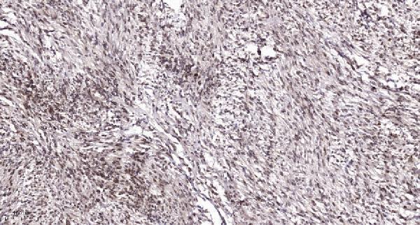
The catalog shows human paraffin-section IHC images from lung, rectal, and colon cancer tissue, plus an IF image from A431 cells (catalog: IHC and IF image captions).
M01600-1 is listed for IHC and IF/ICC, with paraffin-section IHC images from human lung and rectal cancer tissue and an IF image from A431 cells (catalog: M01600-1 applications and image captions). A01600-1 is listed for IHC and IF/ICC in human, mouse, and rat, with a paraffin-section IHC image from human colon cancer tissue (catalog: A01600-1 applications, reactivity, IHC caption).
Which to pick: For human tissue IHC, choose mouse monoclonal M01600-1 if its lung and rectal cancer paraffin-section examples match your intended approach; those captions specify EDTA pH 8 retrieval but do not report the fixative (catalog: M01600-1 host, clone, IHC captions). For IF/ICC, M01600-1 has an A431 IF image, while A01600-1 lists IF/ICC without an IF image (catalog: M01600-1 IF caption; A01600-1 applications, IF image captions). For work across human, mouse, and rat, choose rabbit polyclonal A01600-1 based on its listed reactivity and IHC/IF applications; its colon cancer IHC caption specifies a paraffin section and Tris-EDTA pH 9 retrieval but does not report the fixative (catalog: A01600-1 dilution notes, reactivity, applications, IHC caption).