ATG14 / Beclin 1-associated autophagy-related key regulator · IHC design guide

Design Immunohistochemistry for ATG14

Plan chromogenic ATG14 IHC in paraffin sections with the catalog antibody’s 0.5–1 μg/mL range (datasheet PB9481). Assess cytoplasmic and nuclear staining across tissues (HPA tissue IHC), allowing for stimulus-linked cytoplasmic foci (UniProt).

Evidence assembled Oct 2026 · For research use; verify linked source records and product datasheet before use
Immunohistochemistry protocol sheet for ATG14 (IHC for ATG14): expected localisation Cytoplasmic and nuclear tissue staining (HPA tissue IHC), antibody PB9481, validated IHC image, and IHC protocol steps
Printable ATG14 IHC protocol sheet — expected localisation Cytoplasmic and nuclear tissue staining (HPA tissue IHC), antibody PB9481, controls and protocol steps. Open the full ATG14 IHC guide →

ATG14 Immunohistochemistry Experimental Design Guide

Expected localisation, validated protocols, controls and antibodies — the at-a-glance facts below, then the full design guide.

Must know before staining
Expected localisation Cytoplasmic and nuclear tissue staining (HPA tissue IHC)
Staining pattern Cytoplasmic and nuclear staining across tissues (HPA tissue IHC)
Antigen retrieval Citrate pH 6 HIER, heat-mediated (datasheet PB9481)
Positive control ⓘ Adipose tissue+4 more · see all
Negative control ⓘ None in HPA (detected in all 45 tissues); use no-primary + isotype controls
Important caveats
Reasons your staining may differ from the expected pattern.
Fixation Keep fixation consistent across sections (standard IHC practice; not target-specific). Selected-image fixative and duration unreported (datasheet PB9481); verify before use.
Caveat Autophagy stimuli can shift ATG14 into cytoplasmic foci (UniProt)
Regulation Stimulus effects on intensity are unreported (UniProt)
Isoform / epitope 2 isoforms; epitope coverage is unreported (UniProt)
Section 1

Recommended ATG14 IHC & IF Protocols

The catalog antibody uses heat-mediated citrate retrieval at pH 6 (datasheet: PB9481). One published kidney-tissue IHC protocol is available for comparison (PMC7241494).

Recommended immunohistochemistry (IHC-P) protocol parameters
SampleParaffin-embedded Rat Spleen tissue; fixative not specified (datasheet PB9481)
FixationImage fixative and duration unreported (datasheet PB9481); verify before use.
Sectioning4–5 µm sections on charged slides (standard)
DeparaffinisationXylene, graded ethanol series to water (standard)
Antigen retrievalHeat retrieval: Citrate pH 6, 20 min (datasheet PB9481)
Peroxidase block3% H2O2, 10 min, room temperature (standard)
Blocking10% goat serum (datasheet PB9481)
Primary antibodyRabbit anti-ATG14, 0.5-1μg/ml (datasheet PB9481)
Primary incubationOvernight at 4 °C (datasheet PB9481)
DetectionStreptavidin-biotin complex (SABC), DAB chromogen (datasheet PB9481)
CounterstainHematoxylin, blue, dehydrate and mount (standard)
Expected resultATG14-positive staining in adipocytes of adipose tissue (HPA tissue IHC: Medium). HPA tissue profile: Cytoplasmic and nuclear expression in all tissues. No signal in the no-primary control.
💡Decision noteStart with citrate retrieval at pH 6 for the catalog antibody (datasheet: PB9481); the published kidney-tissue protocol used microwave retrieval with Tri-EDTA (PMC7241494).
Section 2

What Is the Expected ATG14 Staining Pattern?

ATG14 is cytoplasmic and associates with endoplasmic reticulum, preautophagosomal and autophagosome membranes; it has no transmembrane segment (UniProt Q6ZNE5 localization and topology). In tissue IHC, expect cytoplasmic and nuclear staining across tissues, with medium staining in several listed cell populations (HPA tissue IHC). HPA rates this tissue pattern Approved, with medium consistency between staining and RNA data (HPA tissue IHC).

What am I looking at on my slide?
Cytoplasmic staining in adipocytes, respiratory epithelial cells or glial cells.These populations have medium staining in the HPA tissue survey (HPA tissue IHC). Read a positive result in its tissue and cell context: HPA also reports nuclear staining across tissues, while UniProt places ATG14 in the cytoplasm and on autophagic membranes (HPA tissue IHC; UniProt Q6ZNE5 localization).
Cytoplasmic foci accompany a more diffuse cytoplasmic pattern.ATG14 is cytosolic under nutrient-rich conditions and becomes predominantly focal after autophagy stimuli in the cited localization work (UniProt Q6ZNE5 localization). A single paraffin section cannot establish the stimulus or identify an individual focus as an autophagosome; interpret foci alongside tissue staining and controls.
Staining is confined to an extracellular deposit or luminal material, with no convincing cellular pattern.That distribution does not match the cellular cytoplasmic and nuclear tissue pattern (HPA tissue IHC) or the intracellular locations described for ATG14 (UniProt Q6ZNE5 localization). Treat it as suspect and review morphology, controls and chromogen deposition before assigning ATG14 positivity (general IHC practice).
Strong staining dominates spleen red-pulp cells or bone-marrow hematopoietic cells.Both populations are listed as low, so a strong result is discordant with the HPA examples; low does not mean absent (HPA tissue IHC). Check for cross-reactivity or endogenous detection activity with appropriate controls before calling the cells ATG14-high (general IHC practice).
No staining appears in a section expected to provide a positive reference.Medium staining is reported for adipocytes, bronchial respiratory epithelial cells and several other listed populations (HPA tissue IHC). A blank result in one of these calls for a run-level check of the reference section, IHC-validated antibody and detection controls; it alone does not establish absent ATG14 (general IHC practice).
💡Expected ATG14 appearanceCall a convincing positive when the expected cells show discernible medium cytoplasmic staining, with possible nuclear staining (HPA tissue IHC); extracellular-only deposits or strong staining where HPA reports low levels warrant control review (HPA tissue IHC; general IHC practice).
How each factor affects the staining
Tissue context and evidence strengthHPA reports expression across tissues, low tissue RNA specificity and an Approved IHC pattern with medium staining–RNA consistency (HPA tissue IHC). Use the named cell populations as references without treating any listed tissue as an absolute positive or negative control.
Autophagy state and membrane associationATG14 may be cytosolic or form cytoplasmic foci after autophagy stimuli; it associates with several intracellular membranes despite lacking a transmembrane segment (UniProt Q6ZNE5 localization and topology). Do not infer a treatment condition from puncta in a single tissue section.
Isoforms and modificationsUniProt lists 2 isoforms and phosphorylation sites at residues 29, 416 and 429 (UniProt Q6ZNE5 isoforms and modified residues). The supplied records do not map the IHC antibody epitope, so they cannot establish isoform or phosphorylation dependence.
Assay-specific antibody evidenceCAB037242 has Approved IHC status, whereas CAB080659 has Supported ICC status (HPA antibodies). These ratings apply to their listed assays; an ICC image is not, by itself, validation of a paraffin-section staining pattern.
Endogenous detection activityFor enzyme-based chromogenic IHC, tissue activity can contribute color independently of primary-antibody binding (general IHC practice). A detection control helps distinguish this contribution from cellular ATG14 staining; the HPA low-staining examples are not proven ATG14-negative tissues.
Why is my staining missing, weak or wrong?
SituationLikely causeNext action
Reference tissue is blank, including cells reported at medium intensity (HPA tissue IHC).The section, staining run or detection chain may have failed; the blank slide cannot identify which step (general IHC practice).Confirm the expected cell population is present, review the catalog antibody's IHC-P instructions, and check the run's positive and detection controls (general IHC practice).
Color covers the section broadly and obscures cell boundaries.Diffuse background can arise from nonspecific binding, detection reagents or chromogen handling (general IHC practice).Compare the primary-omission and detection controls; review blocking, reagent concentration, washes and chromogen development for this run (general IHC practice).
Strong color appears in low-staining spleen red pulp or marrow cells (HPA tissue IHC).Cross-reactivity or endogenous enzyme activity is possible; HPA's low rating is not proof that these cells contain no ATG14 (HPA tissue IHC; general IHC practice).Inspect morphology and primary-omission controls, then compare staining with a medium-staining HPA reference population processed in the same run (HPA tissue IHC; general IHC practice).
Only nuclear staining is visible.HPA reports nuclear as well as cytoplasmic tissue staining, while UniProt describes cytoplasmic and membrane-associated ATG14 (HPA tissue IHC; UniProt Q6ZNE5 localization). Nuclear color alone leaves the expected cytoplasmic component unconfirmed.Check cytoplasmic visibility against counterstain and background, then compare an HPA medium-staining cell population and run controls before interpreting the nuclear signal (HPA tissue IHC; general IHC practice).
Distinct dots appear, but their identity is uncertain.UniProt describes ATG14 foci after autophagy stimuli, yet morphology alone cannot assign each dot to an isolation membrane or autophagosome (UniProt Q6ZNE5 localization).Score cellular localization and control quality first. If organelle identity is essential, seek independent localization evidence suited to that question (general IHC practice).
What should IF/ICC show?HPA reports supported vesicle localization and uncertain plasma-membrane localization in ICC-IF images from U2OS cells (HPA subcellular ICC-IF).Treat vesicular signal as the better-supported IF observation; interpret plasma-membrane signal cautiously. Use the separate IF/ICC guide for that assay, since CAB080659's Supported ICC status does not validate an IHC-P protocol (HPA subcellular ICC-IF; HPA antibodies).

Sample controls for ATG14 IHC & IF

🧪Run adipose tissue first and expect staining in adipocytes (Medium; HPA: adipose tissue, adipocytes). HPA detects ATG14 in all 45 scored tissues, so there is no validated negative tissue; use no-primary and isotype controls, and treat any unstained neighboring cells as background comparators rather than proven ATG14-negative cells (HPA: no negative tissues; standard IHC practice).
Positive control tissue: Adipose tissue (Adipocytes, HPA Medium)
Negative control tissue: None in HPA: ATG14 is detected in all 45 scored tissues. Use a no-primary (secondary-only) and an isotype control instead.
ICC-IF cell lines (HPA subcellular resource): HPA ICC-IF images show ATG14 in U2OS, with annotated localisation: Vesicles (supported), Plasma membrane (uncertain) (HPA subcellular).
Technical controls: Include a secondary-only slide, a control with rabbit immunoglobulin matched to the primary antibody’s clonality, and ATG14-knockout tissue if available (caption: rabbit primary; standard IHC control practice). Quench endogenous peroxidase for chromogenic detection and assess background around adipocyte lipid vacuoles (standard IHC practice).
⚠️Feasibility: No target-specific fixation window or fixation effect is reported, and the selected PB9481 paraffin-section caption does not state a fixative (PB9481 caption: fixative unreported). The caption uses citrate retrieval at pH 6 for 20 minutes, but does not establish that retrieval is required; the supplied evidence does not establish whether frozen sections or IF are easier (PB9481 caption). In adipose tissue, empty lipid vacuoles can complicate interpretation of cellular staining (standard IHC practice).

HPA tissue IHC evidence for ATG14

Comprehensive Human Protein Atlas IHC scoring per tissue (reliability: Approved — Medium consistency between antibody staining and RNA expression data.). Rows are taken directly from the HPA tissue chart — click any row's HPA link to view the source.

Positive expression · recommended positive controls

TissueCell typeLevelEvidenceSource
Adipose tissue Adipocytes Medium Protein (IHC) HPA →
Adrenal gland Glandular cells Medium Protein (IHC) HPA →
Appendix Glandular cells Medium Protein (IHC) HPA →
Breast Adipocytes Medium Protein (IHC) HPA →
Bronchus Respiratory epithelial cells Medium Protein (IHC) HPA →

Undetected expression · recommended negative controls

TissueCell typeLevelEvidenceSource
None in HPA: ATG14 is detected in all 45 scored tissues. Use a no-primary (secondary-only) and an isotype control instead.
Section 3

Advanced ATG14 IHC Tips

Troubleshoot ATG14 staining in paraffin sections by checking retrieval, compartment, tissue context and detection controls before scoring chromogenic IHC.

How should I retrieve ATG14 in paraffin sections when staining is weak?
Start with heat-mediated retrieval in citrate buffer at pH 6 for 20 minutes (datasheet PB9481). That condition was used for the catalog antibody in a paraffin-embedded rat spleen section, followed by 1 μg/ml primary antibody overnight at 4°C (datasheet PB9481). If staining remains weak, compare a modestly adjusted heating time on adjacent sections while keeping antibody concentration and detection constant; inspect morphology and background before choosing a condition (standard IHC practice). Include a no-primary control and the same reference section in each comparison, because weak ATG14 signal in spleen red pulp cells is plausible (HPA: Low in spleen red pulp cells).
Can I choose a fixative based on the published ATG14 staining patterns?
The selected paraffin-section caption does not state its fixative, so ATG14-specific fixation sensitivity is unknown (datasheet PB9481: fixative not stated). HPA reports cytoplasmic and nuclear staining across tissues, but those patterns do not establish which fixation conditions preserve the epitope (HPA: tissue IHC profile). For a new fixation workflow, compare matched specimens processed with the intended fixative and fixation time, then apply the same pH 6 citrate retrieval and detection conditions (datasheet PB9481; standard IHC practice). Judge signal alongside morphology, background and a no-primary control; record the fixation conditions so differences between runs can be interpreted (standard IHC practice).
Should diffuse nuclear or membrane staining count as ATG14-positive?
Evaluate cytoplasmic and vesicular staining first: ATG14 is reported in cytoplasm, endoplasmic reticulum membrane and autophagosome-related membranes (UniProt Q6ZNE5: subcellular location). Its distribution can shift from cytosol under nutrient-rich conditions to cytoplasmic foci after autophagy stimuli (UniProt Q6ZNE5: PubMed 19050071). HPA describes cytoplasmic and nuclear tissue staining, while its cellular imaging supports vesicles and assigns plasma membrane staining uncertain status (HPA: tissue IHC profile; HPA: subcellular location). Score nuclear or sharply outlined membrane signal separately and compare it with no-primary controls, morphology and the retrieval condition before attributing it to ATG14 (standard IHC practice).
Could isoforms or epitope masking explain inconsistent ATG14 staining?
ATG14 has 2 annotated isoforms, and the supplied caption does not identify the PB9481 epitope (UniProt Q6ZNE5: isoforms; datasheet PB9481). The record also lists phosphoserine at positions 29 and 416 and phosphothreonine at 429, but their effects on this antibody's tissue staining are unknown (UniProt Q6ZNE5: modified residues). If sections disagree, verify the antibody's stated immunogen or epitope before assigning staining to an isoform, and compare adjacent sections under identical retrieval conditions (standard IHC practice). Report staining as ATG14 unless isoform recognition has been established independently; do not infer isoform selectivity from a punctate pattern alone (standard IHC practice).
How can IF help check ATG14 localisation alongside the IHC result?
Use IF as a separate localisation check rather than treating the paraffin-section caption as an IF protocol; that caption documents chromogenic IHC in rat spleen (datasheet PB9481). For a cell-type comparison, multiplex ATG14 with CD31 in an endothelial-cell region, since HPA reports medium ATG14 staining in cerebral-cortex endothelial cells (HPA: tissue IHC profile; standard IF practice: CD31 marker). Choose a spectrally separated far-red fluorophore and include unstained tissue to assess autofluorescence before interpreting puncta (standard IF practice). ATG14 has no annotated transmembrane segment and is reported at cytoplasmic and autophagic membranes, so optimise permeabilisation for access to the cytoplasmic epitope while recognising that this antibody's exact epitope side is unspecified (UniProt Q6ZNE5: topology and location; datasheet PB9481).
What should I check when ATG14 DAB staining looks diffuse?
Check no-primary and secondary-only sections to distinguish tissue or detection background from primary-antibody signal (standard IHC practice). The documented workflow used 10% goat serum, a biotinylated goat anti-rabbit secondary, a streptavidin–biotin complex and DAB (datasheet PB9481). In that detection format, evaluate endogenous biotin and endogenous peroxidase with appropriate controls or blocking steps, particularly when diffuse colour appears outside intact cellular structures (standard IHC practice). Compare adjacent sections processed with the same pH 6 citrate retrieval and primary concentration, then shorten development or adjust blocking only after the controls identify the source of background (datasheet PB9481; standard IHC practice).
How should I score ATG14 staining across tissue sections? ⚠ ANSWER MARKED FOR VERIFICATION
Define the cell population and compartment before scoring, because the supplied tissue profile includes cytoplasmic and nuclear staining and variable signal among cell types (HPA: tissue IHC profile). For chromogenic sections, record the percentage of positive cells and staining intensity, then calculate an H-score using one fixed scoring rubric across the cohort (standard IHC practice). If the biological question concerns ATG14-positive foci, count foci per cell or stained area per mm² within viable tissue, using consistent image settings and thresholds (UniProt Q6ZNE5: stimulus-associated cytoplasmic foci; standard image analysis practice). Normalise counts to the number of eligible cells or viable tissue area, and score background-control sections alongside samples (standard IHC practice).
How do I distinguish convincing ATG14 signal from section artefacts?
A convincing result should follow intact-cell boundaries and a plausible cytoplasmic or vesicular distribution; autophagy stimulation can produce cytoplasmic foci (UniProt Q6ZNE5: subcellular location and PubMed 19050071). Interpret intensity against the sampled cell type: HPA reports medium staining in several cell populations but low staining in spleen red pulp cells (HPA: tissue IHC profile). Treat colour concentrated at section edges, in necrotic areas or over damaged tissue as suspect, and check no-primary and peroxidase controls for detection artefacts (standard IHC practice). Nuclear staining warrants separate reporting because HPA describes it in tissue IHC while UniProt's localisation record emphasises cytoplasmic and autophagic membranes (HPA: tissue IHC profile; UniProt Q6ZNE5: subcellular location).
Boster reagents

Best ATG14 / Beclin 1-associated autophagy-related key regulator IHC Antibodies

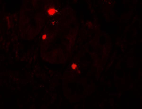
Two anti-ATG14 antibodies have image data: paraffin-section IHC in rat spleen and human lung cancer, and IF in human small intestine and U2OS cells (catalog image captions).

Real IHC data IHC analysis of ATG14L using anti-ATG14L antibody (PB9481). ATG14L was detected in paraffin-embedded section of Rat Spleen Tissue. Heat mediated antigen retrieval was performed in citrate buffer (pH6, epitope retrieval solution) for 20 mins. The tissue section was blocked with 10% goat serum. The tissue section was then incubated with 1μg/ml rabbit anti-ATG14L Antibody (PB9481) overnight at 4°C. Biotinylated goat anti-rabbit IgG was used as secondary antibody and incubated for 30 minutes at 37°C. The tissue section was developed using Strepavidin-Biotin-Complex (SABC)(Catalog # SA1022) with DAB as the chromogen.
Anti-ATG14L Antibody ®
Cat # PB9481
Real IF data Immunofluorescence of ATG14 in human small intestine tissue with ATG14 antibody at 20 μg/mL.
Anti-ATG14 Antibody
Cat # A03546

PB9481 is listed for IHC and IF, with paraffin-section IHC images from rat spleen and human lung cancer and an IF image from U2OS cells (catalog applications and image captions). A03546 is listed for IF, with an image from human small intestine tissue (catalog applications and image caption).

Which to pick: Choose PB9481 for chromogenic IHC on paraffin sections: its rat spleen and human lung cancer captions report citrate retrieval at pH 6 for 20 minutes and antibody at 1 μg/mL; the fixative is unreported (PB9481 IHC image captions). For IF/ICC, PB9481 has a U2OS cell IF image, while A03546 has a human small intestine IF image and no listed IHC application (catalog applications and image captions). For species selection, PB9481 lists human and rat reactivity; A03546 lists human, mouse and rat reactivity, though its supplied IF image documents human tissue only (catalog reactivity and A03546 image caption).

Each figure is that product's own IHC / IF validation image from its datasheet.

References

  1. UniProt Consortium. UniProt entry Q6ZNE5 (BAKOR_HUMAN, Beclin 1-associated autophagy-related key regulator).
  2. Human Protein Atlas. ATG14 tissue IHC expression (reliability: Approved).
  3. Human Protein Atlas. ATG14 subcellular location (ICC-IF): Localized to the plasma membrane and vesicles..
  4. Human Protein Atlas. ATG14 antibody validation summary (2 antibodies).
  5. miR-25-3p promotes proliferation and inhibits autophagy of renal cells in polycystic kidney mice by regulating ATG14-Beclin 1. Renal failure 2020 — PMC7241494.
  6. Long non-coding RNA MLLT4 antisense RNA 1 induces autophagy to inhibit tumorigenesis of cervical cancer through modulating the myosin-9/ATG14 axis. Scientific reports 2024 — PMC10944523.
  7. PubMed PMID:10048485 — UniProt-cited evidence.
  8. PubMed PMID:14702039 — UniProt-cited evidence.
  9. PubMed PMID:12508121 — UniProt-cited evidence.