ATG16L1 / Autophagy-related protein 16-1 · IHC design guide

Design Immunohistochemistry for ATG16L1

Plan ATG16L1 staining in paraffin sections using the catalog antibody at 2–5 μg/mL (datasheet A00526-3). Use cell-specific positive and negative controls and assess cytoplasmic, membranous and ciliary staining (HPA tissue IHC).

Evidence assembled Oct 2026 · For research use; verify linked source records and product datasheet before use
Immunohistochemistry protocol sheet for ATG16L1 (IHC for ATG16L1): expected localisation Cytoplasmic and membranous tissue staining (HPA tissue IHC), antibody A00526-3, validated IHC image, and IHC protocol steps
Printable ATG16L1 IHC protocol sheet — expected localisation Cytoplasmic and membranous tissue staining (HPA tissue IHC), antibody A00526-3, controls and protocol steps. Open the full ATG16L1 IHC guide →

ATG16L1 Immunohistochemistry Experimental Design Guide

Expected localisation, validated protocols, controls and antibodies — the at-a-glance facts below, then the full design guide.

Must know before staining
Expected localisation Cytoplasmic and membranous tissue staining (HPA tissue IHC)
Staining pattern Ciliary staining plus cytoplasmic/membranous tissue signal (HPA tissue IHC)
Antigen retrieval EDTA pH 8.0 HIER, heat-mediated (datasheet A00526-3)
Positive control ⓘ Bronchus+4 more · see all
Negative control ⓘ Lymph node+2 more · see all
Important caveats
Reasons your staining may differ from the expected pattern.
Fixation Keep fixation consistent across sections (standard IHC practice; not target-specific)
Caveat Starvation can shift ATG16L1 complexes to phagophores (UniProt)
Regulation Abundance regulation unreported (UniProt)
Isoform / epitope 5 isoforms; antibody epitope coverage needs validation (UniProt)
Section 1

Recommended ATG16L1 IHC & IF Protocols

The catalog antibody's IHC-P protocol is accompanied by a published FFPE tumor-tissue method using CC1 retrieval and DAB detection (PMC10517947: IHC methods).

Recommended immunohistochemistry (IHC-P) protocol parameters
SampleParaffin-embedded human the renal pelvis is squamous metaplasia tissue; fixative not specified (datasheet A00526-3)
FixationImage fixative and duration unreported (datasheet A00526-3); verify before use.
Sectioning4–5 µm sections on charged slides (standard)
DeparaffinisationXylene, graded ethanol series to water (standard)
Antigen retrievalHeat retrieval: EDTA pH 8.0 (datasheet A00526-3); 20 min, 95–100 °C (standard)
Peroxidase block3% H2O2, 10 min, room temperature (standard)
Blocking10% goat serum (datasheet A00526-3)
Primary antibodyRabbit anti-ATG16L1, 2-5 μg/ml (datasheet A00526-3)
Primary incubationOvernight at 4 °C (datasheet A00526-3)
DetectionStreptavidin-biotin complex (SABC), DAB chromogen (datasheet A00526-3)
CounterstainHematoxylin, blue, dehydrate and mount (standard)
Expected resultATG16L1-positive staining in ciliated cells (tip of cilia) of bronchus (HPA tissue IHC: High). HPA tissue profile: Membranous and cytoplasmic expression in several tissue types. No signal in the no-primary control.
💡Decision noteStart with heat-mediated EDTA retrieval at pH 8.0 for the catalog antibody (datasheet: A00526-3); use CC1 when reproducing the published method (PMC10517947: IHC methods).
Section 2

What Is the Expected ATG16L1 Staining Pattern?

ATG16L1 is mainly cytoplasmic, with recruitment to preautophagosomal, endosomal and lysosomal membranes; it has no transmembrane segment (UniProt Q676U5). In paraffin-section IHC, expect membranous and cytoplasmic staining in several tissues (HPA: tissue profile; reliability Supported). Strong examples include bronchial and fallopian-tube ciliated cells, kidney distal tubules and round or early spermatids (HPA: High in each listed cell type). Interpret staining by cell type and compartment together.

What am I looking at on my slide?
Cytoplasmic staining in kidney distal tubules, with stronger signal than nearby weakly stained cells.This fits a documented high-staining cell population (HPA: kidney distal tubules High). Compare cells within the same section before interpreting a faint deposit elsewhere as ATG16L1; HPA reports low tissue specificity at the RNA level, so weak staining outside a high-staining population is not automatically aberrant (HPA: RNA low tissue specificity).
Signal at bronchial cilia tips or along fallopian-tube cilia axonemes.These are documented high-staining sites (HPA: bronchus ciliated-cell cilia tips High; fallopian-tube cilia axoneme High). Assess the ciliated cells themselves, rather than treating a thin apical deposit in isolation as proof of specificity (general IHC practice). UniProt also describes discrete ciliary-axoneme puncta, qualified as By similarity (UniProt Q676U5).
Dominant nuclear staining with little corresponding cytoplasmic signal.A dominant nuclear pattern conflicts with the reported cytoplasmic and membrane distribution (UniProt Q676U5; HPA: membranous and cytoplasmic tissue profile). Treat it as suspect, then compare a known high-staining cell population and the negative reagent control before assigning target localisation (general IHC practice).
Equally strong staining in lymph-node germinal-center cells or splenic red-pulp cells.Those populations were not detected in the HPA tissue survey (HPA: germinal-center cells Not detected; splenic red-pulp cells Not detected). Consider cross-reactivity or endogenous detection activity; a discordant result warrants control review, not an absolute claim that these cells can never express ATG16L1 (general IHC practice; HPA: observed tissue pattern).
Diffuse deposit across cells and extracellular spaces, or no signal in kidney distal tubules.Widespread deposit obscures the reported cell and compartment pattern (HPA: tissue profile). Conversely, absent staining in an HPA high-staining population makes this run difficult to interpret (HPA: kidney distal tubules High). Check detection controls and the validated antibody's IHC-P instructions before drawing a biological conclusion (general IHC practice).
💡Expected ATG16L1 appearanceCall a result positive when identifiable HPA high-staining cells show clear cytoplasmic or documented ciliary staining, with possible membranous signal; dominant nuclear staining or uniform diffuse deposit is suspect (HPA: tissue profile and High examples; UniProt Q676U5 localisation; general IHC practice).
How each factor affects the staining
Cell population and anatomical siteIntensity is cell specific: kidney distal tubules and round or early spermatids are High, while liver cholangiocytes and skeletal-muscle myocytes are Low (HPA: tissue IHC). Choose and score the named cell population; a whole-tissue average can conceal the reported pattern (general IHC practice).
Compartment and recruitment stateUniProt places ATG16L1 in cytoplasm and at preautophagosomal, endosomal and lysosomal membranes (UniProt Q676U5). Starvation-associated phagophore recruitment and activation-associated endolysosomal recruitment are described in the UniProt record; routine tissue IHC alone does not establish which pathway produced a particular deposit (UniProt Q676U5; general IHC practice).
Antibody evidenceThe HPA tissue profile has Supported reliability based on agreement between staining and RNA data (HPA: reliability description). HPA063900 has IHC Supported status; HPA012577 has no supplied IHC status (HPA: antibody validation). Apply validation status to its specified antibody, not automatically to a different catalog antibody (general IHC practice).
Molecular formsFive isoforms are listed, with no signal peptide, propeptide or annotated cleavage into shorter chains; the listed chain spans residues 1–607 (UniProt Q676U5). The supplied record gives no antibody epitope, so it cannot establish which isoforms a particular IHC antibody recognizes (UniProt Q676U5; supplied evidence scope).
IF/ICC Q: What localisation should an IF image show?A: Predominantly cytosolic signal is the supported HPA ICC-IF location; equatorial segment, mid piece and principal piece are additional approved locations in sperm (HPA: subcellular ICC-IF). Interpret IF on its separate guide page; these ICC-IF observations do not supply an IHC-P protocol (HPA: subcellular ICC-IF; supplied application scope).
Why is my staining missing, weak or wrong?
SituationLikely causeNext action
No visible signal in kidney distal tubules.A high-staining reference population has failed to show its reported pattern (HPA: kidney distal tubules High); the image alone does not identify the technical cause.Verify the section contains distal tubules, then check the IHC-validated antibody's IHC-P instructions, reagent integrity and detection controls (general IHC practice). Do not infer a target-specific fixation effect from this result (supplied evidence scope).
Strong stain spreads through cells that HPA lists as not detected.Cross-reactivity or endogenous detection activity is possible; HPA reports germinal-center and splenic red-pulp cells as Not detected (HPA: tissue IHC; general IHC practice).Compare a high-staining cell population and run the appropriate negative reagent and endogenous-activity controls for the chosen chromogenic system (general IHC practice).
Main signal appears nuclear.Nuclear dominance disagrees with cytoplasmic and membrane localisation (UniProt Q676U5; HPA: tissue profile); specificity is unresolved.Check staining controls and reassess the same antibody in an HPA high-staining cell population before scoring nuclear signal as ATG16L1 (general IHC practice; HPA: High examples).
A uniform haze hides cell boundaries.Diffuse background prevents cell-level interpretation and can reflect nonspecific detection or inadequate blocking (general IHC practice).Review blocking, antibody concentration, washes and chromogen development using the catalog antibody's IHC-P instructions; retain a control section to locate the background source (general IHC practice).
Apical staining is visible but seems absent from the rest of a ciliated cell.Cilia-tip or axoneme staining is documented in the relevant high-staining cells (HPA: bronchus cilia tips High; fallopian-tube cilia axoneme High); a thin deposit may be hard to assign in a section (general IHC practice).Confirm the ciliated-cell morphology and focal plane, then compare cytoplasmic staining and controls before calling the apical deposit specific (general IHC practice; HPA: membranous and cytoplasmic tissue profile).
Staining is weak in a chosen low-staining population.Low staining is the documented observation for some populations, including liver cholangiocytes and skeletal-muscle myocytes (HPA: tissue IHC).Assess an HPA high-staining population in the same run and score the named cells separately; weak signal in a Low population alone does not show assay failure (HPA: tissue IHC; general IHC practice).

Sample controls for ATG16L1 IHC & IF

🧪Run bronchus first and score staining at the ciliated-cell cilia tips, where ATG16L1 is High (HPA: bronchus, ciliated cells, High). Use lymph-node germinal-center cells as the negative tissue (HPA: lymph node, germinal center cells, Not detected); on the bronchus slide, assess nonciliated cells for background, but do not assume they are ATG16L1-negative without validation (HPA: bronchus row specifies ciliated cells only).
Positive control tissue: Bronchus (Ciliated cells (tip of cilia), HPA High)
Negative control tissue: Lymph node (HPA Not detected)
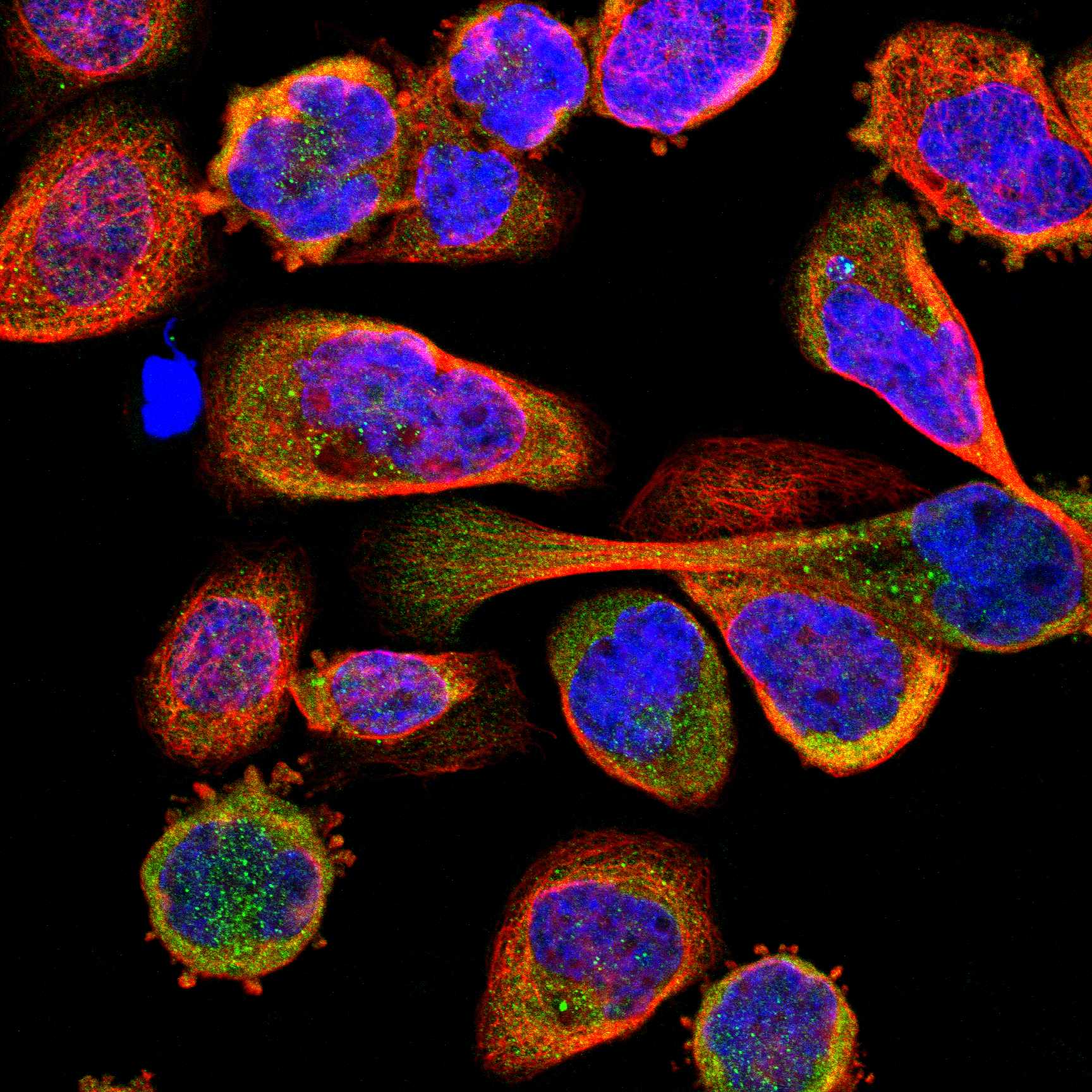
ICC-IF cell lines (HPA subcellular resource): HPA ICC-IF images show ATG16L1 in A-431, Hep-G2, Sperm, with annotated localisation: Cytosol (supported) (HPA subcellular).
Technical controls: Include a no-primary, secondary-only control, a rabbit IgG isotype control matched to the catalog antibody’s clonality if known, and an identically processed ATG16L1-knockout specimen if available (selected-SKU caption: rabbit primary; standard IHC practice). Quench endogenous peroxidase for chromogenic detection and block endogenous biotin if using an avidin–biotin system (selected-SKU caption: biotinylated secondary and SABC–DAB; standard IHC practice).
⚠️Feasibility: A target-specific fixation window or fixation effect is unreported, and the selected-SKU paraffin-section caption does not state a fixative (selected-SKU caption: fixative not stated). The caption uses heat retrieval in EDTA at pH 8.0 for renal-pelvis tissue, but does not establish that retrieval condition as necessary for bronchus (selected-SKU caption: EDTA, pH 8.0; HPA: bronchus, ciliated cells, High). Neither frozen sections nor IF can be judged easier from the supplied evidence; in bronchus, distinguish cilia-tip staining from signal in neighboring cells and check for endogenous peroxidase or biotin background (HPA: bronchus, ciliated cells at cilia tips; standard IHC practice).

HPA tissue IHC evidence for ATG16L1

Comprehensive Human Protein Atlas IHC scoring per tissue (reliability: Supported — High consistency between antibody staining and RNA expression data.). Rows are taken directly from the HPA tissue chart — click any row's HPA link to view the source.

Positive expression · recommended positive controls

TissueCell typeLevelEvidenceSource
Bronchus Ciliated cells (tip of cilia) High Protein (IHC) HPA →
Fallopian tube Ciliated cells (cilia axoneme) High Protein (IHC) HPA →
Kidney Distal tubules High Protein (IHC) HPA →
Testis Round or early spermatids High Protein (IHC) HPA →
Adrenal gland Glandular cells Medium Protein (IHC) HPA →

Undetected expression · recommended negative controls

TissueCell typeLevelEvidenceSource
Lymph node Germinal center cells Not detected Protein (IHC) HPA →
Soft tissue Peripheral nerve Not detected Protein (IHC) HPA →
Spleen Cells in red pulp Not detected Protein (IHC) HPA →
Section 3

Advanced ATG16L1 IHC Tips

Troubleshoot ATG16L1 staining in paraffin sections by checking retrieval, cellular pattern, controls and scoring before interpreting chromogenic signal.

Which retrieval condition should I try first for weak ATG16L1 staining?
Start with heat-mediated retrieval in EDTA at pH 8.0 for paraffin sections (datasheet A00526-3). This condition was used with the catalog antibody at 2 μg/ml, incubated overnight at 4°C (datasheet A00526-3). If staining is weak, check deparaffinisation and heating consistency, then compare a short retrieval-time series on matched sections (standard IHC practice). Hold primary concentration and DAB development time constant so differences can be attributed to retrieval (standard IHC practice). Assess both intensity and cellular pattern: ATG16L1 has cytoplasmic and membrane-associated locations (UniProt Q676U5 subcellular).
Could fixation explain weak or uneven ATG16L1 staining?
Target-specific fixation sensitivity is unknown: the selected paraffin-section caption does not report its fixative (datasheet A00526-3). Record fixation conditions and processing history for each specimen before comparing staining across cases (standard IHC practice). For an uneven section, inspect morphology and compare well-preserved interior regions with damaged or folded areas under identical retrieval and detection conditions (standard IHC practice). Test matched sections with one processing variable changed at a time, using the same positive control and DAB development time (standard IHC practice). Do not attribute a fixation effect to ATG16L1 without that direct comparison (standard IHC practice).
Should ATG16L1 appear diffuse, punctate or membranous in tissue?
Expect a cellular pattern that can include cytoplasm and membranes, since tissue staining is described as membranous and cytoplasmic (HPA tissue IHC). ATG16L1 also associates with preautophagosomal, endosomal and lysosomal membranes, although it has no transmembrane segment (UniProt Q676U5 subcellular; UniProt Q676U5 topology). Discrete puncta can therefore be plausible, but chromogenic puncta alone do not identify a particular organelle (UniProt Q676U5 subcellular; standard IHC interpretation). Check cell identity and morphology before scoring: strong staining is reported in kidney distal tubules and at cilia in bronchus and fallopian tube (HPA tissue IHC). Compare suspect signal with a negative control processed alongside the section (standard IHC practice).
How should unknown epitope coverage affect interpretation of ATG16L1 staining?
ATG16L1 has 5 annotated isoforms, but the supplied antibody caption does not identify its epitope or establish which isoforms it detects (UniProt Q676U5 isoforms; datasheet A00526-3). Avoid assigning a staining difference to a specific isoform from this IHC result alone (standard IHC interpretation). The protein has annotated phosphorylation sites, including Ser139, Ser269 and Ser287; the caption does not establish whether they affect antibody binding (UniProt Q676U5 modified residues; datasheet A00526-3). If isoform or modification dependence matters, obtain epitope information and validate against independently characterised material before interpreting tissue differences (standard IHC practice). Keep retrieval identical across comparisons (standard IHC practice).
How can I check ATG16L1 localisation with multiplex immunofluorescence?
For a tissue follow-up, pair ATG16L1 with a validated marker of the cell population being assessed, such as a distal-tubule marker when examining kidney (HPA tissue IHC; standard IF practice). Place the weaker signal in a bright, spectrally separated channel and consider a far-red fluorophore where tissue autofluorescence obscures shorter wavelengths (standard IF practice). ATG16L1 lacks a transmembrane segment and is mainly cytosolic, so use permeabilisation that gives antibody access to intracellular epitopes while preserving morphology (UniProt Q676U5 topology; HPA subcellular; standard IF practice). Include single-label and no-primary controls to assess bleed-through and background (standard IF practice). The paraffin IHC caption does not specify IF fixation or validate an IF protocol (datasheet A00526-3).
What should I check when ATG16L1 DAB staining is widespread?
First inspect the no-primary control and verify that endogenous peroxidase was blocked before DAB development (standard chromogenic IHC practice). The catalog tissue example used 10% goat serum, a biotinylated secondary and a streptavidin–biotin detection complex (datasheet A00526-3). With that detection scheme, assess possible endogenous biotin signal using an appropriate detection control and blocking step (standard IHC practice). Reduce nonspecific staining by checking primary concentration, wash stringency and DAB development time while retaining a positive control (standard IHC practice). Diffuse staining is not automatically background because ATG16L1 can be cytoplasmic, and tissue staining can include membranes (UniProt Q676U5 subcellular; HPA tissue IHC).
How should I score heterogeneous ATG16L1 staining across sections? ⚠ ANSWER MARKED FOR VERIFICATION
Define the cell population and compartment before scoring, since reported tissue patterns include cytoplasm, membranes and cilia-associated signal (HPA tissue IHC). For cellular DAB staining, record percent positive cells and intensity using a consistent 0–3 scale; an H-score can summarize their distribution on a 0–300 scale (standard IHC practice). If discrete puncta are the endpoint, report puncta density per mm² of evaluable tissue and specify the segmentation rule (standard image-analysis practice). Normalise cellular measures to the number of eligible cells and area measures to analyzable tissue area, excluding folds and necrosis (standard IHC practice). Use identical retrieval, imaging and scoring thresholds across cases (standard IHC practice).
How can I distinguish credible ATG16L1 staining from artefact?
Credible staining should occupy identifiable cells and a plausible cytoplasmic or membrane-associated compartment (HPA tissue IHC; UniProt Q676U5 subcellular). Strong signal in kidney distal tubules or ciliated cells has tissue-pattern support, whereas an unexpected cell population needs independent validation (HPA tissue IHC; standard IHC interpretation). Treat section-edge concentration, folds, necrotic material and signal reproduced without primary antibody as possible artefacts (standard IHC practice). Check the peroxidase block when brown signal persists in a no-primary control, because DAB reports peroxidase activity (standard chromogenic IHC practice). Finally, ATG16L1 staining shows protein distribution; it does not by itself establish autophagic activity or distinguish canonical from non-canonical pathways (UniProt Q676U5 function; standard IHC interpretation).
Boster reagents

Best ATG16L1 / Autophagy-related protein 16-1 IHC Antibodies

IHC images document human paraffin sections, including a formalin-fixed specimen; IF/ICC images document human cells. Catalog reactivity also includes mouse and rat for selected antibodies (catalog image captions; catalog reactivity).

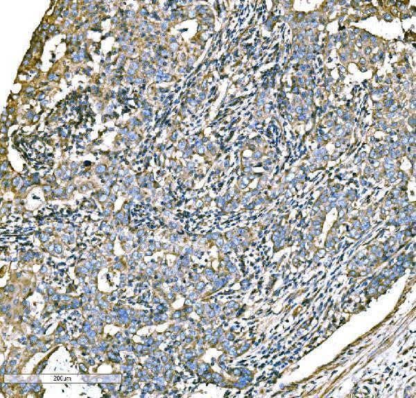
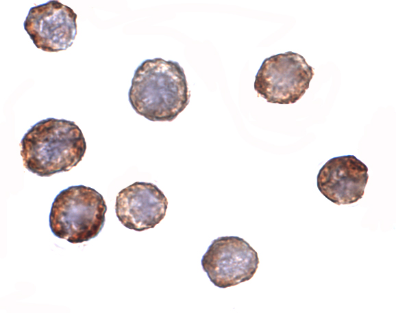
Real IHC data IHC analysis of ATG16L1 using anti-ATG16L1 antibody (A00526-3). ATG16L1 was detected in a paraffin-embedded section of human the renal pelvis is squamous metaplasia tissue. Heat mediated antigen retrieval was performed in EDTA buffer (pH 8.0, epitope retrieval solution). The tissue section was blocked with 10% goat serum. The tissue section was then incubated with 2 μg/ml rabbit anti-ATG16L1 Antibody (A00526-3) overnight at 4°C. Biotinylated goat anti-rabbit IgG was used as secondary antibody and incubated for 30 minutes at 37°C. The tissue section was developed using Strepavidin-Biotin-Complex (SABC) (Catalog # SA1022) with DAB as the chromogen.
Anti-ATG16L1 Antibody ®
Cat # A00526-3
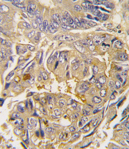
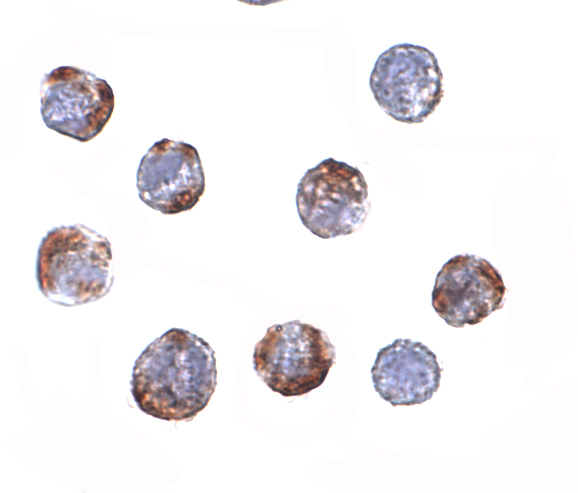
Real IHC data Formalin-fixed and paraffin-embedded human colon carcinoma tissue reacted with Autophagy APG16L antibody (L176), which was peroxidase-conjugated to the secondary antibody, followed by DAB staining. This data demonstrates the use of this antibody for immunohistochemistry; clinical relevance has not been evaluated.
Anti-ATG16L Antibody
Cat # A00526-2
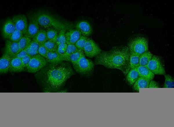
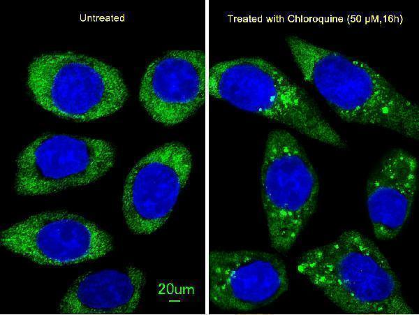
Real IF data Immunocytochemistry of ATG16 in HeLa cells with ATG16 antibody at 2 μg/mL.
Anti-ATG16 ATG16L1 Antibody
Cat # A00526
Real IF data Immunocytochemistry of ATG16 in HeLa cells with ATG16 antibody at 2 μg/mL.
Anti-ATG16 ATG16L1 Antibody
Cat # A00526-1

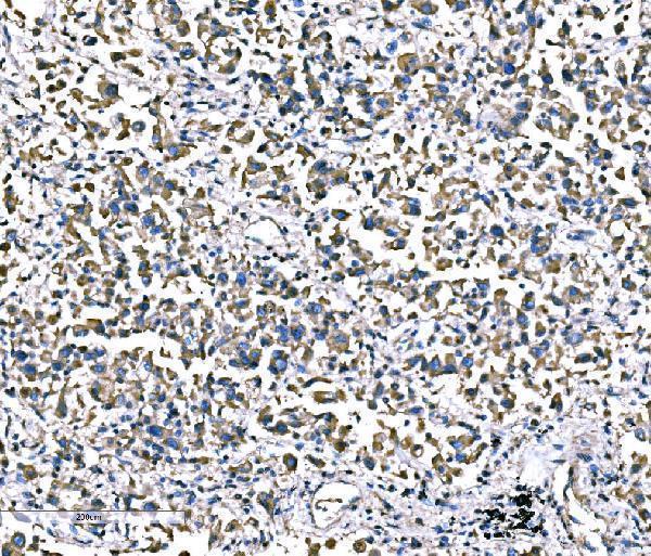
A00526-3 has IHC images from human renal-pelvis squamous metaplasia, lung cancer and esophageal squamous carcinoma paraffin sections; A00526-2 has an IHC image from formalin-fixed, paraffin-embedded human colon carcinoma (catalog IHC captions). A00526 and A00526-1 have ICC images from HeLa cells; both list IF and ICC applications, while only A00526-1 lists mouse and rat reactivity alongside human (catalog figure captions; catalog applications; catalog reactivity).

Which to pick: For tissue IHC, choose A00526-3 for its documented human paraffin-section staining; its caption does not report the fixative. Choose A00526-2 when a paraffin-section, paraffin-embedded example is useful, as its own colon carcinoma caption specifies that preparation (catalog IHC captions). For IF/ICC, A00526 and A00526-1 both list those applications and show HeLa ICC images; A00526-1 offers the broadest listed reactivity—human, mouse and rat—whereas A00526-2 lists human and mouse for IHC-P, with a human-only tissue image (catalog applications; catalog reactivity; catalog figure captions). The selected A00526-3 tissue-IHC caption documents paraffin sections, but does not specify the fixative (selected-SKU IHC image A00526-3).

Each figure is that product's own IHC / IF validation image from its datasheet.

References

  1. UniProt Consortium. UniProt entry Q676U5 (A16L1_HUMAN, Autophagy-related protein 16-1).
  2. Human Protein Atlas. ATG16L1 tissue IHC expression (reliability: Supported).
  3. Human Protein Atlas. ATG16L1 subcellular location (ICC-IF): Mainly localized to the cytosol. In addition localized to the equatorial segment, mid piece and principal piece..
  4. Human Protein Atlas. ATG16L1 antibody validation summary (2 antibodies).
  5. Tumor-intrinsic expression of the autophagy gene Atg16l1 suppresses anti-tumor immunity in colorectal cancer. Nature communications 2023 — PMC10517947.
  6. MicroRNA-20a-mediated loss of autophagy contributes to breast tumorigenesis by promoting genomic damage and instability. Oncogene 2017 — PMC5658668.
  7. MIR106B and MIR93 prevent removal of bacteria from epithelial cells by disrupting ATG16L1-mediated autophagy. Gastroenterology 2014 — PMC3870037.
  8. Paneth cells as a site of origin for intestinal inflammation. Nature 2013 — PMC3862182.
  9. PubMed PMID:15620219 — UniProt-cited evidence.
  10. PubMed PMID:17200669 — UniProt-cited evidence.
  11. PubMed PMID:12975309 — UniProt-cited evidence.