Click image to see more details
-

-
-
-
-
+2
Product Info Summary
| SKU: | A00526-2 |
|---|---|
| Size: | 80 µl |
| Reactive Species: | Human, Mouse |
| Host: | Rabbit |
| Application: | IF, IHC-P, WB |
Customers Who Bought This Also Bought

Product info
Product Name
Anti-ATG16L Antibody
SKU/Catalog Number
A00526-2
Size
80 µl
Form
Liquid
Description
Boster Bio Anti-ATG16L Antibody (Catalog # A00526-2). Tested in IF, WB, IHC-P application(s). This antibody reacts with Human, Mouse.
Storage & Handling
Maintain refrigerated at 2-8°C for up to 2 weeks. For long-term storage, store at -20°C in small aliquots to prevent freeze-thaw cycles.
Cite This Product
Anti-ATG16L Antibody (Boster Biological Technology, Pleasanton CA, USA, Catalog # A00526-2)
Host
Rabbit
Contents
Purified polyclonal antibody supplied in PBS with 0.09% (W/V) sodium azide.
Clonality
Polyclonal
Isotype
Rabbit IgG
Immunogen
This ATG16L antibody is generated from rabbits immunized with a KLH conjugated synthetic peptide between 161-190 amino acids from human ATG16L.
Cross-reactivity
No cross reactivity with other proteins.
Reactive Species
A00526-2 is reactive to ATG16L1 in Human, Mouse
Calculated molecular weight
68.3 kDa
Background of ATG16L1
Macroautophagy is the major inducible pathway for the general turnover of cytoplasmic constituents in eukaryotic cells, it is also responsible for the degradation of active cytoplasmic enzymes and organelles during nutrient starvation. Macroautophagy involves the formation of double-membrane bound autophagosomes which enclose the cytoplasmic constituent targeted for degradation in a membrane bound structure, which then fuse with the lysosome (or vacuole) releasing a single-membrane bound autophagic bodies which are then degraded within the lysosome (or vacuole). The APG12-APG5-APG16L complex is esential for the elongation of autophagic isolation membranes. This complex initially associates in uniform distribution with small vesicle membranes. During membrane elongation, the complex partitions, with a great concentration building on the outer side of the isolation membrane. Upon completion of the formation of the autophagosome, the APG12-APG5-APG16L dissociates from the membrane.
Antibody Validation
Boster validates all antibodies on WB, IHC, ICC, Immunofluorescence, and ELISA with known positive control and negative samples to ensure specificity and high affinity, including thorough antibody incubations.
Application & Images
Applications
A00526-2 is guaranteed for IF, IHC-P, WB Boster Guarantee
Recommend Dilution
IF: 1:25
WB: 1:1000
IHC-P: 1:10-1:50
Validation Images & Assay Conditions
Click image to see more details
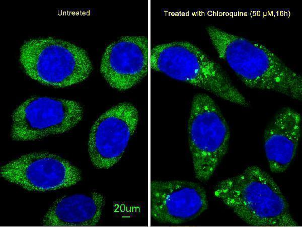
Immunofluorescent analysis of U251 cells, using ATG16L Antibody. U251 cells (right) were treated with Chloroquine (50 μM,16h). A00526-2 was diluted at 1:25 dilution. Alexa Fluor 488-conjugated goat anti-rabbit lgG at 1:400 dilution was used as the secondary antibody (green). DAPI was used to stain the cell nuclear (blue).

Click image to see more details
Cos7, HEK293, MEF, and Hela cells, left to right, respectively. Data courtesy of Drs. Jiefei Geng and Dan Klionsky, University of Michigan.

Click image to see more details
Anti-ATG16L Antibody at 1:1000 dilution + Jurkat whole cell lysate
Lysates/proteins at 20 µg per lane.
Secondary
Goat Anti-Rabbit IgG, (H+L), Peroxidase conjugated at 1/10000 dilution.
Predicted band size : 68 kDa
Blocking/Dilution buffer: 5% NFDM/TBST.

Click image to see more details
APG16L Antibody (Cat. #A00526-2) western blot analysis in NCI-H460 cell line lysates (35ug/lane). This demonstrates the APG16L antibody detected the APG16L protein (arrow).

Click image to see more details
Western blot of APG16L (L92) Pab (Cat. #A00526-2) in mouse brain tissue lysate.

Click image to see more details
Formalin-fixed and paraffin-embedded human colon carcinoma tissue reacted with Autophagy APG16L antibody (L176), which was peroxidase-conjugated to the secondary antibody, followed by DAB staining. This data demonstrates the use of this antibody for immunohistochemistry; clinical relevance has not been evaluated.
Specific Publications For Anti-ATG16L Antibody (A00526-2)
Loading publications
Recommended Resources
Here are featured tools and databases that you might find useful.
- Boster's Pathways Library
- Protein Databases
- Bioscience Research Protocol Resources
- Data Processing & Analysis Software
- Photo Editing Software
- Scientific Literature Resources
- Research Paper Management Tools
- Molecular Biology Software
- Primer Design Tools
- Bioinformatics Tools
- Phylogenetic Tree Analysis
Customer Reviews
Have you used Anti-ATG16L Antibody?
Share your experimental results or join a short interview to earn up to $1,000 in product credits or other rewards.
0 Reviews For Anti-ATG16L Antibody
Customer Q&As
Have a question?
Find answers in Q&As, reviews.
Can't find your answer?
Submit your question
