This website uses cookies to ensure you get the best experience on our website.


- Table of Contents
Plan paraffin-section ATG4A IHC around the reported cytoplasmic tissue pattern, which awaits external verification (HPA tissue IHC). Human colon glandular cells show high staining (HPA tissue IHC); the catalog antibody A06539-2 has a starting range of 0.5–1 μg/mL (datasheet A06539-2).

Expected localisation, validated protocols, controls and antibodies — the at-a-glance facts below, then the full design guide.
| Expected localisation | General cytoplasmic tissue staining (HPA tissue IHC) | |
| Staining pattern | Cytoplasmic staining across multiple cell types (HPA tissue IHC) | |
| Antigen retrieval | EDTA pH 8.0 HIER, heat-mediated (datasheet A06539-2) | |
| Positive control | Appendix+4 more · see all | |
| Negative control | Adipose tissue+1 more · see all |
| Fixation | Keep fixation consistent across paraffin sections. (standard IHC practice; not target-specific) | |
| Caveat | Tissue staining awaits external verification (HPA tissue IHC) | |
| Regulation | No expression regulation annotated (UniProt) | |
| Isoform / epitope | Four isoforms; no propeptide annotated (UniProt) |
The catalog antibody’s IHC-P protocol (datasheet: EDTA pH 8.0) is accompanied by two published paraffin-section IHC protocols (PMC5129932; PMC12234470).
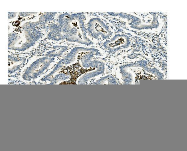
| Sample | Paraffin-embedded human rectal cancer tissue; fixative not specified (datasheet A06539-2) |
| Fixation | Image fixative and duration unreported (datasheet A06539-2); verify before use. |
| Sectioning | 4–5 µm sections on charged slides (standard) |
| Deparaffinisation | Xylene, graded ethanol series to water (standard) |
| Antigen retrieval | Heat retrieval: EDTA pH 8.0 (datasheet A06539-2); 20 min, 95–100 °C (standard) |
| Peroxidase block | 3% H2O2, 10 min, room temperature (standard) |
| Blocking | 10% goat serum (datasheet A06539-2) |
| Primary antibody | Rabbit anti-ATG4A, 0.5-1μg/ml (datasheet A06539-2) |
| Primary incubation | Overnight at 4 °C (datasheet A06539-2) |
| Detection | Streptavidin-biotin complex (SABC), DAB chromogen (datasheet A06539-2) |
| Counterstain | Hematoxylin, blue, dehydrate and mount (standard) |
| Expected result | ATG4A-positive staining in glandular cells of appendix (HPA tissue IHC: High). HPA tissue profile: General cytoplasmic expression. No signal in the no-primary control. |
ATG4A is a cytoplasmic protein with no transmembrane segment (UniProt Q8WYN0). In paraffin-section IHC, expect cytoplasmic staining in glandular, epithelial, hematopoietic, germinal-center, and kidney tubular cells reported as High by HPA (HPA: tissue IHC). Treat this as a provisional pattern: HPA describes general cytoplasmic expression, but rates its tissue IHC evidence Uncertain, pending external verification (HPA: tissue IHC reliability).
| Distinct cytoplasmic staining in colon or appendix glandular cells, with visible cell boundaries. | This fits the reported compartment and cell types (UniProt Q8WYN0: Cytoplasm; HPA: High in colon and appendix glandular cells). Score the fraction and intensity in the intended cells, and compare sections processed in the same run (general IHC practice). The HPA tissue pattern remains Uncertain pending external verification (HPA: tissue IHC reliability). |
| Predominantly nuclear or sharply membrane-restricted staining, with little cytoplasmic signal. | That distribution conflicts with the annotated cytoplasmic location and absence of a transmembrane segment (UniProt Q8WYN0). Review the counterstain, morphology, and detection controls before assigning it to ATG4A (general IHC practice). HPA tissue IHC does not validate either pattern as target-specific (HPA: tissue IHC reliability Uncertain). |
| Strong signal in adipocytes or ovarian stromal cells while nearby expected-positive cells are weak. | HPA reports ATG4A as Not detected in those cell types (HPA: adipocytes; ovarian stroma). Investigate cross-reactivity or endogenous detection activity with appropriate controls (general IHC practice). A negative HPA observation is a comparison point, not proof that every specimen of that tissue must remain unstained (HPA: tissue IHC reliability Uncertain). |
| Uniform haze across cells, stroma, and empty areas, obscuring cell boundaries. | A non-cellular distribution cannot support cell-specific scoring (general IHC practice). Check the no-primary control, wash quality, blocking, and chromogen development in the same run (general IHC practice). It does not resemble HPA's reported general cytoplasmic expression (HPA: tissue IHC profile). |
| No staining in colon glandular cells or kidney tubular cells despite preserved morphology. | Both are reported High in HPA tissue IHC (HPA: colon glandular cells; kidney tubular cells). First check that the assay's positive control worked, then review antibody dilution, retrieval, and detection steps (general IHC practice). Absence alone cannot establish true ATG4A loss because HPA rates its tissue IHC evidence Uncertain (HPA: tissue IHC reliability). |
| Tissue and cell selection | HPA reports High staining in appendix, colon, duodenum, and gallbladder glandular cells; kidney tubular cells; esophageal squamous cells; bone-marrow hematopoietic cells; and lymph-node germinal-center cells (HPA: tissue IHC). Choose an identifiable reported cell population for a positive comparison, while retaining HPA's Uncertain reliability qualification (HPA: tissue IHC reliability). |
| Compartment and topology | UniProt places ATG4A in the cytoplasm and annotates no transmembrane segment (UniProt Q8WYN0). Use a cytoplasmic scoring region in IHC; a purely nuclear or membrane-rim pattern needs independent confirmation (UniProt Q8WYN0; general IHC practice). Topology alone does not predict how paraffin processing affects staining. |
| Isoforms and antibody recognition | UniProt lists 4 ATG4A isoforms (UniProt Q8WYN0). The supplied sources do not map an IHC epitope to those isoforms, so a staining difference cannot be assigned to one isoform from this evidence. Keep antibody identity consistent across compared sections (general IHC practice). |
| IHC evidence strength | HPA labels tissue IHC reliability Uncertain, pending external verification (HPA: tissue IHC reliability). Its antibody HPA036374 has Uncertain IHC status, while HPA064759 has Approved ICC status (HPA: antibody validation). ICC approval does not upgrade the tissue IHC result; corroborate an important IHC finding independently. |
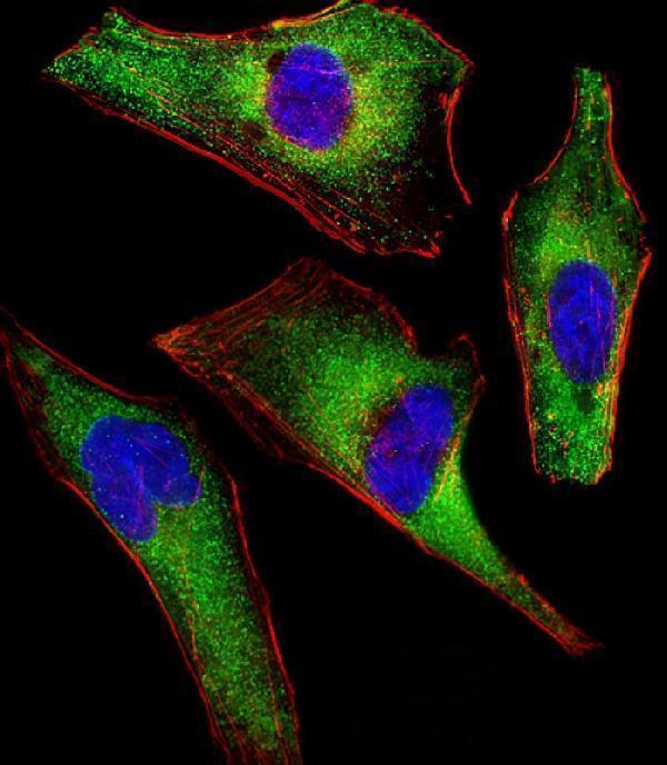
| IF/ICC Q&A | Q: Should IF show the same pattern as paraffin IHC? A: HPA reports mainly microtubules and additional actin filaments in ICC-IF, with Approved locations; UniProt gives the broader Cytoplasm annotation (HPA: subcellular ICC-IF; UniProt Q8WYN0). Interpret IF on its own guide page and do not use filament staining as a required IHC criterion. |
| Situation | Likely cause | Next action |
|---|---|---|
| Reported High tissue is blank, including the run's positive-control section. | The run may have failed at antibody application, retrieval, detection, or chromogen development (general IHC practice). | Check reagent application and expiry, the recorded dilution, retrieval settings, and detection sequence; repeat with a documented positive-control section (general IHC practice). Colon glands or kidney tubules are reported High, although HPA tissue IHC reliability is Uncertain (HPA: tissue IHC). |
| Only the study specimen is blank; the positive-control section stains. | The specimen may differ biologically or technically from the control; absence alone does not distinguish those possibilities (general IHC practice). | Confirm target-cell preservation and section quality, then repeat alongside the positive control using the same run conditions (general IHC practice). Compare like cell types; HPA reports Low staining in several tissues and Uncertain tissue IHC reliability (HPA: tissue IHC). |
| Nuclear or membrane-rim staining dominates the slide. | The observed compartment conflicts with cytoplasmic ATG4A and its lack of a transmembrane segment (UniProt Q8WYN0). | Check localization against morphology and the no-primary control; seek independent confirmation before scoring it as ATG4A (general IHC practice). Do not reinterpret the HPA ICC-IF filament pattern as nuclear or membrane-rim IHC staining (HPA: subcellular ICC-IF). |
| Adipocytes or ovarian stromal cells stain more strongly than expected-positive cells. | These cell types are Not detected in the HPA tissue observations; cross-reactivity or detection background is possible (HPA: tissue IHC; general IHC practice). | Inspect the no-primary control and compare cell-specific signal with a reported High population in the same staining run (general IHC practice; HPA: tissue IHC). Treat the discrepancy as a reason to verify the assay, not as proof of a biological change. |
| Brown signal appears in the no-primary control. | Signal without primary antibody points to a detection or tissue-background contribution rather than primary-dependent staining (general IHC practice). | Review endogenous enzyme blocking, secondary detection, chromogen timing, and wash steps; correct the source before scoring the test slide (general IHC practice). HPA's ATG4A tissue pattern does not validate control-positive chromogen (HPA: tissue IHC profile). |
| Diffuse haze or uneven edge staining makes cellular scoring unreliable. | Nonuniform handling, inadequate washing, or excessive detection development can produce spatial background (general IHC practice). | Compare the no-primary control, inspect section coverage and wash steps, and adjust development using run controls (general IHC practice). Score only interpretable, cell-associated cytoplasmic signal; HPA reports general cytoplasmic expression but Uncertain IHC reliability (HPA: tissue IHC). |


Comprehensive Human Protein Atlas IHC scoring per tissue (reliability: Uncertain — Pending external verification.). Rows are taken directly from the HPA tissue chart — click any row's HPA link to view the source.
| Tissue | Cell type | Level | Evidence | Source |
|---|---|---|---|---|
| Appendix | Glandular cells | High | Protein (IHC) | HPA → |
| Bone marrow | Hematopoietic cells | High | Protein (IHC) | HPA → |
| Colon | Glandular cells | High | Protein (IHC) | HPA → |
| Duodenum | Glandular cells | High | Protein (IHC) | HPA → |
| Esophagus | Squamous epithelial cells | High | Protein (IHC) | HPA → |
Troubleshoot ATG4A staining in paraffin sections by checking retrieval, controls, cellular localisation and scoring before interpreting chromogenic signal.
The catalog includes ATG4A antibodies with human tissue IHC images (catalog IHC captions) and an IF image in human cells (M06539-1 IF caption); listed reactivity also includes mouse and rat (A06539 catalog).
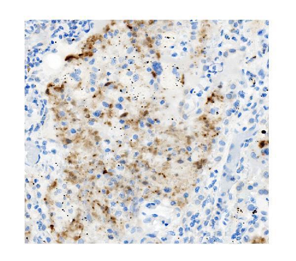
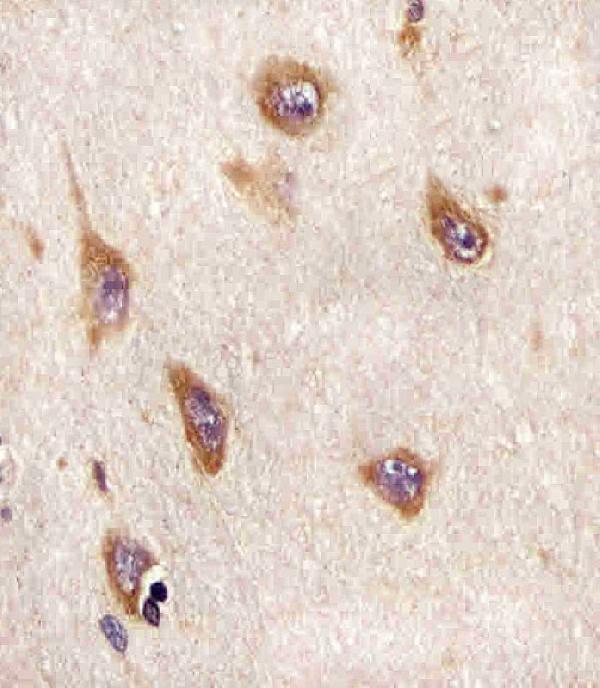
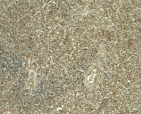
A06539-2 shows IHC in human rectal cancer paraffin sections (A06539-2 IHC caption); M06539-1 shows IHC in formaldehyde-fixed human brain paraffin sections (M06539-1 IHC caption). A06539 shows IHC in human spleen tissue (A06539 IHC caption), while M06539-1 also shows IF in paraformaldehyde-fixed, permeabilized HeLa cells (M06539-1 IF caption).
Which to pick: For human paraffin-section IHC, A06539-2 has an image using EDTA retrieval and 1 μg/mL primary antibody; its fixative is unreported (A06539-2 IHC caption). For IF/ICC, choose mouse monoclonal M06539-1: IF is listed at 1:25 and illustrated in HeLa cells; its separate IHC image documents paraffin-section brain sections with citrate retrieval (M06539-1 catalog; M06539-1 IF and IHC captions). For mouse or rat studies, A06539 lists those species as reactive, but its pictured IHC validation is in human spleen, so verify IHC performance in the intended species (A06539 catalog; A06539 IHC caption). The selected A06539-2 tissue-IHC caption documents paraffin sections, but does not specify the fixative (selected-SKU IHC image A06539-2).