This website uses cookies to ensure you get the best experience on our website.


- Table of Contents
Plan ATG4C paraffin IHC around general cytoplasmic staining (HPA tissue IHC). Start catalog antibody A09728 at 5 μg/mL (datasheet), and interpret intensity cautiously because tissue staining and RNA show low consistency (HPA tissue IHC).

Expected localisation, validated protocols, controls and antibodies — the at-a-glance facts below, then the full design guide.
| Expected localisation | Cytoplasmic staining in tissue (HPA tissue IHC) | |
| Staining pattern | General cytoplasmic staining (HPA tissue IHC) | |
| Antigen retrieval | Citrate pH 6.0 HIER, 95–98 °C, 20 min (rule: cytoplasmic / membrane antigen) | |
| Positive control | Adipose tissue+4 more · see all | |
| Negative control | None in HPA (detected in all 45 tissues); use no-primary + isotype controls |
| Fixation | Keep fixation consistent across sections (standard IHC practice; not target-specific). Selected-image fixative and duration unreported (datasheet A09728); verify before use. | |
| Caveat | Antibody staining and RNA show low consistency (HPA tissue IHC) | |
| Regulation | No tissue regulation annotated (UniProt) | |
| Isoform / epitope | No isoforms or processing variants annotated (UniProt) |
The catalog antibody IHC-P protocol is followed by four published ATG4C IHC workflows (PMC4369745; PMC12234470; PMC6617611; PMC7219755).
| Sample | Tissue sections; selected-image fixative not specified (standard IHC workflow) |
| Fixation | Image fixative and duration unreported (datasheet A09728); verify before use. |
| Sectioning | 4–5 µm sections on charged slides (standard) |
| Deparaffinisation | Xylene, graded ethanol series to water (standard) |
| Antigen retrieval | Heat-induced epitope retrieval in citrate buffer, pH 6.0, 20 min at 95–98 °C (standard rule: cytoplasmic / membrane antigen) |
| Peroxidase block | 3% H2O2, 10 min, room temperature (standard) |
| Blocking | 10% normal serum of the secondary host, 30 min, room temperature (standard) |
| Primary antibody | Rabbit anti-ATG4C, 5 μg/mL (datasheet A09728) |
| Primary incubation | Overnight at 4 °C (standard) |
| Detection | HRP-polymer secondary, DAB chromogen 5–10 min (standard) |
| Counterstain | Hematoxylin, blue, dehydrate and mount (standard) |
| Expected result | ATG4C-positive staining in adipocytes of adipose tissue (HPA tissue IHC: Medium). HPA tissue profile: General cytoplasmic expression. No signal in the no-primary control. |
Expect ATG4C staining chiefly in the cytoplasm of cells with an HPA IHC signal, including adipocytes, glandular cells, hematopoietic cells, neurons and endothelial cells (HPA tissue IHC: general cytoplasmic expression; listed cell types). ATG4C is cytoplasmic and has no annotated transmembrane segment (UniProt Q96DT6: location and topology). Treat the pattern as provisional: HPA rates its IHC evidence Approved but reports low agreement with RNA data and pending external verification (HPA tissue IHC: reliability).
| Cytoplasmic chromogen signal in adipocytes or listed glandular, hematopoietic, neuronal or endothelial cells. | This fits the reported compartment and cell distribution. HPA records Medium staining in these listed examples, so score the named cells rather than calling every cell in the section positive (HPA tissue IHC: profile and positive entries; UniProt Q96DT6: cytoplasm). |
| Predominantly nuclear-only, surface-rim or extracellular signal in an IHC section. | This is discordant with the reported cytoplasmic IHC pattern and warrants an artefact check (HPA tissue IHC: profile; UniProt Q96DT6: topology). Nuclear signal needs assay context: HPA separately approves nucleoplasmic localisation by ICC-IF (HPA subcellular: approved locations). |
| Strong colour in cells outside the reported stained cell populations, especially where expected cells remain clear. | Consider cross-reactivity or endogenous detection activity before assigning ATG4C to those cells (general IHC practice). HPA lists specific stained cell types and cautions that its antibody staining has low agreement with RNA data (HPA tissue IHC: positive entries and reliability). |
| Diffuse colour across stroma, empty spaces and many unrelated cell types. | A nonselective background pattern does not establish ATG4C localisation. Check the detection controls and compare cellular signal with background using the same scoring threshold (general IHC practice); the reported IHC profile is cytoplasmic (HPA tissue IHC: profile). |
| No cellular signal in a section chosen from an HPA Medium-staining tissue. | First assess assay performance with the reported positive cell population and controls (general IHC practice). Absence alone is inconclusive because HPA's IHC pattern has low agreement with RNA data and awaits external verification (HPA tissue IHC: reliability). |
| Compartment and topology | UniProt places ATG4C in the cytoplasm and annotates no transmembrane segment; HPA describes general cytoplasmic IHC expression (UniProt Q96DT6: location and topology; HPA tissue IHC: profile). Evaluate signal within cells rather than expecting a cell-surface outline. |
| Choice of reference tissue and cells | HPA reports Medium staining in, among others, adipocytes, adrenal glandular cells, bone marrow hematopoietic cells, cortical neurons and colon endothelial cells (HPA tissue IHC: positive entries). Its Low entries include lung alveolar cells and bronchial respiratory epithelium (HPA tissue IHC: low entries). |
| Strength of the tissue evidence | HPA calls the IHC result Approved, while flagging low agreement between antibody staining and RNA expression, a splice or transcript discrepancy, and pending external verification (HPA tissue IHC: reliability). Treat its intensity map as a comparison point, not a definitive expression boundary. |
| Antibody validation scope | The supplied HPA record lists one rabbit polyclonal antibody, HPA007049, as Approved for IHC and ICC; it does not list an Enhanced IHC result (HPA antibodies: HPA007049). A matching image alone therefore cannot establish that every stained cell contains ATG4C. |
| IF/ICC Q&A: is nuclear staining expected? | For ICC-IF, HPA approves both nucleoplasm and cytosol and lists images from A-431, U-251MG and U2OS (HPA subcellular: approved locations and image lines). For paraffin IHC, use the separately reported general cytoplasmic profile when interpreting a section (HPA tissue IHC: profile). |
| Situation | Likely cause | Next action |
|---|---|---|
| A reference section is blank. | The assay may have failed, or the sampled area may lack the HPA-listed positive cells; a blank section alone cannot distinguish these possibilities (general IHC practice; HPA tissue IHC: positive entries). | Locate the reported cell population, review counterstain and detection controls, then repeat with an HPA Medium-staining tissue if needed (general IHC practice; HPA tissue IHC: positive entries). |
| Only nuclei stain in paraffin IHC. | This conflicts with HPA's general cytoplasmic IHC profile, although HPA separately approves nucleoplasmic ICC-IF localisation (HPA tissue IHC: profile; HPA subcellular: approved locations). | Check control sections and whether colour is genuinely intranuclear before assigning an IHC result; record the assay-specific difference (general IHC practice; HPA subcellular: ICC-IF locations). |
| Unexpected cells stain more strongly than the reported population. | Cross-reactivity or endogenous detection activity is possible (general IHC practice); HPA also warns of low agreement between staining and RNA data (HPA tissue IHC: reliability). | Compare the same cell types across appropriate reagent controls and an HPA-listed tissue, then report any discordance rather than extending the cell-type claim (general IHC practice; HPA tissue IHC: positive entries). |
| The whole section has diffuse chromogen. | Nonselective reagent binding or endogenous detection activity can obscure cellular localisation (general IHC practice). This appearance differs from the reported general cytoplasmic profile (HPA tissue IHC: profile). | Review blocking, washes and detection controls, and assess whether cell-associated signal remains distinguishable from background (general IHC practice). |
| Dark staining obscures cell boundaries. | Excess primary antibody or chromogen development can reduce readable contrast (general IHC practice); the reported HPA positives are described as Medium, not as a required saturation level (HPA tissue IHC: positive entries). | Optimize the catalog antibody's IHC-P dilution and development time using its IHC instructions, then score discernible cytoplasmic signal in identified cells (general IHC practice; HPA tissue IHC: profile). |
| A Low-staining cell population appears negative. | A weak or absent visible signal is compatible with a Low HPA entry; examples include lung alveolar cells and parathyroid glandular cells (HPA tissue IHC: low entries). | Check an HPA Medium-staining cell population in parallel before judging assay failure, and report the Low population separately (general IHC practice; HPA tissue IHC: positive and low entries). |
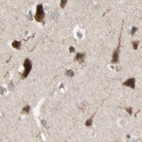

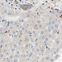


Comprehensive Human Protein Atlas IHC scoring per tissue (reliability: Approved — Low consistency between antibody staining and RNA expression data. Caution, Splice and/or transcript discrepancy exists. Pending external verification.). Rows are taken directly from the HPA tissue chart — click any row's HPA link to view the source.
| Tissue | Cell type | Level | Evidence | Source |
|---|---|---|---|---|
| Adipose tissue | Adipocytes | Medium | Protein (IHC) | HPA → |
| Adrenal gland | Glandular cells | Medium | Protein (IHC) | HPA → |
| Appendix | Glandular cells | Medium | Protein (IHC) | HPA → |
| Bone marrow | Hematopoietic cells | Medium | Protein (IHC) | HPA → |
| Breast | Glandular cells | Medium | Protein (IHC) | HPA → |
| Tissue | Cell type | Level | Evidence | Source |
|---|---|---|---|---|
| None in HPA: ATG4C is detected in all 45 scored tissues. Use a no-primary (secondary-only) and an isotype control instead. | ||||
Use compartment, cell type and control stains to evaluate ATG4C signal; the supplied tissue and subcellular references warrant careful interpretation (UniProt Q96DT6; HPA tissue IHC; HPA subcellular).
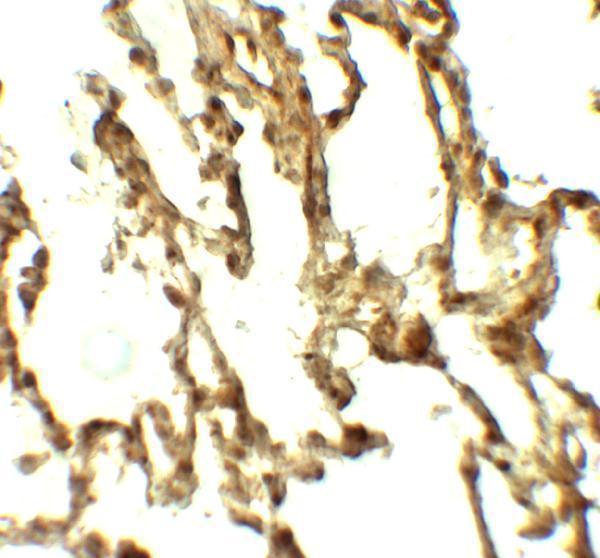
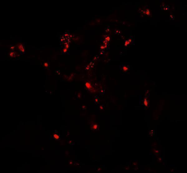
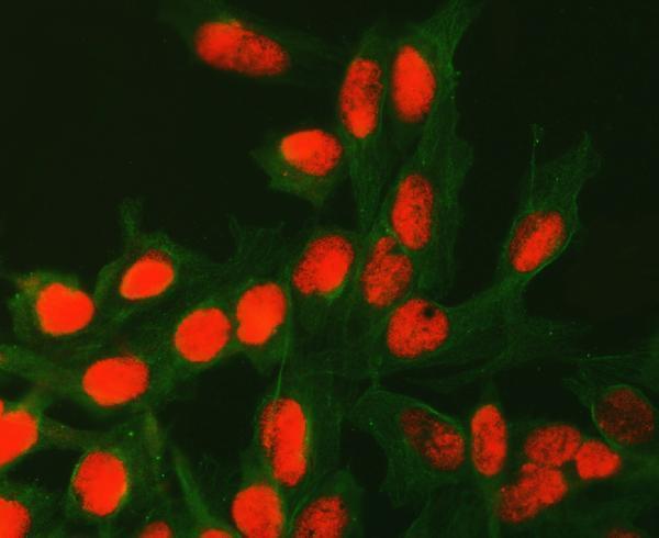
A09728 has human lung IHC and IF images (catalog image captions); A09728-2 has a human U20S IF/ICC image (catalog IF image caption; catalog reactivity).
A09728 will render with human lung IHC data at 5 μg/mL and has a human lung IF image (A09728 image captions). A09728-2 will render with IF/ICC data from a U20S cell at 5 μg/mL (A09728-2 IF image caption).
Which to pick: Choose A09728 for paraffin-section IHC: IHC-P is listed, and its own IHC caption shows human lung staining at 5 μg/mL; the fixative is unreported (A09728 catalog applications; A09728 IHC image caption). Choose A09728-2 for human IF/ICC because both applications are listed and its own image shows U20S cells (A09728-2 catalog applications; A09728-2 IF image caption; catalog reactivity). For mouse or rat work, A09728 lists reactivity with both species, while A09728-2 lists human only; both have a rabbit host and no clone stated (catalog reactivity; catalog host; catalog clone).