This website uses cookies to ensure you get the best experience on our website.


- Table of Contents
Plan chromogenic IHC on paraffin sections with the IHC-validated antibody at a starting concentration of 5 μg/mL (datasheet: A09793). Use duodenal glandular cells or testicular Leydig cells as positive tissue controls, and assess mainly cytoplasmic staining (HPA tissue IHC).

Expected localisation, validated protocols, controls and antibodies — the at-a-glance facts below, then the full design guide.
| Expected localisation | Tissue: mainly cytoplasmic (HPA tissue IHC); mitochondrial matrix after CASP3 cleavage (UniProt) | |
| Staining pattern | Mainly cytoplasmic in glandular and Leydig cells (HPA tissue IHC) | |
| Antigen retrieval | Citrate pH 6.0 HIER, 95–98 °C, 20 min (rule: cytoplasmic / membrane antigen) | |
| Positive control | Duodenum+4 more · see all | |
| Negative control | Adipose tissue+4 more · see all |
| Fixation | Matched tissue-IHC evidence does not establish the fixation claim. Validate the specimen-specific method before use. (selected-SKU IHC image A09793) | |
| Caveat | Staining has medium consistency with RNA expression (HPA tissue IHC) | |
| Regulation | Expression regulation is unreported (UniProt) | |
| Isoform / epitope | 2 isoforms; CASP3 cleavage removes residues 1–63, so epitope position matters (UniProt) |
Compare the catalog antibody’s IHC-P protocol with four published ATG4D tissue IHC methods (PMC12397559; PMC12234470; PMC5554887; PMC4398399).
| Sample | Tissue sections; selected-image fixative not specified (standard IHC workflow) |
| Fixation | Image fixative and duration unreported (datasheet A09793); verify before use. |
| Sectioning | 4–5 µm sections on charged slides (standard) |
| Deparaffinisation | Xylene, graded ethanol series to water (standard) |
| Antigen retrieval | Heat-induced epitope retrieval in citrate buffer, pH 6.0, 20 min at 95–98 °C (standard rule: cytoplasmic / membrane antigen) |
| Peroxidase block | 3% H2O2, 10 min, room temperature (standard) |
| Blocking | 10% normal serum of the secondary host, 30 min, room temperature (standard) |
| Primary antibody | Rabbit anti-ATG4D, 5 μg/mL (datasheet A09793) |
| Primary incubation | Overnight at 4 °C (standard) |
| Detection | HRP-polymer secondary, DAB chromogen 5–10 min (standard) |
| Counterstain | Hematoxylin, blue, dehydrate and mount (standard) |
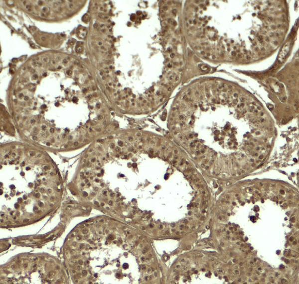
| Expected result | ATG4D-positive staining in glandular cells of duodenum (HPA tissue IHC: High). HPA tissue profile: Mainly cytoplasmic expression in several different tissue types. No signal in the no-primary control. |
In paraffin-section IHC, expect mainly cytoplasmic ATG4D staining, especially in duodenal and small-intestinal glandular cells and testicular Leydig cells (HPA tissue IHC: High; profile: mainly cytoplasmic). ATG4D has no transmembrane segment, and UniProt also places it in the mitochondrial matrix after CASP3 cleavage during oxidative stress or cell death (UniProt Q86TL0 topology and subcellular location). HPA rates its tissue IHC evidence Approved, with medium consistency against RNA expression (HPA tissue IHC).
| Cytoplasmic staining is strong in duodenal or small-intestinal glandular cells, or testicular Leydig cells. | This matches the expected cell types, compartment and High staining level (HPA tissue IHC). Compare signal with neighboring cells and a negative control on the same run (general IHC practice). |
| Staining is predominantly nuclear, with little cytoplasmic signal in a paraffin section. | Recheck specificity and detection conditions: the HPA tissue IHC profile is mainly cytoplasmic (HPA tissue IHC). Nuclear signal alone is not proof of artefact, because HPA reports nucleoplasmic localisation by ICC-IF (HPA subcellular). |
| Strong staining appears in adipocytes, cardiomyocytes or skeletal myocytes. | These cell types are reported as Not detected in HPA tissue IHC (HPA tissue IHC). Consider cross-reactivity or endogenous detection activity, and compare with a no-primary control (general IHC practice). |
| Brown signal is diffuse across cells, stroma or the entire section without a cell-specific pattern. | This does not reproduce the mainly cytoplasmic, cell-type-dependent tissue profile (HPA tissue IHC). Review blocking, washes and detection background with a no-primary control (general IHC practice). |
| No staining appears in duodenal or small-intestinal glandular cells or testicular Leydig cells. | These are High-staining reference cell types, so an absent signal calls for a run check (HPA tissue IHC). Review section integrity, antigen retrieval and detection controls before interpreting the specimen as negative (general IHC practice). |
| Compartment and processing | UniProt places full-length ATG4D in the cytoplasm and reports mitochondrial-matrix import after CASP3 cleavage during oxidative stress or cell death (UniProt Q86TL0). A mitochondrial pattern should therefore be interpreted in context; the HPA paraffin-section profile remains mainly cytoplasmic (HPA tissue IHC). |
| Sequence and isoforms | ATG4D has no annotated transmembrane segment and has two isoforms (UniProt Q86TL0). UniProt lists full-length residues 1–474 and a mitochondrial chain spanning residues 64–474 (UniProt Q86TL0); the supplied evidence does not identify the antibody epitope or establish which forms it detects. |
| Tissue controls and validation | Duodenal and small-intestinal glandular cells and testicular Leydig cells provide High-staining comparisons; adipocytes, cardiomyocytes and skeletal myocytes are reported as Not detected (HPA tissue IHC). The tissue IHC reliability is Approved with medium RNA-to-staining consistency, and HPA067683 is IHC Approved (HPA tissue IHC; HPA antibody validation). |
| IF/ICC interpretation | What if IF shows nuclear or mitochondrial signal? HPA reports nucleoplasm as approved and mitochondria as supported in ICC-IF, with U2OS images; HPA067683 is ICC Supported (HPA subcellular; HPA antibody validation). Those ICC-IF observations do not replace the mainly cytoplasmic paraffin-section IHC expectation (HPA tissue IHC). |
| Retrieval and fixation evidence | Target-specific fixation effects are not established by the supplied assay evidence. Verify with a matched IHC source before attributing a result to fixation. |
| Situation | Likely cause | Next action |
|---|---|---|
| Known-positive glandular or Leydig cells show no chromogenic signal. | The run may have failed, or retrieval or detection may be inadequate (general IHC practice); these cells are High in HPA tissue IHC (HPA tissue IHC). | Check the positive section and detection controls, then optimize retrieval within the assay workflow (general IHC practice). Do not score the test section as ATG4D-negative until controls work. |
| The whole section develops a similar brown haze. | Non-specific background or endogenous detection activity can obscure cell-specific staining (general IHC practice); HPA describes a mainly cytoplasmic tissue pattern (HPA tissue IHC). | Run a no-primary control; review blocking, wash stringency and chromogen development, then reassess whether the expected cell types stand out (general IHC practice; HPA tissue IHC). |
| Adipocytes, cardiomyocytes or myocytes stain as strongly as expected positive cells. | Cross-reactivity or endogenous detection activity is possible (general IHC practice); HPA reports these cell types as Not detected (HPA tissue IHC). | Compare a no-primary control and the HPA High-staining reference cells in the same run; interpret unexpected positivity cautiously (general IHC practice; HPA tissue IHC). |
| The signal is chiefly nuclear in paraffin-section IHC. | This differs from the mainly cytoplasmic tissue profile, although nucleoplasmic localisation is reported in ICC-IF (HPA tissue IHC; HPA subcellular). | Check the negative control and compare cytoplasmic staining in HPA High-staining cell types before assigning the nuclear signal to ATG4D (general IHC practice; HPA tissue IHC). |
| Punctate signal is interpreted as mitochondrial ATG4D. | Mitochondrial-matrix import is reported after CASP3 cleavage during oxidative stress or cell death, and mitochondrial localisation is supported in ICC-IF (UniProt Q86TL0; HPA subcellular). IHC puncta alone do not establish organelle identity. | Report the observed pattern as punctate unless an appropriate colocalisation experiment establishes mitochondrial localisation (general IF practice). Keep the tissue IHC expectation anchored to HPA’s mainly cytoplasmic profile (HPA tissue IHC). |
| A weakly stained test section is called negative after comparison with another tissue. | HPA reports Medium staining in several cell types and Low staining in others, so expected intensity depends on the sampled cell type (HPA tissue IHC). | Identify and score the relevant cells, compare an appropriate HPA High-staining reference, and verify run controls before concluding that weak signal is absent (HPA tissue IHC; general IHC practice). |


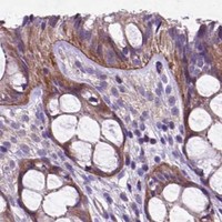


Comprehensive Human Protein Atlas IHC scoring per tissue (reliability: Approved — Medium consistency between antibody staining and RNA expression data.). Rows are taken directly from the HPA tissue chart — click any row's HPA link to view the source.
| Tissue | Cell type | Level | Evidence | Source |
|---|---|---|---|---|
| Duodenum | Glandular cells | High | Protein (IHC) | HPA → |
| Small intestine | Glandular cells | High | Protein (IHC) | HPA → |
| Testis | Leydig cells | High | Protein (IHC) | HPA → |
| Adrenal gland | Glandular cells | Medium | Protein (IHC) | HPA → |
| Appendix | Glandular cells | Medium | Protein (IHC) | HPA → |
| Tissue | Cell type | Level | Evidence | Source |
|---|---|---|---|---|
| Adipose tissue | Adipocytes | Not detected | Protein (IHC) | HPA → |
| Heart muscle | Cardiomyocytes | Not detected | Protein (IHC) | HPA → |
| Ovary | Ovarian stroma cells | Not detected | Protein (IHC) | HPA → |
| Skeletal muscle | Myocytes | Not detected | Protein (IHC) | HPA → |
| Smooth muscle | Smooth muscle cells | Not detected | Protein (IHC) | HPA → |
Troubleshoot ATG4D staining in paraffin sections by checking retrieval, tissue context, compartment, controls, and scoring before interpreting chromogenic signal.
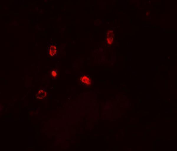
ATG4D antibodies have tissue IHC data in human testis (A09793 image caption) and cell IF data in U251 cells (M09793 image caption); catalog reactivity includes human, mouse and rat (A09793 catalog).
A09793 will render with human testis IHC at 5 μg/mL (A09793 IHC image caption); its catalog lists IHC-P and IF applications (A09793 catalog). M09793 will render with U251 cell IF at 1:25 (M09793 IF image caption); its catalog lists IF, but not IHC-P (M09793 catalog).
Which to pick: Choose A09793 for paraffin-section IHC: it lists IHC-P (A09793 catalog) and has a human testis IHC image at 5 μg/mL (A09793 IHC image caption); the caption does not report the fixative (A09793 IHC image caption). For cell IF, choose monoclonal M09793, shown in U251 cells (M09793 catalog; M09793 IF image caption). For mouse or rat work, A09793 is the listed cross-species option (A09793 catalog reactivity), although its supplied IHC image shows human tissue (A09793 IHC image caption).