This website uses cookies to ensure you get the best experience on our website.


- Table of Contents
Plan chromogenic ATG9A IHC on paraffin sections using the 2 μg/mL condition shown for catalog antibody A03757-3 (datasheet). Compare cytoplasmic staining with tissue examples while accounting for the HPA profile’s pending external verification (HPA tissue IHC).

Expected localisation, validated protocols, controls and antibodies — the at-a-glance facts below, then the full design guide.
| Expected localisation | Cytoplasmic tissue staining (HPA tissue IHC) | |
| Staining pattern | Cytoplasmic staining across several tissue cell types (HPA tissue IHC) | |
| Antigen retrieval | EDTA pH 8.0 HIER, heat-mediated (datasheet A03757-3) | |
| Positive control | Adrenal gland+4 more · see all | |
| Negative control | Adipose tissue+4 more · see all |
| Fixation | Keep fixation consistent across paraffin sections (standard IHC practice; not target-specific); Matched tissue-IHC evidence does not establish the fixation claim. Validate the specimen-specific method before use. (selected-SKU IHC image A03757-3) | |
| Caveat | Tissue staining awaits external verification (HPA tissue IHC) | |
| Regulation | Starvation redistributes ATG9A to PAS (UniProt) | |
| Isoform / epitope | 3 isoforms; epitope map unknown across cytoplasmic and luminal regions (UniProt) |
Compare the catalog antibody’s IHC-P protocol (datasheet A03757-3) with four published ATG9A IHC workflows (PMC4076416; PMC9296391; PMC10520003; PMC12314380).
| Sample | Paraffin-embedded human glioma tissue; fixative not specified (datasheet A03757-3) |
| Fixation | Image fixative and duration unreported (datasheet A03757-3); verify before use. |
| Sectioning | 4–5 µm sections on charged slides (standard) |
| Deparaffinisation | Xylene, graded ethanol series to water (standard) |
| Antigen retrieval | Heat retrieval: EDTA pH 8.0 (datasheet A03757-3); 20 min, 95–100 °C (standard) |
| Peroxidase block | 3% H2O2, 10 min, room temperature (standard) |
| Blocking | 10% goat serum (datasheet A03757-3) |
| Primary antibody | Rabbit anti-ATG9A, 2-5μg/ml (datasheet A03757-3) |
| Primary incubation | Overnight at 4 °C (datasheet A03757-3) |
| Detection | Streptavidin-biotin complex (SABC), DAB chromogen (datasheet A03757-3) |
| Counterstain | Hematoxylin, blue, dehydrate and mount (standard) |
| Expected result | ATG9A-positive staining in glandular cells of adrenal gland (HPA tissue IHC: Medium). HPA tissue profile: Cytoplasmic expression in several tissues. No signal in the no-primary control. |
ATG9A is a membrane protein that cycles mainly between the trans-Golgi network and endosomal system (UniProt Q7Z3C6 localization; UniProt Q7Z3C6 topology). In paraffin-section IHC, expect cytoplasmic staining in selected glandular and respiratory epithelial cells, among other reported sites (HPA tissue IHC). Treat intensity and distribution as provisional: HPA describes its tissue IHC reliability as Uncertain, pending external verification (HPA tissue IHC).
| Cytoplasmic staining in adrenal or duodenal glandular cells, or bronchial respiratory epithelium. | These are reported Medium-staining cell populations (HPA tissue IHC). A granular or perinuclear component is compatible with ATG9A trafficking through the trans-Golgi network and endosomes (UniProt Q7Z3C6 localization). Chromogenic IHC may not resolve individual vesicles (general IHC practice). |
| Predominantly nuclear staining, especially without a matching cytoplasmic pattern. | A nuclear-dominant result does not match the reported cytoplasmic tissue profile or membrane-associated locations (HPA tissue IHC; UniProt Q7Z3C6 localization). Review the staining against a control section before interpreting it as ATG9A; the location alone cannot identify the source of artefact (general IHC practice). |
| Strong staining in a cell population reported as Not detected, such as adipocytes in adipose tissue. | That result conflicts with the specified HPA cell-level observation (HPA tissue IHC). Cross-reactivity or endogenous chromogenic activity is possible (general IHC practice). HPA tissue IHC is Uncertain, so investigate the discrepancy with controls rather than treating the reference image as an absolute negative (HPA tissue IHC). |
| Diffuse color across cells and surrounding tissue, with little separation between compartments. | This obscures the reported cytoplasmic pattern (HPA tissue IHC). Broad background can reflect nonspecific antibody binding, detection reagent activity, or inadequate washing (general IHC practice). Compare the primary-antibody omission control and inspect whether the apparent signal tracks tissue structures or spreads uniformly (general IHC practice). |
| No staining in adrenal or duodenal glandular cells, or bronchial respiratory epithelium. | Each is listed as Medium in HPA tissue IHC (HPA tissue IHC). Check whether the expected cell population is present and whether the run controls worked (general IHC practice). An absent result could reflect assay performance or biological variation; HPA labels its tissue IHC reliability Uncertain (HPA tissue IHC). |
| Cell population and reference strength | HPA reports Medium staining in selected glandular, respiratory epithelial, and neural compartments, but Not detected in specified populations including adipocytes and alveolar cells (HPA tissue IHC). Score the named cells within each tissue, since an entire organ is not uniformly positive or negative (HPA tissue IHC). |
| Membrane location and trafficking | ATG9A has four annotated transmembrane segments and chiefly cycles between the trans-Golgi network and endosomal system (UniProt Q7Z3C6 topology; UniProt Q7Z3C6 localization). These facts support a cytoplasmic compartment interpretation, but they do not establish which epitope a particular antibody detects (UniProt Q7Z3C6 topology). |
| Condition-dependent redistribution | Amino acid starvation or rapamycin can redistribute ATG9A toward the phagophore assembly site, while only a small portion transiently occupies autophagic membranes during induction (UniProt Q7Z3C6 localization). This may affect punctate patterns in experimental models; it is not a basis for assigning an autophagy state from routine tissue IHC alone (UniProt Q7Z3C6 localization). |
| Validation limits across methods | HPA rates the listed antibody HPA059551 Uncertain for IHC and Supported for ICC, and calls its tissue IHC profile Uncertain (HPA antibody validation; HPA tissue IHC). HPA ICC-IF localizes ATG9A to vesicles, but that result does not independently validate a chromogenic tissue pattern (HPA subcellular ICC-IF; HPA antibody validation). |
| Situation | Likely cause | Next action |
|---|---|---|
| Expected cytoplasmic staining is absent in a Medium-staining reference population. | The target cells may be missing from the section, or the IHC run may have failed (HPA tissue IHC; general IHC practice). | Confirm cell identity and inspect the run control; review the validated antibody's own IHC-P instructions for retrieval and dilution (general IHC practice). Do not infer an ATG9A-specific fixation effect from HPA staining levels (HPA tissue IHC). |
| Signal is predominantly nuclear. | The compartment conflicts with reported cytoplasmic tissue staining and membrane localization (HPA tissue IHC; UniProt Q7Z3C6 localization). | Compare the result with a primary-antibody omission control and a reported cytoplasmic reference population; reassess specificity if the nuclear pattern persists (general IHC practice; HPA tissue IHC). |
| A reported Not detected population stains strongly. | Cross-reactivity or endogenous chromogenic activity may contribute (general IHC practice); HPA's negative observation has Uncertain overall IHC reliability (HPA tissue IHC). | Check an omission control and the exact cell population named by HPA before calling the tissue positive; use independent validation if the discrepancy matters to the study (general IHC practice; HPA tissue IHC). |
| Color is diffuse across the section. | Nonspecific binding, detection background, or inadequate washing may mask cellular contrast (general IHC practice). | Inspect omission and run controls, then review blocking, detection, washing, and primary-antibody conditions under the chosen IHC workflow (general IHC practice). Reassess whether cytoplasmic signal separates from background (HPA tissue IHC). |
| Puncta are hard to distinguish in chromogenic tissue IHC. | ATG9A occupies vesicle-related compartments, but section thickness and chromogenic resolution can blur small structures (UniProt Q7Z3C6 localization; general IHC practice). | Score the broader cytoplasmic distribution and the correct cell type first (HPA tissue IHC). Do not require countable vesicles for a positive chromogenic result (general IHC practice). |
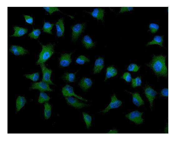
| IF/ICC Q: Should ATG9A appear in vesicles? | HPA reports a supported vesicular main location in ICC-IF, while the listed antibody's IHC status is Uncertain (HPA subcellular ICC-IF; HPA antibody validation). | IF/ICC A: Vesicular localization is supported for ICC-IF (HPA subcellular ICC-IF). Interpret it within an IF/ICC design; it does not by itself validate paraffin-section IHC staining (HPA antibody validation). |
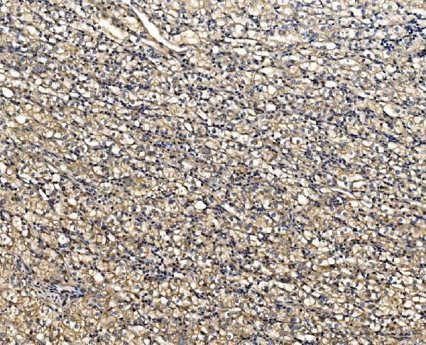
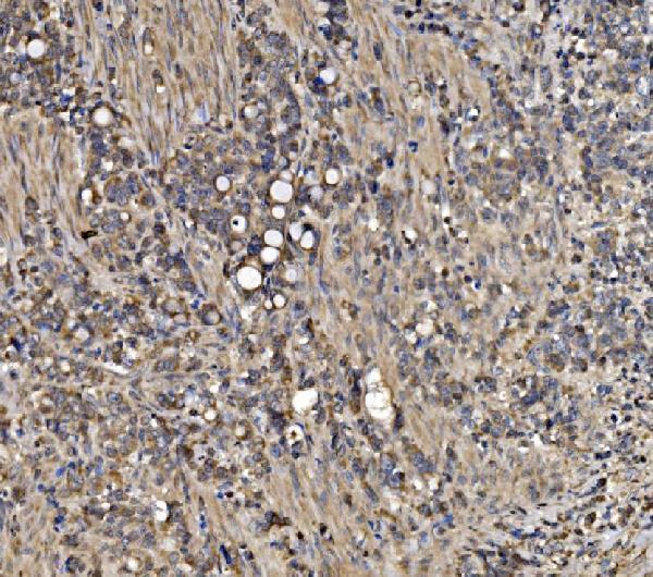
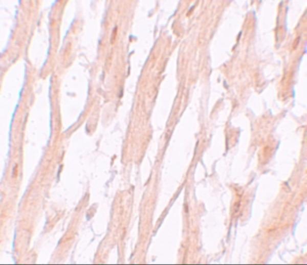
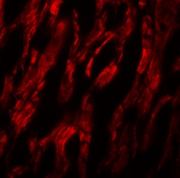




Comprehensive Human Protein Atlas IHC scoring per tissue (reliability: Uncertain — Pending external verification.). Rows are taken directly from the HPA tissue chart — click any row's HPA link to view the source.
| Tissue | Cell type | Level | Evidence | Source |
|---|---|---|---|---|
| Adrenal gland | Glandular cells | Medium | Protein (IHC) | HPA → |
| Appendix | Glandular cells | Medium | Protein (IHC) | HPA → |
| Bronchus | Respiratory epithelial cells | Medium | Protein (IHC) | HPA → |
| Cerebellum | Cells in granular layer | Medium | Protein (IHC) | HPA → |
| Cerebral cortex | Neuropil | Medium | Protein (IHC) | HPA → |
| Tissue | Cell type | Level | Evidence | Source |
|---|---|---|---|---|
| Adipose tissue | Adipocytes | Not detected | Protein (IHC) | HPA → |
| Bone marrow | Hematopoietic cells | Not detected | Protein (IHC) | HPA → |
| Breast | Adipocytes | Not detected | Protein (IHC) | HPA → |
| Caudate | Glial cells | Not detected | Protein (IHC) | HPA → |
| Cervix | Glandular cells | Not detected | Protein (IHC) | HPA → |
Troubleshoot ATG9A staining in paraffin sections by checking retrieval, staining controls, vesicular localisation and the cells being scored (datasheet A03757-3; UniProt Q7Z3C6; HPA tissue IHC).
Validated anti-ATG9A antibodies include IHC images from human heart, glioma and lung cancer tissue, plus IF images from human heart tissue and A549 cells (catalog image captions).
A03757-3 has paraffin-section IHC images from human glioma and mouse brain, while A03757 has human heart IHC and IF images (catalog image captions). M03757 has paraffin-section IHC in human lung cancer and IF in Hepg2 cells; A03757-2 has IF/ICC in A549 cells (catalog image captions).
Which to pick: For tissue IHC, choose A03757-3 when cross-species use matters: human, mouse and rat reactivity is listed, and its own paraffin-section IHC captions show human glioma and mouse brain with EDTA retrieval at pH 8.0; the fixative is unreported (catalog reactivity; A03757-3 IHC captions). For IF/ICC, choose A03757-2, which lists both applications and shows A549 cell IF at 5 μg/mL (catalog applications; A03757-2 IF caption). M03757 is a rabbit monoclonal option for IHC or IF/ICC, with listed human, mouse and rat reactivity; its own IHC caption shows paraffin-embedded human lung cancer, with fixative unreported (catalog: clone FGF-1, applications and reactivity; M03757 IHC caption).