This website uses cookies to ensure you get the best experience on our website.


- Table of Contents
Plan chromogenic IHC for ATP1B2 using retinal cell subsets and nerve fibers as expected staining patterns (HPA tissue IHC). This guide covers fixation consistency, antibody dilution, detection, and interpretation alongside the catalog antibody’s paraffin-section protocol (datasheet A07027-2).

Expected localisation, validated protocols, controls and antibodies — the at-a-glance facts below, then the full design guide.
| Expected localisation | Retinal cell membranes and cytoplasm; retinal and brain fibers (HPA tissue IHC) | |
| Staining pattern | Retinal cell subsets: membranous and cytoplasmic; nerve fibers (HPA tissue IHC) | |
| Antigen retrieval | EDTA pH 8.0 HIER, heat-mediated (datasheet A07027-2) | |
| Positive control | Retina+1 more · see all | |
| Negative control | Adrenal gland+4 more · see all |
| Fixation | Keep fixation consistent across sections (standard IHC practice; not target-specific). Selected-image fixative and duration unreported (datasheet A07027-2); verify before use. | |
| Caveat | Retinal staining is limited to cell subsets; field choice matters (HPA tissue IHC) | |
| Regulation | Brain/retina group-enriched RNA (HPA RNA) | |
| Isoform / epitope | No isoforms; epitope side: cytoplasmic 1–39 vs external 68–290 (UniProt) |
The catalog antibody’s IHC-P protocol (datasheet: A07027-2) is followed by two published paraffin-section IHC protocols (PMC5555477; PMC12203461).
| Sample | Paraffin-embedded human glioma tissue; fixative not specified (datasheet A07027-2) |
| Fixation | Image fixative and duration unreported (datasheet A07027-2); verify before use. |
| Sectioning | 4–5 µm sections on charged slides (standard) |
| Deparaffinisation | Xylene, graded ethanol series to water (standard) |
| Antigen retrieval | Heat retrieval: EDTA pH 8.0 (datasheet A07027-2); 20 min, 95–100 °C (standard) |
| Peroxidase block | 3% H2O2, 10 min, room temperature (standard) |
| Blocking | 10% goat serum (datasheet A07027-2) |
| Primary antibody | Rabbit anti-ATP1B2, 2-5 μg/ml (datasheet A07027-2) |
| Primary incubation | Overnight at 4 °C (datasheet A07027-2) |
| Detection | HRP-conjugated secondary, DAB chromogen (datasheet A07027-2) |
| Counterstain | Hematoxylin, blue, dehydrate and mount (standard) |
| Expected result | ATP1B2-positive staining in inner nuclear layer of retina (HPA tissue IHC: High). HPA tissue profile: Membranous and cytoplasmic expression in subsets of cells in retina. Additional expression in nerve fibers in retina and brain. No signal in the no-primary control. |
ATP1B2 is a cell-membrane protein with a cytoplasmic N terminus and extracellular C terminus (UniProt P14415 topology). In paraffin-section IHC, expect membranous and cytoplasmic staining in subsets of retinal cells, including strong staining in the inner nuclear layer, plus staining of nerve fibers in retina and brain (HPA tissue IHC: Supported; HPA: Retina inner nuclear layer High).
| Strong, selective staining in the retinal inner nuclear layer, with membranous and some cytoplasmic signal. | This fits the reported retinal pattern (HPA tissue IHC: Supported; HPA: inner nuclear layer High). Some cytoplasmic staining is expected in retinal cell subsets, so score it alongside its location and cell distribution (HPA tissue IHC). |
| Moderate staining in cerebral-cortex neuropil or staining along retinal and brain nerve fibers. | These are reported structures, although cortex neuropil is less intense than the listed retinal positive (HPA: cerebral-cortex neuropil Medium; HPA tissue IHC: nerve fibers). Compare the same anatomical structures across sections before calling a difference in intensity. |
| Predominantly nuclear staining, or uniform staining across cells and structures beyond the reported distribution. | Nuclear localisation conflicts with the membrane assignment (UniProt P14415 subcellular location). An unusually widespread pattern needs review for background or nonspecific binding (general IHC practice); do not dismiss reported retinal cytoplasmic staining as artefact (HPA tissue IHC). |
| Clear signal in a cell type listed as not detected, such as adrenal glandular cells. | That differs from the listed HPA result for that cell type (HPA: adrenal glandular cells Not detected). Check specificity and endogenous detection activity before interpreting it as ATP1B2; the HPA entry does not classify every cell in the organ. |
| Diffuse staining throughout the section, or no staining in the retinal inner nuclear layer. | Diffuse signal makes the reported cell and structure pattern hard to identify (HPA tissue IHC). An absent retinal positive is inconsistent with the listed High result (HPA: inner nuclear layer High); first assess section quality and the IHC detection workflow (general IHC practice). |
| Tissue and cell selection | Retinal inner nuclear layer is the listed High positive; cerebral-cortex neuropil is Medium (HPA tissue IHC). Listed negatives refer to specified cell types, such as adrenal glandular cells, not entire tissues (HPA tissue IHC). |
| Membrane topology and epitope location | ATP1B2 spans residues 40–67; residues 1–39 are cytoplasmic and 68–290 extracellular (UniProt P14415 topology). The supplied record gives no antibody epitope, so it cannot predict which side an antibody recognizes or whether retrieval is needed. |
| Glycosylation and processing | Seven glycosylation sites are annotated in the extracellular region, at residues 96, 118, 153, 159, 193, 197 and 238 (UniProt P14415 glycosylation; topology). No cleavage or shedding is annotated in the supplied record; staining cannot establish either event (UniProt P14415 processing). |
| Strength and scope of validation | HPA rates the tissue IHC pattern Supported, citing consistency between staining and RNA expression (HPA tissue IHC: Supported). The listed antibody HPA010698 has IHC Supported status (HPA antibodies); that does not validate every reagent or every tissue. |
| What about IF/ICC? | The HPA subcellular summary says Membrane, but gives no main location or ICC-IF image cell lines, and the listed antibody has no ICC status (HPA subcellular; HPA antibodies). Use the membrane assignment as an interpretation guide, not as evidence of an IF/ICC protocol or validated cell-line result. |
| Situation | Likely cause | Next action |
|---|---|---|
| Retinal inner nuclear layer has no detectable signal. | A listed High positive is missing (HPA: retinal inner nuclear layer High); section quality or an IHC workflow step may have failed (general IHC practice). | Confirm the anatomical layer and tissue preservation, then review reagent activity, detection and the catalog antibody's IHC-P instructions (general IHC practice). Do not assign an ATP1B2-specific fixation effect from this result. |
| Signal is broadly nuclear. | A dominant nuclear pattern conflicts with the recorded cell-membrane localisation (UniProt P14415 subcellular location). Background or nonspecific antibody binding is possible (general IHC practice). | Compare with a no-primary control and the retinal positive pattern; review blocking and detection conditions (general IHC practice; HPA: retinal inner nuclear layer High). |
| A listed negative cell type stains strongly. | The result differs from the HPA listing for that specific cell type (HPA: adrenal glandular cells Not detected). Nonspecific binding or endogenous chromogenic activity could contribute (general IHC practice). | Identify the stained cell type precisely; inspect no-primary and detection-only controls, then compare the signal with a known-positive section (general IHC practice; HPA: retinal inner nuclear layer High). |
| Brown precipitate or diffuse color obscures cell boundaries. | Nonspecific background or detection activity may prevent assessment of the reported membranous and cytoplasmic retinal pattern (general IHC practice; HPA tissue IHC). | Check no-primary controls, blocking, washes, chromogen development and counterstain; adjust using the detection system's instructions (general IHC practice). Reassess whether the expected structures remain distinguishable (HPA tissue IHC). |
| Cortex neuropil appears weaker than retinal inner nuclear layer. | That difference can agree with the listed Medium cortex neuropil and High retinal inner nuclear-layer levels (HPA tissue IHC). | Score each listed structure against its own expected level, using comparable staining conditions (HPA tissue IHC; general IHC practice). Investigate only if its signal or anatomical distribution is otherwise inconsistent. |
| Cytoplasmic staining is seen in retinal cell subsets. | HPA explicitly reports cytoplasmic as well as membranous expression in subsets of retinal cells (HPA tissue IHC); compartment alone does not make this an artefact. | Check whether the signal remains confined to the reported retinal subsets and accompanies a plausible membrane pattern (HPA tissue IHC; UniProt P14415 subcellular location). Use controls if it becomes diffuse or widespread (general IHC practice). |

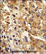
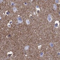

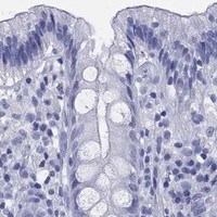

Comprehensive Human Protein Atlas IHC scoring per tissue (reliability: Supported — High consistency between antibody staining and RNA expression data. Antibody staining in cells/structures not annotated, view images.). Rows are taken directly from the HPA tissue chart — click any row's HPA link to view the source.
| Tissue | Cell type | Level | Evidence | Source |
|---|---|---|---|---|
| Retina | Inner nuclear layer | High | Protein (IHC) | HPA → |
| Cerebral cortex | Neuropil | Medium | Protein (IHC) | HPA → |
| Tissue | Cell type | Level | Evidence | Source |
|---|---|---|---|---|
| Adrenal gland | Glandular cells | Not detected | Protein (IHC) | HPA → |
| Appendix | Glandular cells | Not detected | Protein (IHC) | HPA → |
| Bone marrow | Hematopoietic cells | Not detected | Protein (IHC) | HPA → |
| Breast | Adipocytes | Not detected | Protein (IHC) | HPA → |
| Bronchus | Respiratory epithelial cells | Not detected | Protein (IHC) | HPA → |
Troubleshoot ATP1B2 staining in paraffin sections by checking retrieval, compartment, tissue context, and controls before comparing signal intensity.
Two anti-ATP1B2 antibodies have human paraffin-section IHC images (catalog captions). Neither has an IF/ICC image or listed IF/ICC application (catalog applications and image records).
A07027-2 has an IHC image from a paraffin-embedded human glioma section; its listed reactivity is human, mouse and rat (A07027-2 caption; catalog reactivity). A07027-1 has an IHC-P image from formalin-fixed, paraffin-embedded human hepatocarcinoma and lists human reactivity (A07027-1 caption; catalog applications and reactivity).
Which to pick: For human tissue IHC, choose A07027-2 for a paraffin-section workflow with documented EDTA retrieval and 2 μg/ml primary antibody; its caption does not report the fixative (A07027-2 caption). Choose A07027-1 when documented fixation matters; it is a rabbit polyclonal antibody with a listed IHC-P dilution of 1:50–1:100 (A07027-1 caption; catalog dilution record). Neither SKU lists IF/ICC validation; A07027-2 lists mouse and rat reactivity, but its supplied tissue IHC image documents human tissue only (catalog applications and reactivity; A07027-2 caption). The selected A07027-2 tissue-IHC caption documents paraffin sections, but does not specify the fixative (selected-SKU IHC image A07027-2).