This website uses cookies to ensure you get the best experience on our website.


- Table of Contents
Plan chromogenic IHC on paraffin sections using antibody A10401-2 at 2–5 μg/ml (datasheet A10401-2). Interpret cytoplasmic and nuclear staining against the broad tissue pattern, with high signal in colon glandular cells and no detected signal in liver cholangiocytes (HPA tissue IHC).

Expected localisation, validated protocols, controls and antibodies — the at-a-glance facts below, then the full design guide.
| Expected localisation | Cytoplasmic and nuclear tissue staining (HPA tissue IHC) | |
| Staining pattern | Widespread cytoplasmic and nuclear signal (HPA tissue IHC) | |
| Antigen retrieval | EDTA pH 8.0 HIER, heat-mediated (datasheet A10401-2) | |
| Positive control | Adrenal gland+4 more · see all | |
| Negative control | Liver |
| Fixation | Keep fixation consistent across sections. (standard IHC practice; not target-specific) | |
| Caveat | Liver cholangiocytes show no detected signal (HPA tissue IHC) | |
| Regulation | Low tissue specificity (HPA tissue RNA) | |
| Isoform / epitope | 2 isoforms; epitope differences unspecified (UniProt) |
The catalog antibody protocol uses EDTA pH 8.0 heat retrieval (datasheet A10401-2). The published IHC protocol below examines ATP6V1A in human invasive ductal carcinoma (PMC8721851).
| Sample | Paraffin-embedded human adenocarcinoma of the right colon tissue; fixative not specified (datasheet A10401-2) |
| Fixation | Image fixative and duration unreported (datasheet A10401-2); verify before use. |
| Sectioning | 4–5 µm sections on charged slides (standard) |
| Deparaffinisation | Xylene, graded ethanol series to water (standard) |
| Antigen retrieval | Heat retrieval: EDTA pH 8.0 (datasheet A10401-2); 20 min, 95–100 °C (standard) |
| Peroxidase block | 3% H2O2, 10 min, room temperature (standard) |
| Blocking | 10% goat serum (datasheet A10401-2) |
| Primary antibody | Rabbit anti-ATP6V1A, 2-5 μg/ml (datasheet A10401-2) |
| Primary incubation | Overnight at 4 °C (datasheet A10401-2) |
| Detection | HRP-conjugated secondary, DAB chromogen (datasheet A10401-2) |
| Counterstain | Hematoxylin, blue, dehydrate and mount (standard) |
| Expected result | ATP6V1A-positive staining in glandular cells of adrenal gland (HPA tissue IHC: High). HPA tissue profile: Ubiquitous cytoplasmic and nuclear expression. No signal in the no-primary control. |
ATP6V1A should appear mainly in the cytoplasm, including vesicle-associated regions, with some nuclear staining possible (UniProt P38606 localization; HPA: ubiquitous cytoplasmic and nuclear IHC expression; HPA: vesicles and cytosol in ICC-IF, additional nucleoplasm). Expect staining across many cell types, with strong examples in adrenal glandular and cerebellar Purkinje cells (HPA: High). The subunit has no transmembrane segment (UniProt P38606 topology). HPA rates its tissue IHC profile Approved, with medium staining–RNA consistency and external verification pending (HPA: reliability).
| Cytoplasmic staining, sometimes granular, in adrenal glandular cells or cerebellar Purkinje cells (HPA: High). | This matches reported IHC-positive cells (HPA: High). A granular component is compatible with vesicle localization (HPA: ICC-IF vesicles; UniProt P38606 localization). Chromogenic IHC may not resolve individual vesicles; judge the cell and overall compartment before interpreting fine puncta. |
| Nuclear signal accompanies cytoplasmic staining, or appears alongside a vesicular pattern (HPA: tissue IHC profile; HPA: ICC-IF nucleoplasm). | Do not reject nuclear signal solely because ATP6V1A functions in a proton pump: HPA reports nuclear IHC expression and additional nucleoplasmic ICC-IF localization (HPA: tissue IHC profile; HPA: ICC-IF nucleoplasm). A nuclear-only pattern needs comparison with a known-positive control and the negative reagent control. |
| Strong signal is confined to cell borders or extracellular deposits, with little cytoplasmic signal. | This is discordant with the reported cytoplasmic and vesicular distribution (HPA: tissue IHC profile; HPA: ICC-IF vesicles and cytosol). Check whether the apparent border signal follows tissue folds or chromogen deposits; repeat with controls before assigning a membrane-specific ATP6V1A pattern (UniProt P38606 localization). |
| Cholangiocytes stain strongly in liver, despite little signal in the expected positive control. | HPA reports ATP6V1A as not detected in liver cholangiocytes (HPA: Not detected). Consider antibody cross-reactivity or endogenous detection activity; inspect the reagent-negative control and verify cell identity. This comparison applies to cholangiocytes, not to every cell in liver. |
| Diffuse color covers cells and empty tissue spaces, or known-positive adrenal glandular cells remain unstained (HPA: High). | Diffuse staining outside cells suggests background from the detection workflow; weak or absent staining in a reported positive population suggests a failed or insufficiently sensitive run. Neither finding alone establishes ATP6V1A absence. Compare matched control sections before interpreting the specimen (HPA: adrenal glandular cells High). |
| Compartment and resolution | ATP6V1A is cytosolic and associated with vesicles and lysosome membranes (UniProt P38606 localization); ICC-IF resolves vesicles more directly than chromogenic paraffin IHC (HPA: ICC-IF vesicles; standard microscopy practice). Interpret IHC granularity conservatively. |
| Cell and tissue choice | HPA reports High staining in adrenal glandular cells, cerebellar Purkinje cells, and several other populations, but Not detected in liver cholangiocytes (HPA: tissue IHC). Use the named cell population when selecting or reading controls; an entire organ is not uniformly positive or negative. |
| Strength of evidence | The tissue IHC profile is Approved, with medium consistency against RNA and external verification pending (HPA: reliability). CAB006910 is IHC Approved; HPA035084 has ICC Supported status without an IHC status in this payload (HPA: antibody validation). Match claims to the assay and antibody actually used. |
| Topology and epitope | ATP6V1A has no transmembrane segment or signal peptide and is the catalytic V1 subunit (UniProt P38606 topology, processing, function). These facts support an intracellular interpretation but do not locate the catalog antibody's epitope or predict which antigen retrieval condition will work. |
| Isoforms and modifications | UniProt lists two isoforms and three modified residues, including phosphorylation sites (UniProt P38606 isoforms, modified residues). Without an antibody epitope or isoform-specific validation, differences in staining cannot be assigned to an isoform or modification. |
| Fixation and retrieval evidence | No ATP6V1A-specific fixation sensitivity or retrieval condition is established by the supplied UniProt and HPA records. Antigen retrieval is a general paraffin-IHC optimization step; compare conditions using the same positive control before attributing a signal change to target biology. |
| Situation | Likely cause | Next action |
|---|---|---|
| Known-positive adrenal glandular cells are blank or very weak (HPA: High). | The run may lack sufficient usable signal; the HPA tissue result alone cannot identify which preparation or detection step failed. | Check that the positive section and detection reagents worked, then optimize the catalog antibody dilution and antigen retrieval using matched sections. Record each condition; no ATP6V1A-specific dilution or retrieval recipe is supplied. |
| Color is widespread, including empty spaces or section edges. | Chromogen precipitate, insufficient washing, or nonspecific detection can produce diffuse background (standard IHC practice). This pattern does not track the reported cellular localization (HPA: tissue IHC profile). | Inspect a reagent-negative section, remove visible precipitate, and review washing and blocking. Reassess whether signal remains inside cells after background is reduced; use the same exposure and scoring criteria across sections. |
| Liver cholangiocytes show strong staining (HPA: Not detected). | Possible cell misidentification, cross-reactivity, or endogenous detection activity; HPA's finding is specific to cholangiocytes, and its IHC evidence has medium RNA consistency (HPA: reliability). | Confirm the stained cell type on the counterstained section. Compare a reagent-negative control and a reported positive cell population, then repeat or seek independent antibody evidence if the discordance persists. |
| The slide appears exclusively membranous or extracellular. | That distribution conflicts with cytoplasmic IHC and vesicle/cytosol ICC-IF findings (HPA: tissue IHC profile; HPA: ICC-IF localization). Tissue folds or localized background may mimic borders (standard IHC practice). | Review morphology at higher magnification, including intact cells away from edges. Check a reagent-negative control and compare with a reported positive tissue before scoring the border signal as ATP6V1A. |
| Nuclear staining seems unexpected. | Nuclear staining is reported in tissue IHC, and nucleoplasm is an additional ICC-IF location (HPA: tissue IHC profile; HPA: ICC-IF nucleoplasm). Nuclear signal alone does not prove artefact. | Score nuclear and cytoplasmic compartments separately. Compare distribution in a reported positive cell type and inspect reagent-negative staining; investigate a nuclear-only pattern that lacks the expected cytoplasmic component. |
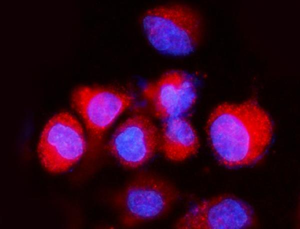
| IF/ICC puncta and paraffin IHC staining look different. | HPA reports vesicles and cytosol in ICC-IF, while its tissue IHC profile describes broader cytoplasmic and nuclear expression (HPA: ICC-IF localization; HPA: tissue IHC profile). The methods reveal different levels of spatial detail. | Use the ICC-IF finding as localization context, not an IHC protocol setting. Judge the paraffin section against IHC cell-type and compartment expectations; consult the separate IF/ICC guide for that assay. |
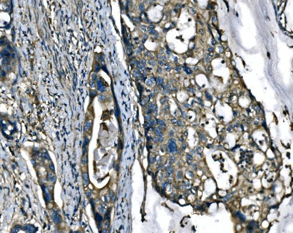
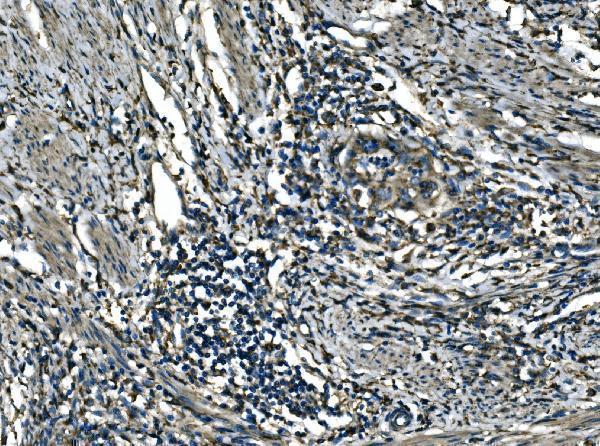

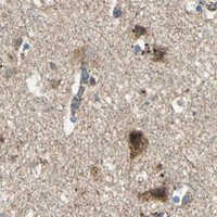


Comprehensive Human Protein Atlas IHC scoring per tissue (reliability: Approved — Medium consistency between antibody staining and RNA expression data. Pending external verification.). Rows are taken directly from the HPA tissue chart — click any row's HPA link to view the source.
| Tissue | Cell type | Level | Evidence | Source |
|---|---|---|---|---|
| Adrenal gland | Glandular cells | High | Protein (IHC) | HPA → |
| Bone marrow | Hematopoietic cells | High | Protein (IHC) | HPA → |
| Bronchus | Respiratory epithelial cells | High | Protein (IHC) | HPA → |
| Cerebellum | Purkinje cells | High | Protein (IHC) | HPA → |
| Cerebral cortex | Neuronal cells | High | Protein (IHC) | HPA → |
| Tissue | Cell type | Level | Evidence | Source |
|---|---|---|---|---|
| Liver | Cholangiocytes | Not detected | Protein (IHC) | HPA → |
Troubleshoot ATP6V1A staining in paraffin sections by checking retrieval, compartment patterns, controls and scoring before interpreting differences between samples.
Anti-ATP6V1A antibodies have human paraffin-section IHC and human cell IF images (catalog image captions); listed reactivity covers human, mouse, rat, and, for one SKU, monkey (catalog reactivity).
A10401-2 is listed for IHC and IF, with IHC images of human paraffin sections and an IF image of A549 cells (catalog applications; A10401-2 image captions). A10401-1 is listed for ICC/IF and has a HeLa cell IF image (catalog applications; A10401-1 image caption).
Which to pick: Choose A10401-2 for tissue IHC: its own caption documents staining of human paraffin sections, but does not report the fixative (A10401-2 IHC image caption). For IF/ICC, either SKU is listed, with A10401-1 imaged in HeLa cells and A10401-2 in A549 cells (catalog applications; respective IF image captions). Both are rabbit antibodies; A10401-1 additionally lists monkey reactivity, while neither catalog entry establishes IHC staining in species beyond human (catalog host, reactivity, and IHC image captions).