This website uses cookies to ensure you get the best experience on our website.


- Table of Contents
ATP6V1A antibody (A10401-2) performs well in ELISA, Flow Cytometry, IF, IHC, ICC, WB and shows reactivity with Human, Mouse, Rat.
| Reactive Species | Host | Application |
|---|---|---|
| Human, Mouse, Rat | Rabbit | ELISA, Flow Cytometry, IF, IHC, ICC, WB |
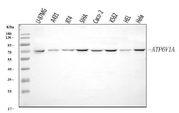
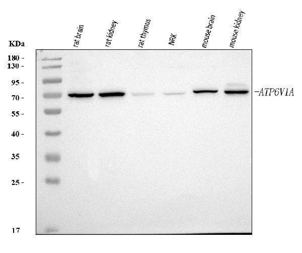
ATP6V1A antibody was positively detected by WB in:
human U-87MG whole cell, human A431 whole cell, human RT4 whole cell, human SiHa whole cell, human Caco-2 whole cell, human K562 whole cell, human HEL whole cell, human Hela whole cell, rat brain tissue, rat kidney tissue, rat thymus tissue, rat NRK whole cell, mouse brain tissue, mouse kidney tissue
Suggested blocking solution with 5% non-fat milk or BSA; (*)Recommended protein loading: 20-40 ug per lane
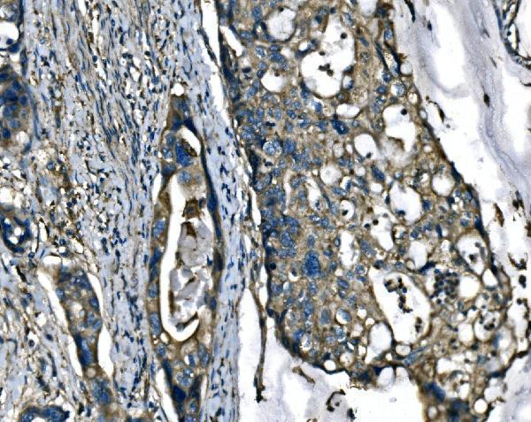
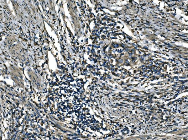
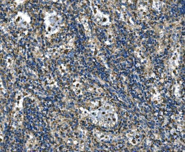
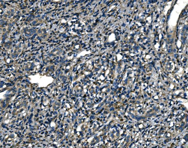
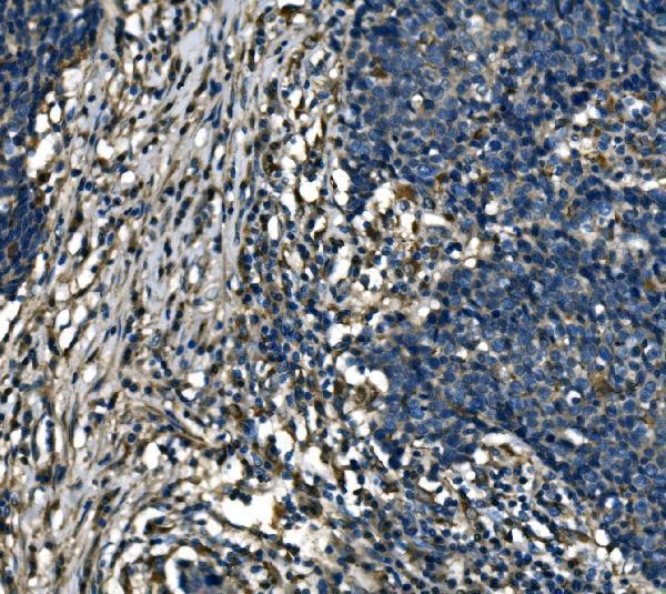
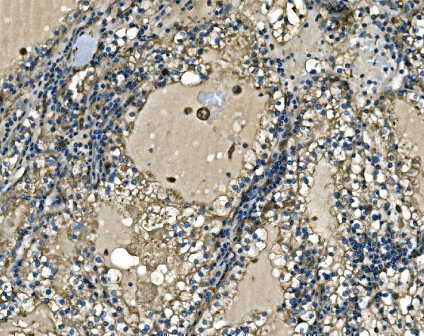
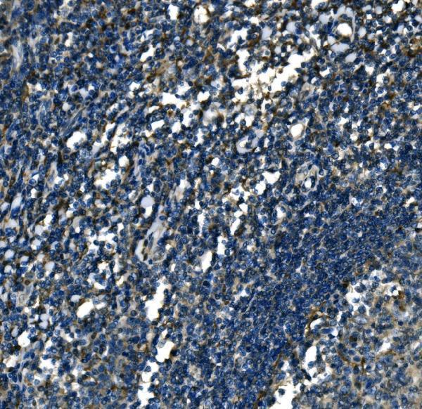
ATP6V1A antibody was positively detected by IHC in:
human adenocarcinoma of the right colon tissue, human cervical cancer tissue, human lienal rupture tissue, human liver cancer tissue, human lung cancer tissue, human renal cell carcinoma tissue, human tonsil tissue
Use TE buffer pH 9.0 for antigen retrieval; (*) citrate buffer pH 6.0 is an alternative.
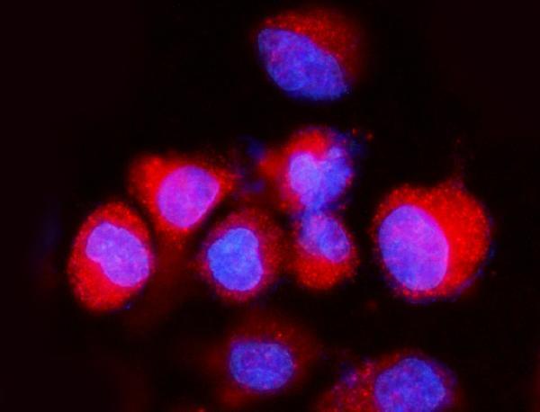
ATP6V1A antibody was positively detected by ICC/IF in:
A549 cell
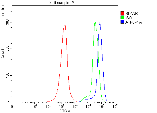
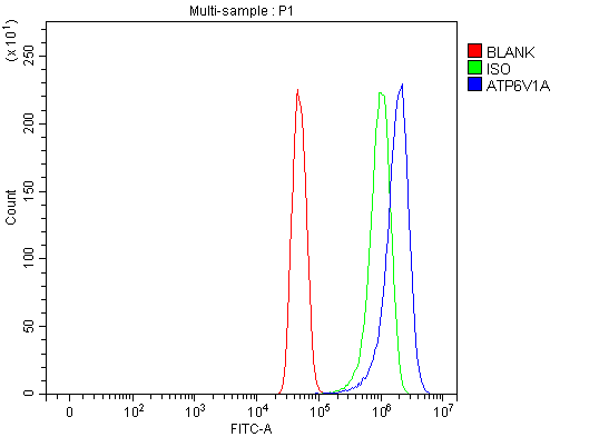
ATP6V1A antibody was positively detected by FC in:
JK cell, U251 cell
* It is recommended to optimize the amount of this reagent in each experimental system to achieve the best results.
Note: For flow cytometry (FCM), validated for fixed cells only. Not validated for FACS or surface flow cytometry using live cells.
68.3 kDa
The results generated by this homology analysis tool for commonly used animal species are shown below. For unlisted species, please refer to the homology analysis between the immunogen sequence and the corresponding species.
A homology of ≥90% is recommended. Generally, higher homology indicates a greater potential for cross-species reactivity. However, protein expression levels may vary among different species, tissues, and cell types. Therefore, the homology analysis results are for reference only and do not guarantee antibody cross-reactivity or experimental performance.








Submit a review and receive $370 in product credits, a $185 Amazon gift card, or our carefully curated gift bundles.
We promise Anti-ATP6V1A Antibody Picoband® performs excellently in relevant applications for ELISA, Flow Cytometry, IF, IHC, ICC, WB. If it doesn't perform as promised, we will refund the product. Learn more



We promise all of our products perform as described in datasheets.
This product is for Research Use Only. Not for use in diagnostic procedures.