This website uses cookies to ensure you get the best experience on our website.
- Table of Contents
Facts about Aurora kinase A.
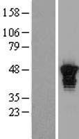
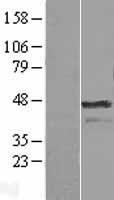
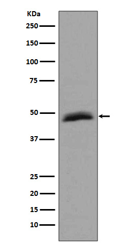
Required for initial activation of CDK1 in centrosomes. Phosphorylates numerous target proteins, such as ARHGEF2, BORA, BRCA1, CDC25B, DLGP5, HDAC6, KIF2A, LATS2, NDEL1, PARD3, PPP1R2, PLK1, RASSF1, TACC3, p53/TP53 and TPX2.
Human | |
---|---|
Gene Name: | AURKA |
Uniprot: | O14965 |
Entrez: | 6790 |
Belongs to: |
---|
protein kinase superfamily |
AIK; AIKAurA; ARK1; ARK-1; ARK1EC 2.7.11.1; AURA; AURKA; Aurora A; aurora kinase AhARK1; aurora/IPL1-like kinase; Aurora/IPL1-related kinase 1; Aurora-related kinase 1; Breast tumor-amplified kinase; breast-tumor-amplified kinase; BTAK; BTAKAURORA2; EC 2.7.11; IPL1-related kinase; serine/threonine kinase 6; serine/threonine protein kinase 15; Serine/threonine-protein kinase 15; serine/threonine-protein kinase 6; Serine/threonine-protein kinase aurora-A; STK15; STK15serine/threonine kinase 15; STK6; STK6MGC34538; STK7
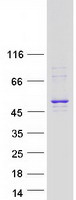
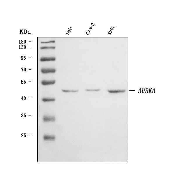
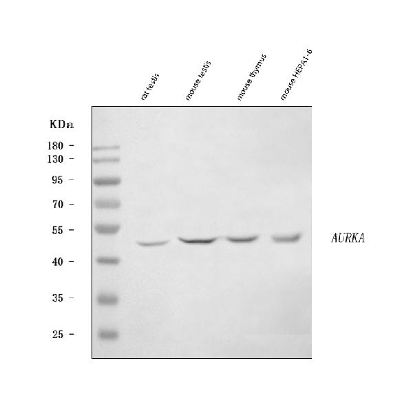
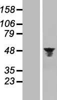
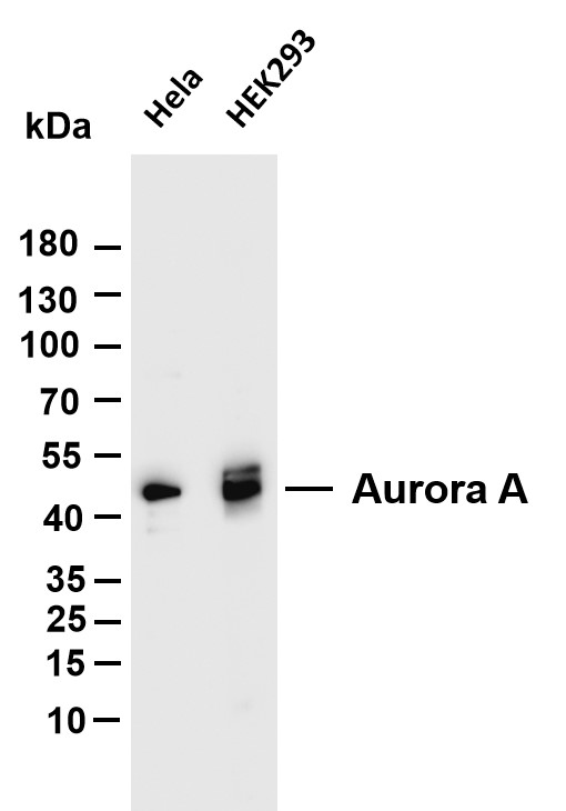
Mass (kDA):
45.809 kDA
Human | |
---|---|
Location: | 20q13.2 |
Sequence: | 20; NC_000020.11 (56369389..56392337, complement) |
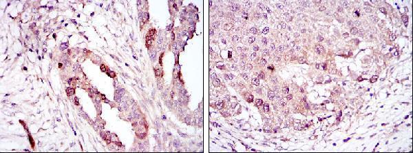
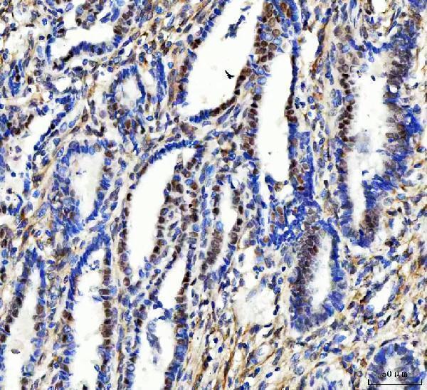
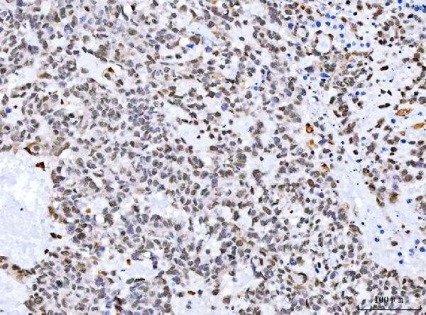
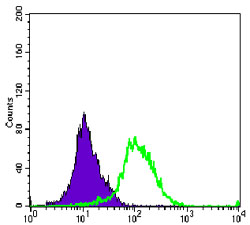
Highly expressed in testis and weakly in skeletal muscle, thymus and spleen. Also highly expressed in colon, ovarian, prostate, neuroblastoma, breast and cervical cancer cell lines.
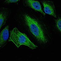
Cytoplasm, cytoskeleton, microtubule organizing center, centrosome. Cytoplasm, cytoskeleton, spindle pole. Cytoplasm, cytoskeleton, cilium basal body. Cytoplasm, cytoskeleton, microtubule organizing center, centrosome, centriole. Cell projection, neuron projection. Detected at the neurite hillock in developing neurons (By similarity). Localizes at the centrosome in mitotic cells from early prophase until telophase, but also localizes to the spindle pole MTs from prophase to anaphase (PubMed:9606188, PubMed:17229885, PubMed:21225229). Colocalized with SIRT2 at centrosome (PubMed:22014574). Move
Scientists from all around the world are finding new uses for the AURKA Marker. Scientists can utilize it to test species, application, and special samples. Boster scientists can submit their results to earn product credits. Boster is available in several languages, and is accessible to scientists of any country. In this article, we'll look at four of the most commonly used applications. Read on for more details.
The AURKA protein is involved in a number of pathways in cancer cells. The development of gastric cancer could be accelerated due to the upregulation AURKA. AURKA is also involved in cell proliferation and survival. These findings support the hypothesis that AURKA kinase can influence OS. Despite the importance of this protein, an unproven argument for treating cancer is still in place.
The Anti-AURKA molecule in Boster Bio is an antibody monoclonal that can detect Aurora kinase A in many biological tests. Antibodies against Aurora kinase A react with a variety of animal samples. Boster Bio develops its antibodies against AURKA using rabbit and mouse. Mitotic processes that lead to the formation of spindles including Aurora KinaseA, are controlled by Boster Bio.
Although AURKA is expressed in normal tissues, it's present at higher levels in various types of cancer. These cancers include pancreatic tumors, head and neck squamous cells carcinoma, lung squamous carcinoma stomach adenocarcinoma as well as stomach adenocarcinoma. A higher expression of AURKA in cancer could result in a lower survival rate, which could be beneficial for the development of new drugs.
AURKA is a kinase which is involved in cell proliferation, differentiation and mobility. This enzyme helps to phosphorylate the histone H3 proteins CENP A at Ser7. It is essential for the proper concentration of AURKB at the centromeres and kinetochore function. The enzyme can be inhibited to slow the growth of tumors by attenuating it's activity.
Several different types of tumors have AURKA. The serine/threonine protein kinase binds with many proteins and plays a role in carcinogenesis. Many cancers express AURKA but not all. In addition to lung cancer, AURKA is also expressed in the esophageal cancer. AUBK, AURKB and BRAF, which are AURKA-related indicators, are linked to cancer-related pathways or genes.
TPX2 regulates AURKA protein expression. At the beginning of mitosis this protein complex is released by Ran-GTP. After the breakdown of the nuclear envelope, TPX2 interacts with AURKA and targets it for degradation via b-TRCP. Two other proteins that regulate AURKA activity are PUM2 (or NEDD9). Furthermore, TPX2 interacts with AURKA and makes it phosphorylated at Ser125 as well as Thr288.
The AURKA protein is among the most commonly expressed proteins in mammary epithelial carcinoma cells, and its overexpression could lead to mammary tumours. Although this protein has been the subject of a lot of pharmaceutical company interest, the inhibitors available have not demonstrated any clinical benefits. Combining AURKA inhibitors with agents that target multiple cancer-related AURKA functions and their partners is the most promising way to go.
AURKA is present in three forms in HEK293 cells, AURKA43 corresponding to the first proteolytic cleavage that PMPCB has performed, AURKA38 representing the mature mitochondrial isoform. Monoclonal and polyclonal antibodies against the AURKA marker can be detected in HEK293 cells. Furthermore, the depletion of PMPCB confirms that mitochondrial AURKA38 and AURKA43 are MTS.
Studies have demonstrated that an increase in AURKA copy numbers is linked to the development of cancerous colorectal polyps. In addition, overexpression of AURKA is linked to the survival of breast cancer cells. However, this finding is not fully supported by the absence of information regarding the role of AURKA in human epithelial cancers. It is important to note that in certain instances the AURKA protein may interfere with the the progesterone receptor , or the human growth factor receptor 2.
The AURKA marker has numerous biological applications and is available in various forms. Boster Bio develops monoclonal and polyclonal antibodies against the Aurora kinase A protein. Boster Bio's antibodies were developed with rabbit and mouse models. They are extremely specific and interact with a large range of animal specimens. Aurora Kinase is a protein that plays a significant role in mitotic events.
AURKA protein is found in many types and types of cancers however, it is the most frequent in malignant tumors. AURKA expression is more prominent in cancers of the lung, liver cancer and mesothelioma. In the THCA dataset the presence of high levels AURKB are significantly associated with lower survival rates than those of normal tissues. Both AURKB and AURKA markers have a direct correlation with the overall survival of cancer patients.
There are many roles for the AURKA protein in various types of cancers. The high level of expression and importance in various types of cancers is caused by its involvement in a myriad of cancer-related pathways. It plays a crucial role in tumor progression. AURKA regulates ERa and activates NF-kB.
Although the function of AURKA remains unclear, it has been shown that it is essential in the diagnosis and treatment of T cell lines that are refractory. The gene encoding AURKA is extremely conserved and is found on the surface of many immune cells, including melanoma as well as leukemia. Its regulation by a variety of factors isn't fully known.
Scientists isolated MDAMB-231 cells that were overexpressed with AURKA from this study. The cells were cultivated in serum-free medium, and seeded into six-well ultralow-adhesive culture plates. Within 72 hours the cells had grown densely packed and contained several to hundreds of cells. After about 10 to 15 days the cells were removed and analyzed. These cells displayed characteristic phenotypes of CSCs.
AURKA is a protein that interacts at the endogenous level with the centriolar satellite protein. These interactions were confirmed by immunoprecipitation studies. These results demonstrate a novel regulatory link between AURKA and centriolar satellites. By inhibiting AURKA, cells can rescue ciliogenesis defects. Anti-T antibodies that recognize AURKA are more effective than monoclonal anti-T antibodies.
The Aurora kinase A (AURKA) is a well-known overexpressed protein in a variety of cancers. Its increased expression can be related to gene amplification and mutation and microRNA regulation. It also plays a role in transcriptional modification and microRNA-mediated regulation. Boster Bio produces antibodies that are specific to AURKA in both mouse and rabbit samples. The antibody has been tested in a wide range of biological assays, including immunohistochemistry, Western blotting, and in vitro applications.
AURKA is a phosphorylation enzyme that activates Lats2 at Ser83, which regulates the location of the centrosomal axis. AURKA also regulates the activity of KCTD12, inhibiting cell proliferation and inhibiting tumorigenesis. This protein is found in many forms of cancer such as stomach, esophageal and lung adenocarcinomas. There is not agreement on the exact mechanism of the activity of AURKA in cancer.
In a variety of cancers there is a AURKA protein interacts with ERa and phosphorylates it on Ser315 and Ser305. Increasing AURKA expression predicts poor prognosis in breast cancers. It also affects the AMPK pathway. It also blocks the growth of NSCLC and suppresses Akt signaling pathways.
The AURKA marker has many uses and applications, including the ability to detect fusion proteins within the basal body. It is also used to measure the activity of p53. Researchers must identify the interactions and functions of this protein within cells to understand its full potential. These studies have confirmed the importance of this protein. Below are some examples of how this protein can be used in research.
The AURKA marker plays a crucial role in regulating the FAK/PI3K pathway and enhances EMT. Due to this, it could be used as a biomarker to detect LSCC. This research could have implications for the development and testing of immunotherapies as well as drugs that target this pathway. These regulators could be used in the near future to serve as diagnostic biomarkers or as therapeutic targets for LSCC.
The AURKA proximity interactome has identified a functional and regulating relationship between AURKA and the centriolar satellites. This complex is comprised of 440 proteins and plays a function in a variety biological processes. In mammalian cells, AURKA gets activated by HEF1/NEDD1, which promotes the deacetylation of microtubules in axonemal cells and ciliogenesis.
PMID: 9153231 by Kimura M., et al. Cell cycle-dependent expression and spindle pole localization of a novel human protein kinase, Aik, related to Aurora of Drosophila and yeast Ipl1.
PMID: 9514916 by Shindo M., et al. cDNA cloning, expression, subcellular localization, and chromosomal assignment of mammalian aurora homologues, aurora-related kinase (ARK) 1 and 2.