Click image to see more details
-

-
-
-
-
+4
Product Info Summary
| SKU: | A00246-4 |
|---|---|
| Size: | 100 μg/vial |
| Reactive Species: | Human |
| Host: | Rabbit |
| Application: | ELISA, Flow Cytometry, IHC, WB |
Customers Who Bought This Also Bought

Product info
Product Name
Anti-Aurora A/AURKA Antibody Picoband®
SKU/Catalog Number
A00246-4
Size
100 μg/vial
Form
Lyophilized
Description
Boster Bio Anti-Aurora A/AURKA Antibody Picoband® catalog # A00246-4. Tested in ELISA, Flow Cytometry, IHC, WB applications. This antibody reacts with Human. The brand Picoband indicates this is a premium antibody that guarantees superior quality, high affinity, and strong signals with minimal background in Western blot applications. Only our best-performing antibodies are designated as Picoband, ensuring unmatched performance.
Storage & Handling
At -20°C for one year from date of receipt. After reconstitution, at 4°C for one month. It can also be aliquotted and stored frozen at -20°C for six months. Avoid repeated freezing and thawing.
Cite This Product
Anti-Aurora A/AURKA Antibody Picoband® (Boster Biological Technology, Pleasanton CA, USA, Catalog # A00246-4)
Host
Rabbit
Contents
Each vial contains 4 mg Trehalose, 0.9 mg NaCl, 0.2 mg Na2HPO4.
Clonality
Polyclonal
Isotype
Rabbit IgG
Immunogen
E.coli-derived human Aurora A/AURKA recombinant protein (Position: K23-S403).
Cross-reactivity
No cross-reactivity with other proteins.
Reactive Species
A00246-4 is reactive to AURKA in Human
Observed Molecular Weight
46 kDa
Calculated molecular weight
45.8 kDa
Background of AURKA
AURKA(aurora kinase A), also called ARK1, AurA, AIK , AURORA2 ,BTAK, PPP1R47, STK7, STK15,STK6, is a mitotic centrosomal protein kinase. The main role of AURKA in tumor development is in controlling chromosome segregation during mitosis. Aurora A is a member of a family of mitotic serine/threonine kinases. Cell cycle and Northern blot analyses showed that peak expression of AURKA occurs during the G2/M phase and then decreases. By fluorescence in situ hybridization, AURKA gene is represented by 2 signals in chromosome bands 20q13.2-q13.3 and 1q41-q42. The AURKA gene is overexpressed in many human cancers. Ectopic overexpression of Aurora kinase A in mammalian cells induces centrosome amplification, chromosome instability, and oncogenic transformation, a phenotype characteristic of loss-of-function mutations of p53. Depletion of Ajuba prevented activation of AURKA at centrosomes in late G2 phase and inhibited mitotic entry. Activation of AURKA was independently sufficient to induce rapid ciliary resorption, and AURKA acted in this process through phosphorylation of HDAC6, leading to HDAC6-dependent tubulin deacetylation and destabilization of the ciliary axoneme. Small molecule inhibitors of AURKA and HDAC6 reduced regulated disassembly of cilia.
Antibody Validation
Boster validates all antibodies on WB, IHC, ICC, Immunofluorescence, and ELISA with known positive control and negative samples to ensure specificity and high affinity, including thorough antibody incubations.
Application & Images
Applications
A00246-4 is guaranteed for ELISA, Flow Cytometry, IHC, WB Boster Guarantee
Recommend Dilution
| Application | Dilution | Species |
|---|---|---|
| Western blot | 0.25-0.5 μg/ml | Human |
| Immunohistochemistry(Paraffin-embedded Section) | 2-5 μg/ml | Human |
| Flow Cytometry (Fixed) | 1-3 μg/1x106 cells | Human |
| ELISA | 0.1-0.5 μg/ml | - |
Tested application
Suggested blocking solution with 5% non-fat milk or BSA; (*)Recommended protein loading: 20-40 µg per lane
Use TE buffer pH 9.0 for antigen retrieval; (*) citrate buffer pH 6.0 is an alternative.
Validation Images & Assay Conditions
Click image to see more details
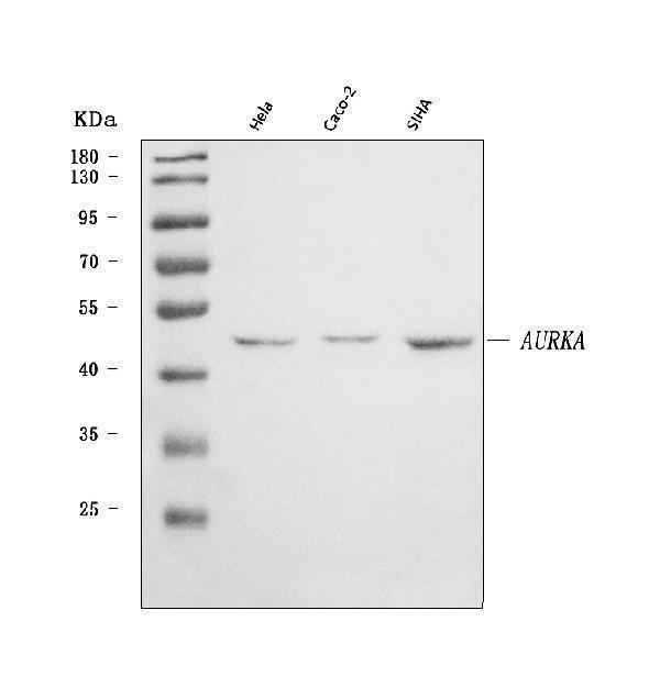
Western blot analysis of Aurora A/AURKA using anti-Aurora A/AURKA antibody (A00246-4).
Electrophoresis was performed on a 5-20% SDS-PAGE gel at 70V (Stacking gel) / 90V (Resolving gel) for 2-3 hours. The sample well of each lane was loaded with 30 ug of sample under reducing conditions.
Lane 1: human Hela whole cell lysates,
Lane 2: human CACO-2 whole cell lysates,
Lane 3: human SiHa whole cell lysates.
After electrophoresis, proteins were transferred to a nitrocellulose membrane at 150 mA for 50-90 minutes. Blocked the membrane with 5% non-fat milk/TBS for 1.5 hour at RT. The membrane was incubated with rabbit anti-Aurora A/AURKA antigen affinity purified polyclonal antibody (Catalog # A00246-4) at 0.5 μg/mL overnight at 4°C, then washed with TBS-0.1%Tween 3 times with 5 minutes each and probed with a goat anti-rabbit IgG-HRP secondary antibody at a dilution of 1:5000 for 1.5 hour at RT. The signal is developed using an Enhanced Chemiluminescent detection (ECL) kit (Catalog # EK1002) with Tanon 5200 system. A specific band was detected for Aurora A/AURKA at approximately 46 kDa. The expected band size for Aurora A/AURKA is at 46 kDa.

Click image to see more details
IHC analysis of Aurora A/AURKA using anti-Aurora A/AURKA antibody (A00246-4).
Aurora A/AURKA was detected in a paraffin-embedded section of human cervical cancer tissue. Heat mediated antigen retrieval was performed in EDTA buffer (pH 8.0, epitope retrieval solution). The tissue section was blocked with 10% goat serum. The tissue section was then incubated with 2 μg/ml rabbit anti-Aurora A/AURKA Antibody (A00246-4) overnight at 4°C. Peroxidase Conjugated Goat Anti-rabbit IgG was used as secondary antibody and incubated for 30 minutes at 37°C. The tissue section was developed using HRP Conjugated Rabbit IgG Super Vision Assay Kit (Catalog # SV0002) with DAB as the chromogen.

Click image to see more details
IHC analysis of Aurora A/AURKA using anti-Aurora A/AURKA antibody (A00246-4).
Aurora A/AURKA was detected in a paraffin-embedded section of human breast cancer tissue. Heat mediated antigen retrieval was performed in EDTA buffer (pH 8.0, epitope retrieval solution). The tissue section was blocked with 10% goat serum. The tissue section was then incubated with 2 μg/ml rabbit anti-Aurora A/AURKA Antibody (A00246-4) overnight at 4°C. Peroxidase Conjugated Goat Anti-rabbit IgG was used as secondary antibody and incubated for 30 minutes at 37°C. The tissue section was developed using HRP Conjugated Rabbit IgG Super Vision Assay Kit (Catalog # SV0002) with DAB as the chromogen.

Click image to see more details
IHC analysis of Aurora A/AURKA using anti-Aurora A/AURKA antibody (A00246-4).
Aurora A/AURKA was detected in a paraffin-embedded section of human ovarian cancer tissue. Heat mediated antigen retrieval was performed in EDTA buffer (pH 8.0, epitope retrieval solution). The tissue section was blocked with 10% goat serum. The tissue section was then incubated with 2 μg/ml rabbit anti-Aurora A/AURKA Antibody (A00246-4) overnight at 4°C. Peroxidase Conjugated Goat Anti-rabbit IgG was used as secondary antibody and incubated for 30 minutes at 37°C. The tissue section was developed using HRP Conjugated Rabbit IgG Super Vision Assay Kit (Catalog # SV0002) with DAB as the chromogen.

Click image to see more details
IHC analysis of Aurora A/AURKA using anti-Aurora A/AURKA antibody (A00246-4).
Aurora A/AURKA was detected in a paraffin-embedded section of human squamous cell carcinoma tissue. Heat mediated antigen retrieval was performed in EDTA buffer (pH 8.0, epitope retrieval solution). The tissue section was blocked with 10% goat serum. The tissue section was then incubated with 2 μg/ml rabbit anti-Aurora A/AURKA Antibody (A00246-4) overnight at 4°C. Peroxidase Conjugated Goat Anti-rabbit IgG was used as secondary antibody and incubated for 30 minutes at 37°C. The tissue section was developed using HRP Conjugated Rabbit IgG Super Vision Assay Kit (Catalog # SV0002) with DAB as the chromogen.

Click image to see more details
IHC analysis of Aurora A/AURKA using anti-Aurora A/AURKA antibody (A00246-4).
Aurora A/AURKA was detected in a paraffin-embedded section of human lung adenocarcinoma tissue. Heat mediated antigen retrieval was performed in EDTA buffer (pH 8.0, epitope retrieval solution). The tissue section was blocked with 10% goat serum. The tissue section was then incubated with 2 μg/ml rabbit anti-Aurora A/AURKA Antibody (A00246-4) overnight at 4°C. Peroxidase Conjugated Goat Anti-rabbit IgG was used as secondary antibody and incubated for 30 minutes at 37°C. The tissue section was developed using HRP Conjugated Rabbit IgG Super Vision Assay Kit (Catalog # SV0002) with DAB as the chromogen.

Click image to see more details
IHC analysis of Aurora A/AURKA using anti-Aurora A/AURKA antibody (A00246-4).
Aurora A/AURKA was detected in a paraffin-embedded section of human renal oncocytoma tissue. Heat mediated antigen retrieval was performed in EDTA buffer (pH 8.0, epitope retrieval solution). The tissue section was blocked with 10% goat serum. The tissue section was then incubated with 2 μg/ml rabbit anti-Aurora A/AURKA Antibody (A00246-4) overnight at 4°C. Peroxidase Conjugated Goat Anti-rabbit IgG was used as secondary antibody and incubated for 30 minutes at 37°C. The tissue section was developed using HRP Conjugated Rabbit IgG Super Vision Assay Kit (Catalog # SV0002) with DAB as the chromogen.

Click image to see more details
Flow Cytometry analysis of HepG2 cells using anti-Aurora A/AURKA antibody (A00246-4).
Overlay histogram showing HepG2 cells stained with A00246-4 (Blue line). To facilitate intracellular staining, cells were fixed with 4% paraformaldehyde and permeabilized with permeabilization buffer. The cells were blocked with 10% normal goat serum. And then incubated with rabbit anti-Aurora A/AURKA Antibody (A00246-4, 1 μg/1x106 cells) for 30 min at 20°C. DyLight®488 conjugated goat anti-rabbit IgG (BA1127, 5-10 μg/1x106 cells) was used as secondary antibody for 30 minutes at 20°C. Isotype control antibody (Green line) was rabbit IgG (1 μg/1x106) used under the same conditions. Unlabelled sample without incubation with primary antibody and secondary antibody (Red line) was used as a blank control.
Specific Publications For Anti-Aurora A/AURKA Antibody Picoband® (A00246-4)
Loading publications
Recommended Resources
Here are featured tools and databases that you might find useful.
- Boster's Pathways Library
- Protein Databases
- Bioscience Research Protocol Resources
- Data Processing & Analysis Software
- Photo Editing Software
- Scientific Literature Resources
- Research Paper Management Tools
- Molecular Biology Software
- Primer Design Tools
- Bioinformatics Tools
- Phylogenetic Tree Analysis
Customer Reviews
Have you used Anti-Aurora A/AURKA Antibody Picoband®?
Share your experimental results or join a short interview to earn up to $1,000 in product credits or other rewards.
0 Reviews For Anti-Aurora A/AURKA Antibody Picoband®
Customer Q&As
Have a question?
Find answers in Q&As, reviews.
Can't find your answer?
Submit your question
