AURKA / Aurora kinase A · IHC design guide

Design Immunohistochemistry for AURKA

Plan paraffin AURKA IHC with the catalog antibody’s documented staining workflow and HPA tissue controls (datasheet A00246-4; HPA tissue IHC). Assess cytoplasmic and nuclear staining in pachytene spermatocytes and tonsillar germinal center cells, both reported at medium intensity (HPA tissue IHC).

Evidence assembled Oct 2026 · For research use; verify linked source records and product datasheet before use
Immunohistochemistry protocol sheet for AURKA (IHC for AURKA): expected localisation Cytoplasmic and nuclear tissue staining (HPA tissue IHC), antibody A00246-4, validated IHC image, and IHC protocol steps
Printable AURKA IHC protocol sheet — expected localisation Cytoplasmic and nuclear tissue staining (HPA tissue IHC), antibody A00246-4, controls and protocol steps. Open the full AURKA IHC guide →

AURKA Immunohistochemistry Experimental Design Guide

Expected localisation, validated protocols, controls and antibodies — the at-a-glance facts below, then the full design guide.

Must know before staining
Expected localisation Cytoplasmic and nuclear tissue staining (HPA tissue IHC)
Staining pattern Pachytene spermatocytes and immune cells: cytoplasmic, nuclear (HPA tissue IHC)
Antigen retrieval EDTA pH 8.0 HIER, heat-mediated (datasheet A00246-4)
Positive control ⓘ Testis+1 more · see all
Negative control ⓘ Adipose tissue+4 more · see all
Important caveats
Reasons your staining may differ from the expected pattern.
Fixation Matched tissue-IHC evidence does not establish the fixation claim. Validate the specimen-specific method before use. (selected-SKU IHC image A00246-4)
Caveat Mitotic cells may show centrosomal or spindle-pole signal (UniProt)
Regulation RNA enhanced in testis and lymphoid tissue (HPA tissue RNA)
Isoform / epitope No annotated isoforms; one 1–403 chain (UniProt)
Section 1

Recommended AURKA IHC & IF Protocols

The catalog antibody protocol uses EDTA pH 8.0 retrieval (datasheet: A00246-4). The published option below describes AURKA staining in colorectal tumor sections (PMC8688726).

Recommended immunohistochemistry (IHC-P) protocol parameters
SampleParaffin-embedded human cervical cancer tissue; fixative not specified (datasheet A00246-4)
FixationImage fixative and duration unreported (datasheet A00246-4); verify before use.
Sectioning4–5 µm sections on charged slides (standard)
DeparaffinisationXylene, graded ethanol series to water (standard)
Antigen retrievalHeat retrieval: EDTA pH 8.0 (datasheet A00246-4); 20 min, 95–100 °C (standard)
Peroxidase block3% H2O2, 10 min, room temperature (standard)
Blocking10% goat serum (datasheet A00246-4)
Primary antibodyRabbit anti-AURKA, 2-5 μg/ml (datasheet A00246-4)
Primary incubationOvernight at 4 °C (datasheet A00246-4)
DetectionHRP-conjugated secondary, DAB chromogen (datasheet A00246-4)
CounterstainHematoxylin, blue, dehydrate and mount (standard)
Expected resultAURKA-positive staining in pachytene spermatocytes of testis (HPA tissue IHC: Medium). HPA tissue profile: Cytoplasmic and nuclear expression most abundant in pachytene spermatocytes and immune cells. No signal in the no-primary control.
💡Decision noteStart with EDTA pH 8.0 heat retrieval for the catalog antibody (datasheet: A00246-4); the published protocol specifies cell conditioning solution without its composition (PMC8688726).
Section 2

What Is the Expected AURKA Staining Pattern?

AURKA staining should be enriched in pachytene spermatocytes in testis and germinal center cells in tonsil, with cytoplasmic and nuclear staining reported in tissue (HPA tissue IHC). In dividing cells, AURKA localizes to centrosomes and spindle poles (UniProt O14965). HPA rates the tissue staining profile Enhanced, while noting medium consistency with RNA expression; interpret weak or unexpected staining cautiously (HPA tissue IHC). AURKA has no transmembrane segment (UniProt O14965 topology).

What am I looking at on my slide?
Medium cytoplasmic and nuclear staining in pachytene spermatocytes or tonsil germinal center cells.This matches the reported positive cell types, intensity, and broad tissue compartments (HPA tissue IHC). Score the relevant cells separately from surrounding cells; a positive region does not imply that every cell in the section should stain (general IHC practice).
A punctate signal near the nucleus or spindle-associated signal in an identifiable mitotic cell.Centrosomal and spindle-pole localization is consistent with AURKA biology (UniProt O14965; HPA subcellular ICC-IF). Chromogenic IHC may not resolve these small structures clearly, so use cell identity and the broader tissue pattern when interpreting a fine punctum (general IHC practice).
Strong, uniform membrane staining dominates the candidate positive cells.Treat this as an unexpected compartment pattern: AURKA has no transmembrane segment, and HPA reports cytoplasmic and nuclear tissue staining (UniProt O14965 topology; HPA tissue IHC). Review the negative control and antibody specificity before assigning it to AURKA (general IHC practice).
Prominent staining appears in HPA-listed cells reported as not detected, such as adipocytes.The pattern conflicts with the HPA observations for adipocytes in adipose tissue and breast (HPA tissue IHC). Cross-reactivity or endogenous detection activity is possible; compare the matched negative control and assess tissue morphology before interpreting it as target signal (general IHC practice).
No staining is visible in pachytene spermatocytes in a testis control section.HPA reports medium staining in these cells, so an entirely blank result warrants a technical review (HPA tissue IHC). Check tissue identity, preservation, retrieval, primary antibody use, and detection controls; a negative result alone does not identify which step failed (general IHC practice).
💡Expected AURKA appearanceCall the IHC result positive when pachytene spermatocytes or tonsil germinal center cells show approximately medium cytoplasmic and nuclear staining (HPA tissue IHC); dominant uniform membrane staining is discordant with the reported pattern and AURKA topology (HPA tissue IHC; UniProt O14965 topology).
How each factor affects the staining
Cell type and tissue contextHPA reports medium staining in pachytene spermatocytes and tonsil germinal center cells, but low staining in several other listed cell types (HPA tissue IHC). Choose the specific positive cell population for assessment rather than treating all tissue cells as equivalent (general IHC practice).
Cell cycle and spatial resolutionAURKA associates with centrosomes and spindle structures during mitosis and moves to the midbody during telophase and cytokinesis (UniProt O14965). The fraction of recognizable dividing cells can therefore affect how often those structures are seen; small structures may be difficult to distinguish in chromogenic sections (general IHC practice).
Evidence tied to the antibodyHPA lists IHC status as Approved for HPA002636 and Enhanced for CAB001454 (HPA antibodies). The tissue profile is also labeled Enhanced, with medium consistency between staining and RNA data (HPA tissue IHC). Keep the antibody-specific status and the tissue-profile rating distinct when weighing an unexpected result.
Topology and processingUniProt lists a single 1–403 chain, no signal peptide, no propeptide, and no transmembrane segment for AURKA (UniProt O14965). These annotations do not support expecting a secreted or membrane-spanning staining pattern; they do not predict how fixation or retrieval will affect this antibody (UniProt O14965; target-specific fixation evidence not supplied).
IF/ICC pattern — interpretation onlyFor the separate IF/ICC guide, HPA reports centrosome and basal-body localization as supported, while mitotic spindle, nucleoplasm, and cytosol are labeled uncertain (HPA subcellular ICC-IF). Use those confidence labels when comparing images; no IF/ICC protocol option is established by this section (HPA subcellular ICC-IF).
Why is my staining missing, weak or wrong?
SituationLikely causeNext action
Known-positive testis cells are blank.The expected medium signal in pachytene spermatocytes was not recovered (HPA tissue IHC); the failed step is undetermined.Confirm the positive cell population, then review the section, primary antibody application, retrieval conditions, and detection controls together (general IHC practice). Do not infer AURKA-specific fixation sensitivity from this result.
Only diffuse haze is visible across the section.A broad haze cannot be assigned to the reported cell-selective pattern (HPA tissue IHC); nonspecific staining or detection background is possible (general IHC practice).Compare a matched no-primary control and inspect blocking, washing, detection reagent, and chromogen development using the established IHC workflow (general IHC practice). Reassess whether the positive cells remain distinguishable.
An apparent positive signal concentrates at tissue edges or damaged areas.Edge or damage-associated staining can reflect a section or detection artifact (general IHC practice); HPA's AURKA profile describes cell types and compartments instead (HPA tissue IHC).Inspect an intact region and compare controls before scoring the affected area (general IHC practice). Record whether the signal still follows pachytene spermatocytes or germinal center cells (HPA tissue IHC).
Strong membrane staining outweighs cytoplasmic and nuclear staining.That distribution conflicts with HPA's tissue pattern and UniProt's absence of a transmembrane segment (HPA tissue IHC; UniProt O14965 topology).Check the negative control and the antibody's IHC validation status; HPA assigns Approved to HPA002636 and Enhanced to CAB001454 (general IHC practice; HPA antibodies). Treat unresolved membrane staining as unexpected.
Cells listed as not detected stain as strongly as the positive control.For example, prominent adipocyte staining conflicts with HPA's not-detected calls in adipose tissue and breast (HPA tissue IHC). Cross-reactivity or endogenous detection activity may contribute (general IHC practice).Compare the same cell type in control sections, inspect the no-primary control, and review the detection system's endogenous-activity controls (general IHC practice). Avoid calling the staining specific solely from color intensity.
IF/ICC shows a diffuse signal but no clear centrosome or basal body.HPA supports centrosome and basal-body localization; it labels cytosolic localization uncertain (HPA subcellular ICC-IF). A diffuse image alone gives limited support for the expected spatial pattern.In the separate IF/ICC analysis, check cell morphology and available localization controls before assigning the diffuse signal to AURKA (general IF practice). Interpret spindle and nucleoplasm signals with HPA's uncertain labels (HPA subcellular ICC-IF).

Sample controls for AURKA IHC & IF

🧪Run testis first and score pachytene spermatocytes, where AURKA staining is Medium (HPA: Testis, pachytene spermatocytes, Medium). Use adipose tissue adipocytes as the negative tissue (HPA: Adipose tissue, adipocytes, Not detected); compare neighboring cells on the testis slide for background, but do not assume they are AURKA-negative because the supplied HPA row identifies only pachytene spermatocytes.
Positive control tissue: Testis (Pachytene spermatocytes, HPA Medium)
Negative control tissue: Adipose tissue (HPA Not detected)
ICC-IF cell lines (HPA subcellular resource): HPA ICC-IF images show AURKA in A-431, CACO-2, U2OS, ASC52telo, RPTEC/TERT1, hTERT-RPE1 (serum starved), KOLF2.1J, with annotated localisation: Mitotic spindle (uncertain), Centrosome (supported), Basal body (supported) (HPA subcellular).
Technical controls: Include a no-primary, secondary-only section and an isotype-matched rabbit IgG control, alongside an AURKA knockout sample or a peptide-block control if the immunizing peptide is available (caption: rabbit primary antibody; standard IHC controls). Quench endogenous peroxidase and inspect testis sections for residual background before scoring DAB staining (caption: peroxidase secondary and DAB; standard chromogenic IHC practice).
⚠️Feasibility: A target-specific fixation window and fixation effect are unreported, and the selected A00246-4 caption does not state the fixative (selected-SKU tissue-IHC caption). The caption uses heat-mediated retrieval in EDTA at pH 8.0, but does not establish that retrieval is required; neither frozen sections nor IF is shown to be easier in the supplied evidence (selected-SKU tissue-IHC caption; HPA: subcellular ICC-IF images). In testis, identify pachytene spermatocytes before scoring because the positive HPA finding is specific to that cell population; IF could help assess centrosomal localization, with supported centrosome localization reported by HPA (HPA: Testis, pachytene spermatocytes, Medium; HPA: centrosome, supported).

HPA tissue IHC evidence for AURKA

Comprehensive Human Protein Atlas IHC scoring per tissue (reliability: Enhanced — Medium consistency between antibody staining and RNA expression data.). Rows are taken directly from the HPA tissue chart — click any row's HPA link to view the source.

Positive expression · recommended positive controls

TissueCell typeLevelEvidenceSource
Testis Pachytene spermatocytes Medium Protein (IHC) HPA →
Tonsil Germinal center cells Medium Protein (IHC) HPA →

Undetected expression · recommended negative controls

TissueCell typeLevelEvidenceSource
Adipose tissue Adipocytes Not detected Protein (IHC) HPA →
Adrenal gland Glandular cells Not detected Protein (IHC) HPA →
Breast Adipocytes Not detected Protein (IHC) HPA →
Caudate Glial cells Not detected Protein (IHC) HPA →
Cerebellum Cells in granular layer Not detected Protein (IHC) HPA →
Section 3

Advanced AURKA IHC Tips

Troubleshoot chromogenic AURKA IHC in paraffin sections by checking retrieval, controls, cell type and mitotic localisation before scoring staining.

What retrieval should I try first when AURKA staining is weak in paraffin sections?
Start with heat-mediated retrieval in EDTA at pH 8.0 (datasheet A00246-4). The selected paraffin-section example used that retrieval before incubation with 2 μg/ml antibody overnight at 4°C, so reproduce the documented conditions before changing several variables (datasheet A00246-4). If staining remains weak, adjust heating duration in small steps while keeping the EDTA buffer and detection conditions constant, and inspect tissue morphology after each change (standard IHC practice). Compare the trial sections with pachytene spermatocytes or germinal center cells as positive controls; both show medium staining in the tissue survey (HPA tissue IHC).
Could fixation explain weak or patchy AURKA staining?
The selected paraffin-section caption does not report a fixative, so target-specific fixation sensitivity is unknown (datasheet A00246-4). Record each specimen’s actual fixative and fixation time, then compare sections processed with the same EDTA pH 8.0 retrieval and antibody conditions before attributing differences to fixation (standard IHC practice; datasheet A00246-4). Check morphology and whether nuclei and cytoplasm remain interpretable across the section, especially where staining fades or intensifies (standard IHC practice). Use a matched positive-control section in every run, because differences in processing and detection can also change apparent staining (standard IHC practice).
How should I assess AURKA localisation in a positive section?
Examine the stained cell and its mitotic state before interpreting an isolated brown focus (UniProt O14965 localisation). AURKA localises to centrosomes from early prophase through telophase, spindle poles from prophase through anaphase, and the midbody during telophase and cytokinesis (UniProt O14965 localisation). The tissue survey also reports cytoplasmic and nuclear staining, most abundant in pachytene spermatocytes and immune cells, so diffuse signal deserves cell-type context (HPA tissue IHC). On a counterstained section, compare focal staining with visible mitotic figures and score diffuse staining separately; DAB alone does not establish centrosomal colocalisation (standard IHC practice; UniProt O14965 localisation).
Does an absent isoform or hidden epitope explain discordant AURKA staining?
The supplied record lists 0 alternative isoforms and one AURKA chain spanning residues 1–403, so an isoform switch is unsupported by this evidence (UniProt O14965 processing and isoforms). Its protein kinase domain spans residues 133–383, and the record includes phosphorylation sites, but the selected antibody’s epitope is not specified (UniProt O14965 domains and modified residues; datasheet A00246-4). Consequently, do not assign weak IHC to phosphorylation or epitope masking without independent antibody-characterisation evidence (standard IHC interpretation). First compare retrieval and staining alongside a positive-control section; if discrepancy persists, seek documented epitope information before making a molecular explanation (standard IHC practice).
How can IF help examine an ambiguous AURKA IHC pattern?
Use IF as a separate localisation check, with a marker identifying the expected cell population and a nuclear counterstain (standard IF practice). Pachytene spermatocytes and germinal center cells are useful populations to examine because the tissue survey reports medium AURKA staining in them (HPA tissue IHC). Choose a fluorophore channel with low tissue autofluorescence, and include single-colour and no-primary controls when multiplexing (standard IF practice). AURKA is an intracellular protein with no transmembrane segment, so permeabilise sufficiently to access cytoplasmic and centrosomal epitopes while preserving cell structure (UniProt O14965 topology and localisation; standard IF practice). Compare puncta with centrosome or spindle markers, since the subcellular survey supports centrosomal localisation (HPA subcellular).
What should I check when AURKA DAB staining is widespread or granular?
Run a no-primary control through the same detection steps to identify signal from secondary reagents, endogenous peroxidase or DAB deposition (standard IHC practice). The selected example used 10% goat serum for blocking, a peroxidase-conjugated secondary and DAB detection; its caption does not specify a peroxidase-block step (datasheet A00246-4). Add a peroxidase block as a general chromogenic IHC step, and compare antibody titrations while keeping retrieval and development time fixed (standard IHC practice). Assess background against the reported cell pattern: pachytene spermatocytes and germinal center cells stain at medium levels, whereas adipocytes in adipose tissue were not detected (HPA tissue IHC).
How should I score AURKA staining across paraffin sections? ⚠ ANSWER MARKED FOR VERIFICATION
Define the cell population and compartment before scoring, then apply the same threshold and exposure-independent DAB assessment to every section (standard chromogenic IHC practice). Record either the percentage of positive cells and staining intensity as an H-score, or positive mitotic profiles per mm² when the question concerns focal mitotic signal (standard IHC quantification practice; UniProt O14965 localisation). Normalise cell-based scores to evaluable cells of the same type and area-based counts to viable tissue area; exclude folds and necrosis consistently (standard IHC quantification practice). Report cytoplasmic or nuclear staining separately from mitotic foci, because the tissue survey describes both cytoplasmic and nuclear expression while AURKA also occupies centrosomes and spindle structures (HPA tissue IHC; UniProt O14965 localisation).
How do I distinguish convincing AURKA positivity from staining artefact?
Look for a reproducible cell-type and compartment pattern across intact tissue, supported by a positive control and absent from the no-primary control (standard IHC interpretation). Medium staining in pachytene spermatocytes and germinal center cells supports those control choices, whereas adipocytes in adipose tissue were reported as not detected (HPA tissue IHC). Centrosomal, spindle-associated or midbody foci in the appropriate mitotic phase fit the recorded localisation, but an isolated DAB dot cannot establish subcellular identity (UniProt O14965 localisation; standard IHC interpretation). Treat staining concentrated at section edges, folds or necrotic regions, or staining that persists without primary antibody, as suspect for processing or endogenous-enzyme artefact (standard IHC practice).
Boster reagents

Best AURKA / Aurora kinase A IHC Antibodies

A00246-4 and A00246-3 have IHC images from human paraffin sections (catalog IHC captions); A00246-3 also has IF data from Caco-2 cells (A00246-3 IF caption).

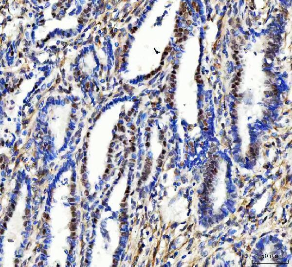
Real IHC data IHC analysis of Aurora A/AURKA using anti-Aurora A/AURKA antibody (A00246-4). Aurora A/AURKA was detected in a paraffin-embedded section of human cervical cancer tissue. Heat mediated antigen retrieval was performed in EDTA buffer (pH 8.0, epitope retrieval solution). The tissue section was blocked with 10% goat serum. The tissue section was then incubated with 2 μg/ml rabbit anti-Aurora A/AURKA Antibody (A00246-4) overnight at 4°C. Peroxidase Conjugated Goat Anti-rabbit IgG was used as secondary antibody and incubated for 30 minutes at 37°C. The tissue section was developed using HRP Conjugated Rabbit IgG Super Vision Assay Kit (Catalog # SV0002) with DAB as the chromogen.
Anti-Aurora A/AURKA Antibody ®
Cat # A00246-4
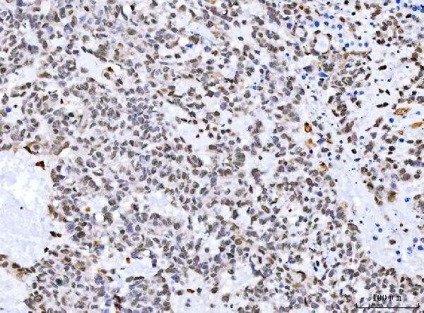
Real IHC data IHC analysis of Aurora A/AURKA using anti-Aurora A/AURKA antibody (A00246-3). Aurora A/AURKA was detected in a paraffin-embedded section of human liver cancer tissue. Heat mediated antigen retrieval was performed in EDTA buffer (pH 8.0, epitope retrieval solution). The tissue section was blocked with 10% goat serum. The tissue section was then incubated with 2 μg/ml rabbit anti-Aurora A/AURKA Antibody (A00246-3) overnight at 4°C. Biotinylated goat anti-rabbit IgG was used as secondary antibody and incubated for 30 minutes at 37°C. The tissue section was developed using Strepavidin-Biotin-Complex (SABC) (Catalog # SA1022) with DAB as the chromogen.
Anti-Aurora A/AURKA Antibody ®
Cat # A00246-3

A00246-4 shows human cervical cancer paraffin-section IHC at 2 μg/ml with EDTA pH 8.0 retrieval; the fixative is unreported (A00246-4 IHC caption). A00246-3 shows human liver cancer paraffin-section IHC at 2 μg/ml with EDTA pH 8.0 retrieval; the fixative is unreported (A00246-3 IHC caption).

Which to pick: For tissue IHC, choose A00246-4 when its human cervical cancer paraffin-section example is relevant (A00246-4 IHC caption). For IF/ICC, choose A00246-3: both applications are listed, and its IF image uses Caco-2 cells (A00246-3 catalog applications; A00246-3 IF caption). For human and mouse IF/ICC work, consider M00246, a rabbit monoclonal listed for both species and applications; IHC is not listed (M00246 catalog).

Each figure is that product's own IHC / IF validation image from its datasheet.

References

  1. UniProt Consortium. UniProt entry O14965 (AURKA_HUMAN, Aurora kinase A).
  2. Human Protein Atlas. AURKA tissue IHC expression (reliability: Enhanced).
  3. Human Protein Atlas. AURKA subcellular location (ICC-IF): Mainly localized to the centrosome, mitotic spindle and basal body. In addition localized to the nucleoplasm and cytosol..
  4. Human Protein Atlas. AURKA antibody validation summary (2 antibodies).
  5. Integrated transcriptomic and immunoinformatics analysis identifies tpx2, bub1b, and ube2c as diagnostic biomarkers and therapeutic targets in endometrial carcinoma. Discover oncology 2025 — PMC12559507.
  6. Clinical analysis of 10 cases with subcutaneous panniculitis-like T-cell lymphoma and tissue AURKA expression. Skin research and technology : official journal of International Society for Bioengineering and the Skin (ISBS) [and] International Society for Digital Imaging of Skin (ISDIS) [and] International Society for Skin Imaging (ISSI) 2024 — PMC11305869.
  7. Aurora kinase A (AURKA) expression in colorectal cancer liver metastasis is associated with poor prognosis. British journal of cancer 2013 — PMC3817339.
  8. Disease Modeling on Tumor Organoids Implicates AURKA as a Therapeutic Target in Liver Metastatic Colorectal Cancer. Cellular and molecular gastroenterology and hepatology 2022 — PMC8688726.
  9. PubMed PMID:9153231 — UniProt-cited evidence.
  10. PubMed PMID:9514916 — UniProt-cited evidence.
  11. PubMed PMID:9771714 — UniProt-cited evidence.