This website uses cookies to ensure you get the best experience on our website.


- Table of Contents
Plan chromogenic B9D1 IHC in paraffin sections using bronchial ciliated cell bodies as a high-staining reference and smooth muscle cells as a reported unstained comparison (HPA tissue IHC). Start the IHC-validated antibody within its 1:50–1:200 IHC dilution range (datasheet: A10722-1).

Expected localisation, validated protocols, controls and antibodies — the at-a-glance facts below, then the full design guide.
| Expected localisation | General cytoplasmic tissue staining (HPA tissue IHC) | |
| Staining pattern | Cytoplasmic staining, including bronchial ciliated cell bodies (HPA tissue IHC) | |
| Antigen retrieval | Citrate pH 6.0 HIER, 95–98 °C, 20 min (rule: cytoplasmic / membrane antigen) | |
| Positive control | Adrenal gland+4 more · see all | |
| Negative control | Caudate+2 more · see all |
| Fixation | Keep tissue fixation consistent across sections. (standard IHC practice; not target-specific) | |
| Caveat | Staining has medium agreement with RNA expression (HPA tissue IHC) | |
| Regulation | Expression regulator not annotated (UniProt) | |
| Isoform / epitope | 2 isoforms; epitope coverage is unspecified (UniProt) |
Compare the catalog antibody’s IHC-P protocol (datasheet) with one published B9d1 paraffin-section protocol (PMC4395494).
| Sample | Paraffin-embedded human stomach tissue; fixative not specified (datasheet A10722-1) |
| Fixation | Image fixative and duration unreported (datasheet A10722-1); verify before use. |
| Sectioning | 4–5 µm sections on charged slides (standard) |
| Deparaffinisation | Xylene, graded ethanol series to water (standard) |
| Antigen retrieval | Heat-induced epitope retrieval in citrate buffer, pH 6.0, 20 min at 95–98 °C (standard rule: cytoplasmic / membrane antigen) |
| Peroxidase block | 3% H2O2, 10 min, room temperature (standard) |
| Blocking | 10% normal serum of the secondary host, 30 min, room temperature (standard) |
| Primary antibody | Rabbit anti-B9D1, 1:50-1:200 (datasheet A10722-1) |
| Primary incubation | Overnight at 4 °C (standard) |
| Detection | HRP-polymer secondary, DAB chromogen 5–10 min (standard) |
| Counterstain | Hematoxylin, blue, dehydrate and mount (standard) |
| Expected result | B9D1-positive staining in glandular cells of adrenal gland (HPA tissue IHC: High). HPA tissue profile: General cytoplasmic expression. No signal in the no-primary control. |
B9D1 localizes to the ciliary basal body, axoneme and transition zone (UniProt Q9UPM9 subcellular location); it has no transmembrane segment (UniProt Q9UPM9 topology). In paraffin tissue sections, expect chiefly cytoplasmic staining, including high staining in bronchial ciliated cell bodies and several glandular cell populations (HPA tissue IHC). Treat this as a reference pattern with limited certainty: HPA rates its tissue IHC reliability Approved, with medium staining–RNA consistency and external verification pending (HPA tissue IHC).
| Cytoplasmic staining in bronchial ciliated cell bodies, with high staining in the same cell population across the section (HPA tissue IHC). | This fits an observed tissue pattern (HPA: High in bronchial ciliated cell bodies). The basal body and transition zone are more precise localization expectations (UniProt Q9UPM9 subcellular location); a diffuse chromogenic cell-body signal alone cannot resolve those small structures (general IHC interpretation). |
| Predominantly nuclear staining in a paraffin section, without the expected cytoplasmic pattern (HPA tissue IHC). | Investigate compartment-specific background or antibody specificity before assigning a tissue IHC result (general IHC interpretation). HPA describes general cytoplasmic tissue expression (HPA tissue IHC); its uncertain nucleoplasm assignment comes from ICC-IF and does not establish a nuclear paraffin-section pattern (HPA subcellular ICC-IF). |
| Strong staining confined to ovarian stroma cells, caudate glial cells or smooth muscle cells (HPA tissue IHC). | These populations are listed as Not detected (HPA tissue IHC). Check for cross-reactivity, endogenous detection activity or misidentified cell types (general IHC troubleshooting). A discrepant result needs validation; the HPA tissue reference remains pending external verification (HPA tissue IHC reliability). |
| Diffuse staining across cells and extracellular spaces, with little distinction between cells expected to differ in signal (general IHC interpretation). | Treat uniform haze as possible background rather than evidence of widespread B9D1 expression (general IHC interpretation). Compare the same run with a no-primary control and with documented high and Not detected populations (general IHC controls; HPA tissue IHC). |
| No visible staining in bronchial ciliated cell bodies or another documented high population (HPA tissue IHC). | First suspect a failed staining run or inadequate assay sensitivity (general IHC troubleshooting). Confirm tissue identity and preservation, then check retrieval, primary-antibody use and detection against the validated IHC-P instructions if available (general IHC practice). Absence alone cannot overturn an Approved reference pattern that awaits external verification (HPA tissue IHC reliability). |
| Subcellular scale and topology | B9D1 is reported at the basal body, axoneme and transition zone, with no transmembrane segment (UniProt Q9UPM9). Score chromogenic tissue IHC at the cell and compartment level; do not demand that a punctum resolve the transition zone (general IHC interpretation). |
| Alternative splicing | Two isoforms are listed (UniProt Q9UPM9). The supplied record gives no antibody epitope or isoform-specific tissue pattern, so do not attribute a difference in staining to one isoform without separate validation (UniProt Q9UPM9; evidence limit). |
| Processing and annotated modifications | The record lists a single chain spanning residues 1–204, no signal peptide or propeptide, and no annotated glycosylation or modified residues (UniProt Q9UPM9). These annotations provide no basis to predict a processed, shed or modification-dependent IHC pattern (evidence limit). |
| Reference-pattern certainty | HPA rates the tissue IHC pattern Approved but reports medium consistency with RNA data and pending external verification; antibody HPA022957 is Approved for IHC and Uncertain for ICC (HPA tissue IHC; HPA antibodies). Use the tissue pattern as a comparison, then verify unexpected results with controls (general IHC practice). |
| IHC versus ICC-IF localization | HPA reports general cytoplasmic tissue staining (HPA tissue IHC), while ICC-IF additionally reports a supported basal body location and an uncertain nucleoplasm location (HPA subcellular ICC-IF). Preserve the application distinction when interpreting a paraffin section (general IHC interpretation). |
| Situation | Likely cause | Next action |
|---|---|---|
| A documented high population stains weakly or not at all (HPA tissue IHC). | The staining run may lack sensitivity, or tissue identification may be wrong (general IHC troubleshooting). | Confirm the cell population and run controls; check retrieval, primary-antibody conditions and chromogenic detection against the applicable IHC-P instructions (general IHC practice). |
| Brown signal appears broadly in cells listed as Not detected (HPA tissue IHC). | Cross-reactivity or endogenous detection activity can mimic target staining (general IHC troubleshooting). | Inspect a no-primary control and the detection-system controls; compare localization with documented high cell populations before scoring the signal (general IHC practice; HPA tissue IHC). |
| The section shows uniform haze or heavy staining outside identifiable cells (general IHC interpretation). | Nonspecific binding, insufficient blocking or excessive detection signal are possible causes (general IHC troubleshooting). | Review blocking, washes and detection exposure using the same controls and tissue type; score cell-associated staining only after background separates from it (general IHC practice). |
| A nuclear-only pattern appears in tissue IHC (HPA tissue IHC comparison). | The tissue reference describes general cytoplasmic expression; ICC-IF nucleoplasm localization is uncertain (HPA tissue IHC; HPA subcellular ICC-IF). | Check counterstain and no-primary control, then seek independent specificity evidence before calling nuclear B9D1 positive in the paraffin section (general IHC practice). |
| Ciliated cell bodies stain, but no discrete transition-zone dot is visible (HPA tissue IHC; UniProt Q9UPM9). | A chromogenic paraffin section may not resolve the small ciliary structure described by UniProt (general IHC interpretation; UniProt Q9UPM9 subcellular location). | Record the supported cell-body and cytoplasmic pattern; reserve a claim of precise subcellular localization for appropriately resolved imaging (HPA tissue IHC; general microscopy practice). |
| Can an ICC-IF image establish the expected paraffin-section pattern? | The applications have different reference observations: tissue IHC is generally cytoplasmic, while ICC-IF includes supported basal body and uncertain nucleoplasm assignments (HPA tissue IHC; HPA subcellular ICC-IF). | Use ICC-IF as localization context only; judge the IHC section against HPA tissue cell-type patterns and IHC controls (HPA tissue IHC; general IHC practice). |


Comprehensive Human Protein Atlas IHC scoring per tissue (reliability: Approved — Medium consistency between antibody staining and RNA expression data. Pending external verification.). Rows are taken directly from the HPA tissue chart — click any row's HPA link to view the source.
| Tissue | Cell type | Level | Evidence | Source |
|---|---|---|---|---|
| Adrenal gland | Glandular cells | High | Protein (IHC) | HPA → |
| Appendix | Lymphoid tissue | High | Protein (IHC) | HPA → |
| Bone marrow | Hematopoietic cells | High | Protein (IHC) | HPA → |
| Breast | Glandular cells | High | Protein (IHC) | HPA → |
| Bronchus | Ciliated cells (cell body) | High | Protein (IHC) | HPA → |
Use compartment-aware controls to optimize B9D1 staining in paraffin sections and assess whether chromogenic signal fits its ciliary localization.
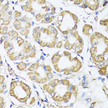
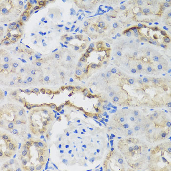
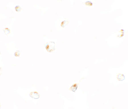
The catalog shows B9D1 IHC in paraffin-embedded human stomach, rat kidney and mouse lung, plus IF/ICC in 293 cells (catalog image captions).
A10722-1 has IHC images from paraffin-embedded human stomach, rat kidney and mouse lung (A10722-1 IHC captions). A10722 has IF/ICC images from 293 cells (A10722 IF/ICC captions).
Which to pick: Choose A10722-1 for tissue IHC: IHC is listed for human, mouse and rat samples (A10722-1 catalog), and its own captions show paraffin-embedded sections stained at 1:100 after microwave retrieval in 10 mM PBS, pH 7.2 (A10722-1 IHC captions); the fixative is unreported (A10722-1 IHC captions). Choose A10722 for IF/ICC in human samples: those applications and human reactivity are listed (A10722 catalog), with pictured 293-cell staining at 5 μg/mL for ICC and 20 μg/mL for IF (A10722 IF/ICC captions). For mouse or rat IF/ICC, A10722-1 lists those species and applications (A10722-1 catalog); clonality is unreported for both SKUs (catalog clone fields).