This website uses cookies to ensure you get the best experience on our website.


- Table of Contents
Plan paraffin-section BAD IHC around widespread cytoplasmic staining (HPA tissue IHC) and a 1:50 catalog-antibody starting dilution (datasheet: M03520 IHC caption). Compare signal across cell types while allowing for phosphorylation-dependent redistribution (UniProt).

Expected localisation, validated protocols, controls and antibodies — the at-a-glance facts below, then the full design guide.
| Expected localisation | Cytoplasm (HPA tissue IHC); mitochondrial outer membrane (UniProt) | |
| Staining pattern | Widespread cytoplasmic staining across cell types (HPA tissue IHC) | |
| Antigen retrieval | EDTA pH 8.0 HIER, heat-mediated (datasheet M03520) | |
| Positive control | Bronchus+4 more · see all | |
| Negative control | Ovary+2 more · see all |
| Fixation | Matched tissue-IHC evidence does not establish the fixation claim. Validate the specimen-specific method before use. (selected-SKU IHC image M03520) | |
| Caveat | Phosphorylation can shift BAD toward the cytoplasm (UniProt) | |
| Regulation | No expression regulator specified (UniProt) | |
| Isoform / epitope | No annotated isoforms; one full-length chain (UniProt) |
The catalog antibody’s IHC protocol is followed by four published BAD IHC protocols, including total BAD and phospho-BAD staining (PMC3787924; PMC5442111; PMC11366015; PMC4145317).
| Sample | Paraffin-embedded human breast cancer tissue; fixative not specified (datasheet M03520) |
| Fixation | Image fixative and duration unreported (datasheet M03520); verify before use. |
| Sectioning | 4–5 µm sections on charged slides (standard) |
| Deparaffinisation | Xylene, graded ethanol series to water (standard) |
| Antigen retrieval | Heat retrieval: EDTA pH 8.0 (datasheet M03520); 20 min, 95–100 °C (standard) |
| Peroxidase block | 3% H2O2, 10 min, room temperature (standard) |
| Blocking | 10% goat serum (datasheet M03520) |
| Primary antibody | Rabbit monoclonal (clone CHC-2) anti-BAD, 1:50 (datasheet M03520) |
| Primary incubation | Overnight at 4 °C (datasheet M03520) |
| Detection | HRP-conjugated secondary, DAB chromogen (datasheet M03520) |
| Counterstain | Hematoxylin, blue, dehydrate and mount (standard) |
| Expected result | BAD-positive staining in respiratory epithelial cells of bronchus (HPA tissue IHC: High). HPA tissue profile: Ubiquitous cytoplasmic expression. No signal in the no-primary control. |
BAD is found at the mitochondrial outer membrane and in the cytoplasm; phosphorylation can shift it toward the cytoplasm (UniProt Q92934 localization; UniProt Q92934 topology: no transmembrane segment). In paraffin IHC, expect cytoplasmic staining in cells such as kidney tubule cells and bronchial respiratory epithelial cells (HPA tissue IHC: High). HPA describes the tissue pattern as ubiquitous cytoplasmic expression, with Approved reliability, medium staining–RNA consistency, and external verification pending (HPA tissue IHC).
| Cytoplasmic staining in kidney tubule cells, bronchial respiratory epithelial cells, or placental trophoblastic cells. | This fits HPA’s High staining calls for those cell types (HPA tissue IHC: High). BAD can also associate with the mitochondrial outer membrane (UniProt Q92934 localization). A chromogenic cytoplasmic signal alone cannot establish precise mitochondrial localization (standard IHC interpretation). |
| Predominantly nuclear staining, with little cytoplasmic signal in a positive control. | A nuclear dominant pattern conflicts with the reported cytoplasmic and mitochondrial locations (UniProt Q92934 localization; HPA tissue IHC: ubiquitous cytoplasmic expression). Treat it as suspect, then compare the staining with an independently validated antibody or an appropriate negative control (standard IHC practice). |
| Strong staining confined to skeletal muscle myocytes, smooth muscle cells, or ovarian stroma cells. | HPA reports BAD as Not detected in those cell types (HPA tissue IHC: Not detected). Such staining warrants checks for cross-reactivity or endogenous detection activity (standard IHC practice). A Not detected call is a reference pattern, not proof that every specimen must be blank (HPA tissue IHC: Approved; external verification pending). |
| Diffuse color over cells and surrounding tissue, without a clear cellular pattern. | Uniform background is difficult to reconcile with HPA’s cell-specific staining calls (HPA tissue IHC). Check blocking, detection-only background, and antibody concentration as general causes of nonspecific chromogenic signal (standard IHC practice); do not score haze as BAD-positive cytoplasm (standard IHC interpretation). |
| No signal in kidney tubule cells or bronchial respiratory epithelial cells used as positive controls. | Both are High reference cell types (HPA tissue IHC: High). First assess section quality, antigen retrieval, primary antibody handling, and detection performance (standard IHC practice). A failed positive control makes a negative result elsewhere uninterpretable; HPA’s Approved assessment does not guarantee staining in every preparation (HPA tissue IHC). |
| Compartment and phosphorylation state | BAD occupies the mitochondrial outer membrane and cytoplasm; upon phosphorylation it locates to the cytoplasm (UniProt Q92934 localization). A less distinctly mitochondrial appearance need not be an IHC failure, but this record gives no threshold for scoring that shift in paraffin sections (UniProt Q92934 localization). |
| Cell type within a tissue | Use the named cells, rather than an entire tissue, as the reference: kidney tubule cells are High, while skeletal muscle myocytes are Not detected (HPA tissue IHC). Low staining is reported in oral squamous epithelial cells and adipocytes, so faint signal there has a different reference level (HPA tissue IHC: Low). |
| Strength of antibody evidence | Three listed antibodies have Approved IHC status (HPA antibodies: HPA028185, HPA062105, CAB004205). The tissue profile has medium agreement with RNA data and awaits external verification (HPA tissue IHC: reliability description); Approved status should therefore inform, not replace, slide-level controls (standard IHC practice). |
| Resolution of the readout | HPA’s ICC-IF summary places BAD mainly at mitochondria (HPA subcellular ICC-IF: Mitochondria, enhanced), while tissue IHC reports ubiquitous cytoplasmic expression (HPA tissue IHC: profile). Chromogenic paraffin IHC may show cytoplasmic color without resolving individual mitochondria (standard IHC interpretation). |
| Retrieval and detection workflow | Retrieval conditions, blocking, and detection can affect chromogenic IHC signal generally (standard IHC practice). No BAD-specific fixation sensitivity or retrieval requirement is established by the supplied UniProt and HPA records; do not infer one from topology, modifications, or tissue staining levels (UniProt Q92934; HPA tissue IHC). |
| Situation | Likely cause | Next action |
|---|---|---|
| Positive-control cytoplasm is blank. | The assay may have failed, or the chosen control may lack the relevant cells (standard IHC practice; HPA tissue IHC: cell-specific calls). | Confirm that kidney tubule or bronchial respiratory epithelial cells are present, then check retrieval, primary antibody, and detection controls (HPA tissue IHC: High; standard IHC practice). |
| The slide looks uniformly brown. | Nonspecific primary binding or detection background can obscure cellular staining (standard IHC practice). | Review a no-primary control, blocking, and antibody concentration; score only a defined cellular pattern (standard IHC practice). |
| Nuclei dominate the staining. | That distribution conflicts with BAD’s reported cytoplasmic and mitochondrial locations (UniProt Q92934 localization). | Compare with a positive tissue control and an independently validated antibody before assigning the nuclear signal to BAD (HPA antibodies: IHC Approved; standard IHC practice). |
| Myocytes or ovarian stroma stain strongly. | Those cells are Not detected in the HPA tissue reference, making cross-reactivity or endogenous activity plausible (HPA tissue IHC: Not detected; standard IHC practice). | Check a no-primary control and repeat with an independently validated antibody; interpret the discrepancy cautiously because HPA verification is pending (standard IHC practice; HPA tissue IHC: reliability description). |
| Expected cytoplasmic signal is faint. | Low reference expression can explain faint staining in adipocytes or oral squamous epithelial cells (HPA tissue IHC: Low); weak assay performance remains possible (standard IHC practice). | Compare the same run with a High reference cell type before changing the interpretation or general IHC conditions (HPA tissue IHC: High; standard IHC practice). |
| IF/ICC shows mitochondrial signal, but paraffin IHC looks broadly cytoplasmic. Is that inconsistent? | HPA reports mainly mitochondrial ICC-IF localization and ubiquitous cytoplasmic tissue IHC; chromogenic IHC has less spatial detail (HPA subcellular ICC-IF; HPA tissue IHC; standard IHC interpretation). | Interpret each readout against its own reference pattern; consult the separate IF/ICC guide for that application (HPA subcellular ICC-IF; HPA tissue IHC). |
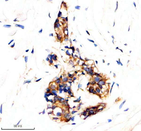
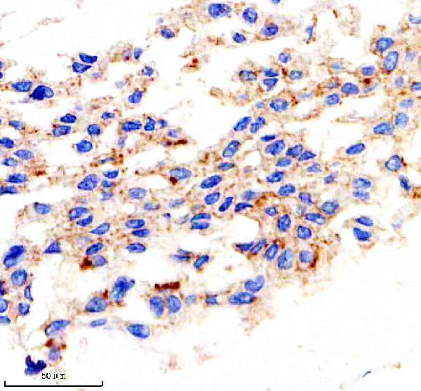
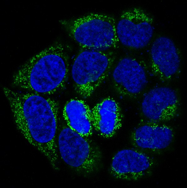
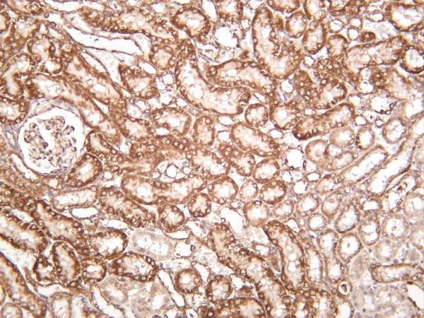
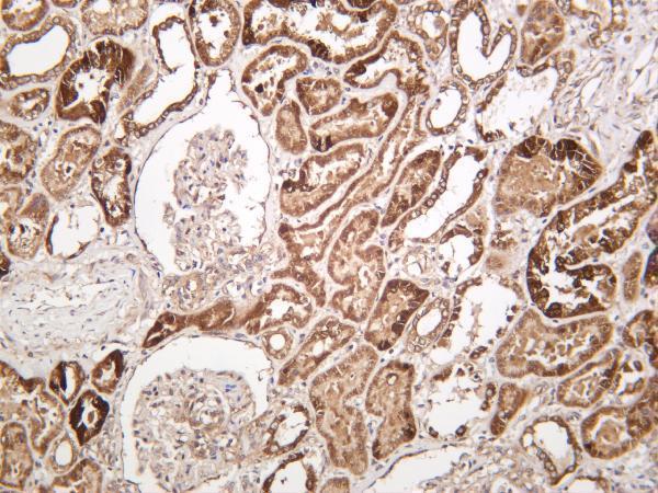
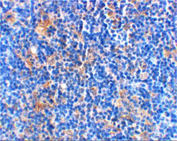
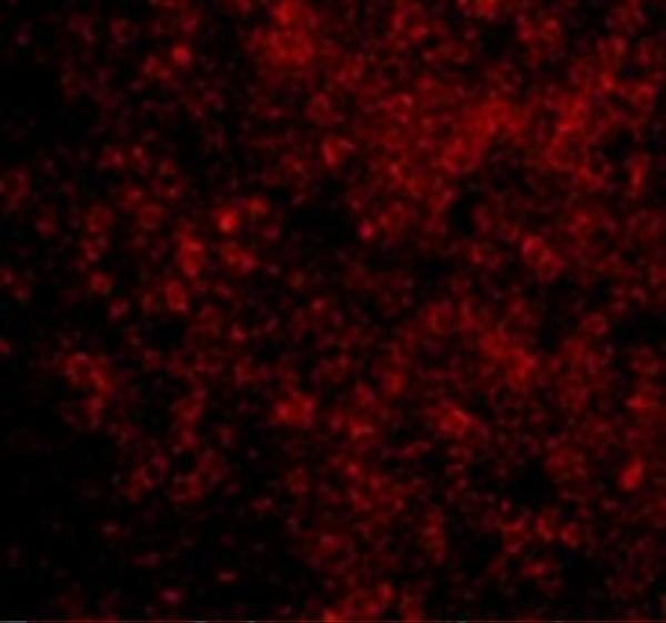


Comprehensive Human Protein Atlas IHC scoring per tissue (reliability: Approved — Medium consistency between antibody staining and RNA expression data. Pending external verification.). Rows are taken directly from the HPA tissue chart — click any row's HPA link to view the source.
| Tissue | Cell type | Level | Evidence | Source |
|---|---|---|---|---|
| Bronchus | Respiratory epithelial cells | High | Protein (IHC) | HPA → |
| Caudate | Neuronal cells | High | Protein (IHC) | HPA → |
| Kidney | Cells in tubules | High | Protein (IHC) | HPA → |
| Nasopharynx | Respiratory epithelial cells | High | Protein (IHC) | HPA → |
| Placenta | Trophoblastic cells | High | Protein (IHC) | HPA → |
Troubleshoot BAD staining in paraffin sections by checking retrieval, compartment, controls and cell-specific scoring before interpreting chromogenic signal.
These anti-BAD antibodies list human, mouse and rat reactivity (catalog: reactivity). IHC images cover human breast cancer, kidney and rat thymus; IF images cover HeLa and rat thymus cells (catalog: image captions).
M03520 has IHC images of human breast cancer, human placenta and mouse small intestine, plus an IF image of HeLa cells (M03520: image captions); M03520-1 has IHC images of human, mouse and rat kidney (M03520-1: image captions). A03520 has IHC and IF images of rat thymus samples (A03520: image captions).
Which to pick: For paraffin-section IHC, start with M03520 at 1:50 after EDTA retrieval at pH 8.0 (M03520: IHC caption); the caption does not report the fixative (M03520: IHC caption). For IF/ICC, choose M03520 because ICC and IF are listed applications and an IF image shows HeLa cells (M03520: applications and IF caption). For cross-species tissue IHC, choose M03520-1: its IHC images show human, mouse and rat kidney, with a listed dilution of 1:200–1:1000 (M03520-1: IHC captions and catalog dilution).