BATF / Basic leucine zipper transcriptional factor ATF-like · IHC design guide

Design Immunohistochemistry for BATF

Plan chromogenic BATF IHC in paraffin sections with the IHC-validated antibody A04089-1 at 1:100–1:300 (datasheet A04089-1). Assess peripheral leukocyte cytoplasm and high staining in testis Sertoli cells (HPA tissue IHC), while considering activation-linked nuclear translocation (UniProt).

Evidence assembled Oct 2026 · For research use; verify linked source records and product datasheet before use
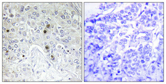
Immunohistochemistry protocol sheet for BATF (IHC for BATF): expected localisation Peripheral leukocyte cytoplasm (HPA tissue IHC), antibody A04089-1, validated IHC image, and IHC protocol steps
Printable BATF IHC protocol sheet — expected localisation Peripheral leukocyte cytoplasm (HPA tissue IHC), antibody A04089-1, controls and protocol steps. Open the full BATF IHC guide →

BATF Immunohistochemistry Experimental Design Guide

Expected localisation, validated protocols, controls and antibodies — the at-a-glance facts below, then the full design guide.

Must know before staining
Expected localisation Peripheral leukocyte cytoplasm (HPA tissue IHC)
Staining pattern Cytoplasmic staining in peripheral leukocytes (HPA tissue IHC)
Antigen retrieval Tris-EDTA pH 8.0 HIER, heat-mediated (datasheet A04089-1)
Positive control ⓘ Testis
Negative control ⓘ Adipose tissue+4 more · see all
Important caveats
Reasons your staining may differ from the expected pattern.
Fixation Keep fixation consistent across sections. (standard IHC practice; not target-specific)
Caveat T-cell activation increases nuclear translocation (UniProt)
Regulation STAT3 raises BATF in damaged HSCs (UniProt)
Isoform / epitope One 1–125 chain; no listed isoforms (UniProt)
Section 1

Recommended BATF IHC & IF Protocols

The catalog antibody protocol is paired with published BATF IHC methods for colorectal cancer, hepatocellular carcinoma, and endometrioid adenocarcinoma samples (PMC11165778; PMC12047759; PMC11630932).

Recommended immunohistochemistry (IHC-P) protocol parameters
SampleParaffin-embedded Human breast cancer tissue; fixative not specified (datasheet A04089-1)
FixationImage fixative and duration unreported (datasheet A04089-1); verify before use.
Sectioning4–5 µm sections on charged slides (standard)
DeparaffinisationXylene, graded ethanol series to water (standard)
Antigen retrievalHeat retrieval: Tris-EDTA pH 8.0 (datasheet A04089-1); 20 min, 95–100 °C (standard)
Peroxidase block3% H2O2, 10 min, room temperature (standard)
Blocking10% normal serum of the secondary host, 30 min, room temperature (standard)
Primary antibodyRabbit anti-BATF, 1:100-1:300 (datasheet A04089-1)
Primary incubationOvernight at 4 °C (standard)
DetectionHRP-polymer secondary, DAB chromogen 5–10 min (standard)
CounterstainHematoxylin, blue, dehydrate and mount (standard)
Expected resultBATF-positive staining in sertoli cells of testis (HPA tissue IHC: High). HPA tissue profile: Cytoplasmic expression in peripheral leukocytes. No signal in the no-primary control.
💡Decision noteStart with heat-mediated Tris-EDTA retrieval at pH 8.0 for the catalog antibody (datasheet A04089-1); the hepatocellular carcinoma study reports different retrieval buffers (PMC12047759).
Section 2

What Is the Expected BATF Staining Pattern?

BATF can occupy the nucleus and cytoplasm, with increased nuclear translocation after T-cell activation (UniProt Q16520). In tissue IHC, HPA reports cytoplasmic staining in peripheral leukocytes and high staining in testicular Sertoli cells (HPA tissue IHC). BATF has no transmembrane segment (UniProt Q16520 topology). HPA rates its tissue staining Approved but reports low consistency between antibody staining and RNA expression (HPA tissue IHC).

What am I looking at on my slide?
Discrete cytoplasmic staining in peripheral leukocytes, with a restrained counterstain.This matches the reported tissue IHC profile (HPA tissue IHC: cytoplasmic expression in peripheral leukocytes). Nuclear BATF can also be biologically plausible, especially after T-cell activation (UniProt Q16520); the slide alone cannot establish activation state or prove that every nuclear signal is specific.
A strong, uniform signal confined to a compartment inconsistent with the expected cellular distribution.Review it as a possible staining artefact before calling BATF positive: BATF is reported in the nucleus and cytoplasm (UniProt Q16520), while HPA tissue IHC describes cytoplasmic peripheral leukocytes (HPA tissue IHC). Compare cell outlines and the counterstain; compartment alone cannot resolve antibody specificity (general IHC practice).
Prominent staining in adipocytes or adrenal glandular cells, or pigment-like signal without cellular definition.HPA reports BATF as not detected in those cell populations (HPA tissue IHC: adipocytes; adrenal glandular cells). Check a no-primary control for endogenous detection activity and review antibody specificity (general IHC practice). An unexpected positive is a reason to investigate, especially given HPA's low staining–RNA consistency (HPA tissue IHC).
Diffuse color across tissue, stroma, and blank areas, obscuring individual cells.Treat the distribution as background rather than a BATF cell pattern (general IHC practice). Examine the no-primary control, blocking, washes, detection reagents, and counterstain (general IHC practice); HPA's reported peripheral-leukocyte pattern does not justify scoring diffuse tissue color as positive (HPA tissue IHC).
No signal in testicular Sertoli cells on a technically adequate slide.HPA records high staining in Sertoli cells, making testis a useful observed positive comparator (HPA tissue IHC: High in Sertoli cells). Recheck section quality, detection controls, and antibody conditions (general IHC practice). HPA's low staining–RNA consistency means one negative section cannot establish that BATF is absent (HPA tissue IHC).
💡Expected BATF appearanceCall a result consistent with BATF when staining is cell-defined and cytoplasmic in peripheral leukocytes (HPA tissue IHC), or high in Sertoli cells without assuming their compartment (HPA: High in Sertoli cells); diffuse color or strong staining in HPA-negative cell populations warrants control-based review (HPA tissue IHC; general IHC practice).
How each factor affects the staining
Cell state and compartmentBATF occurs in nucleus and cytoplasm and shows increased nuclear translocation after T-cell activation (UniProt Q16520). Interpret nuclear signal with cell identity and controls; the activation state is not established by BATF staining alone (general IHC practice).
Observed tissue distributionHPA reports high Sertoli-cell staining, cytoplasmic peripheral-leukocyte expression, and no detection in adipocytes, adrenal glandular cells, or bone-marrow hematopoietic cells (HPA tissue IHC). These are observed IHC patterns, not universal positive or negative guarantees; HPA notes low staining–RNA consistency (HPA tissue IHC).
Antibody evidenceHPA064962 has Approved IHC status; HPA059588 has no listed IHC status (HPA antibodies). HPA's tissue-level Approved rating still carries a low staining–RNA consistency note (HPA tissue IHC). Match interpretations to the IHC-validated antibody and controls used on the slide (general IHC practice).
Processing and topologyUniProt lists one 1–125 BATF chain, no signal peptide or propeptide, no transmembrane segment, and no glycosylation sites (UniProt Q16520). These features provide no basis for expecting a membrane-restricted or secreted staining pattern; they do not establish antigen-retrieval needs or fixation sensitivity (UniProt Q16520 topology; general IHC practice).
IF/ICC Q: What compartment is reported?HPA reports enhanced nucleoplasmic localization by ICC-IF and lists MCF-7, RT-4, and U2OS among image-bearing cell lines (HPA subcellular). This supports an IF/ICC localization check; it does not replace the reported cytoplasmic peripheral-leukocyte pattern in tissue IHC (HPA tissue IHC).
Why is my staining missing, weak or wrong?
SituationLikely causeNext action
Peripheral leukocytes show no defined stain.The slide may lack detectable signal, or the sampled cells may not reproduce HPA's observed pattern (HPA tissue IHC; general IHC practice).Check a positive comparator, section integrity, primary-antibody conditions, retrieval, and detection controls (general IHC practice). Do not infer BATF absence from this slide alone given HPA's low staining–RNA consistency (HPA tissue IHC).
Testicular Sertoli cells are negative.An expected comparator may have failed technically or may vary from HPA's recorded high staining (HPA: High in Sertoli cells; general IHC practice).Verify cell identification and a working detection control, then review the IHC-validated antibody and staining conditions (general IHC practice). Record the discordance without treating HPA's observation as guaranteed in every section (HPA tissue IHC).
Adipocytes or adrenal glandular cells stain strongly.These cells are reported as not detected, so nonspecific binding or endogenous detection activity should be considered (HPA tissue IHC; general IHC practice).Inspect a no-primary control and cell morphology; review blocking, detection chemistry, and antibody specificity before scoring (general IHC practice).
Color is diffuse or extends beyond recognizable cells.Background from staining or detection can obscure a cell-defined IHC pattern (general IHC practice).Compare with a no-primary control; adjust blocking, washes, or detection exposure as needed (general IHC practice). Score only interpretable cells, with HPA's leukocyte pattern as a reference (HPA tissue IHC).
Only nuclear staining appears in tissue.Nuclear BATF is biologically plausible, particularly after T-cell activation, although HPA's tissue profile specifies cytoplasmic peripheral leukocytes (UniProt Q16520; HPA tissue IHC).Confirm which cells stain, compare controls, and report the observed compartment separately from the expected tissue profile (general IHC practice). Do not assign T-cell activation from compartment alone (UniProt Q16520; general IHC practice).
IF/ICC shows a pattern that differs from tissue IHC.HPA reports enhanced nucleoplasmic ICC-IF localization and a cytoplasmic peripheral-leukocyte tissue IHC profile (HPA subcellular; HPA tissue IHC).Interpret each assay with its own cell context and controls (general IHC practice). Use the HPA ICC-IF result to assess IF/ICC localization, and the HPA tissue record to assess the IHC slide (HPA subcellular; HPA tissue IHC).

Sample controls for BATF IHC & IF

🧪Run testis first; Sertoli cells should stain (HPA: High in Sertoli cells). Use adipose tissue as the negative tissue (HPA: Not detected in adipocytes); on the testis slide, assess unstained cells outside the Sertoli compartment as potential internal negatives rather than assuming their BATF status.
Positive control tissue: Testis (Sertoli cells, HPA High)
Negative control tissue: Adipose tissue (HPA Not detected)
ICC-IF cell lines (HPA subcellular resource): HPA ICC-IF images show BATF in MCF-7, RT-4, U2OS, with annotated localisation: Nucleoplasm (enhanced) (HPA subcellular).
Technical controls: Include no-primary (secondary-only) and isotype controls matched to the primary antibody’s host species and antibody class, with a control appropriate to its clonality (standard IHC practice). Use a BATF knockout specimen or immunogen-peptide block as a biological specificity control (caption: peptide-block negative control), and check testis sections for residual endogenous peroxidase signal after quenching (standard chromogenic IHC practice).
⚠️Feasibility: A target-specific fixation window is unreported, and the fixative in the A04089-1 paraffin-section caption is unreported (caption: fixative not stated). That caption uses high-pressure, high-temperature Tris-EDTA at pH 8.0 for retrieval, but does not establish that retrieval is required (caption: Tris-EDTA, pH 8.0); HPA reports nucleoplasmic ICC-IF staining, but the supplied evidence does not show whether IF or frozen sections are easier (HPA: Nucleoplasm, enhanced). Testis-specific artefacts are unreported in the supplied evidence.

HPA tissue IHC evidence for BATF

Comprehensive Human Protein Atlas IHC scoring per tissue (reliability: Approved — Low consistency between antibody staining and RNA expression data.). Rows are taken directly from the HPA tissue chart — click any row's HPA link to view the source.

Positive expression · recommended positive controls

TissueCell typeLevelEvidenceSource
Testis Sertoli cells High Protein (IHC) HPA →

Undetected expression · recommended negative controls

TissueCell typeLevelEvidenceSource
Adipose tissue Adipocytes Not detected Protein (IHC) HPA →
Adrenal gland Glandular cells Not detected Protein (IHC) HPA →
Bone marrow Hematopoietic cells Not detected Protein (IHC) HPA →
Breast Adipocytes Not detected Protein (IHC) HPA →
Bronchus Respiratory epithelial cells Not detected Protein (IHC) HPA →
Section 3

Advanced BATF IHC Tips

Use compartment, cell identity, and matched controls to assess BATF staining in paraffin sections (UniProt Q16520; HPA tissue IHC).

How should I retrieve BATF when nuclear staining is weak?
Use heat-mediated Tris-EDTA at pH 8.0 for BATF antigen retrieval in paraffin sections (datasheet A04089-1). The selected image used high-pressure, high-temperature retrieval and 1:100 primary antibody overnight at 4 °C; these are starting conditions for the same SKU (caption A04089-1). If nuclear signal remains weak, compare shorter and longer heating exposures on matched sections while keeping dilution, detection, and counterstain constant (standard IHC practice). Judge improvement against the immunogen-peptide preabsorbed control, and assess nuclear and cytoplasmic staining separately because BATF can occupy both compartments (caption A04089-1; UniProt Q16520 subcellular location).
Could fixation explain variable BATF staining between blocks?
Target-specific sensitivity of BATF staining to fixative type or fixation duration is unknown from the supplied evidence; the selected paraffin-section caption does not state a fixative (caption A04089-1). Record each block’s fixative, fixation duration, processing history, and section age before comparing chromogenic intensity (standard IHC practice). Run matched sections from the same block together with identical pH 8.0 retrieval and detection conditions to limit technical variation (datasheet A04089-1; standard IHC practice). If differences persist, compare nuclear signal and morphology within matched cell populations, and avoid assigning the difference to a BATF-specific fixation effect without direct validation (standard IHC practice).
Should BATF appear in nuclei, cytoplasm, or both?
Assess nuclear and cytoplasmic staining separately: BATF is reported in both compartments, with increased nuclear translocation after T-cell activation (UniProt Q16520 subcellular location). Enhanced nucleoplasmic localization in cell imaging supports nuclear staining as a useful reference, while tissue IHC reports cytoplasmic expression in peripheral leukocytes (HPA subcellular; HPA tissue IHC). In paraffin sections, inspect a hematoxylin counterstain to distinguish nuclear chromogen from color overlying closely packed cells (standard IHC practice). Compare compartments within the same identified cell type and matched staining run; a cytoplasmic-only result should prompt control review before it is interpreted as altered BATF trafficking (UniProt Q16520 subcellular location; standard IHC practice).
How do I assess whether epitope access affects BATF staining?
BATF has one recorded 1–125 chain, no listed isoforms or transmembrane segment, and a bZIP domain at residues 26–89 (UniProt Q16520 processing, isoforms, topology, and domains). Phosphoserine 43 and phosphothreonine 48 are recorded modifications, but their effects on this antibody’s binding are not established (UniProt Q16520 modified residues; supplied antibody evidence). If staining varies, check the antibody’s stated immunogen or epitope before proposing an isoform or modification-specific explanation (standard IHC practice). Compare matched sections under the specified pH 8.0 retrieval and include the peptide-preabsorbed control; loss of staining with peptide supports binding specificity without identifying the precise tissue epitope (datasheet A04089-1; caption A04089-1).
How can IF help resolve ambiguous BATF IHC staining?
Use IF as a follow-up to chromogenic IHC when cell identity or compartment is unclear, interpreting it alongside the separate IF/ICC guide (standard IHC/IF practice). Multiplex BATF with a marker for the expected cell type, such as a leukocyte marker when assessing peripheral leukocytes, and compare nuclear signal with a DNA counterstain (HPA tissue IHC: cytoplasmic expression in peripheral leukocytes; standard IF practice). Choose spectrally separated fluorophores, placing a weak signal in a far-red channel if tissue autofluorescence interferes, and inspect unstained sections (standard IF practice). BATF has no transmembrane segment and is nuclear and cytoplasmic, so use permeabilisation suited to intracellular epitope access, then check whether nuclear signal agrees with reported nucleoplasmic localization (UniProt Q16520 topology and subcellular location; HPA subcellular).
What should I check when BATF chromogen appears diffuse?
Compare diffuse staining with a no-primary control and the immunogen-peptide preabsorbed control described for this SKU (caption A04089-1; standard IHC practice). Review peroxidase blocking, protein blocking, washes, and chromogen development because residual enzyme activity or nonspecific detection can produce widespread color (standard chromogenic IHC practice). Titrate around the caption’s 1:100 primary dilution on matched sections, changing one variable at a time while preserving the specified pH 8.0 retrieval (caption A04089-1; datasheet A04089-1; standard IHC practice). Inspect nuclear and cytoplasmic signal by cell type; tissue IHC reports cytoplasmic peripheral-leukocyte staining, but its staining-to-RNA consistency is low (HPA tissue IHC).
How should I score BATF staining across paraffin sections? ⚠ ANSWER MARKED FOR VERIFICATION
Define the cell population and compartment before scoring, since BATF is reported in nuclei and cytoplasm and tissue IHC reports cytoplasmic peripheral-leukocyte staining (UniProt Q16520 subcellular location; HPA tissue IHC). For a specified population, report the percentage of positive nuclei and an H-score based on staining intensity; record cytoplasmic scores separately (standard IHC scoring practice). When cell abundance differs between fields, report positive-cell density per mm² and normalize positive counts to the total eligible cells or tissue area assessed (standard IHC quantification practice). Apply the same positivity threshold, retrieval, imaging settings, and exclusion rules across sections, and document the control signal used to set background (datasheet A04089-1; standard IHC practice).
How do I distinguish convincing BATF staining from artefact?
Look for staining in morphologically intact, identified cells and report its compartment: BATF is nuclear and cytoplasmic, and tissue IHC reports cytoplasmic peripheral-leukocyte expression (UniProt Q16520 subcellular location; HPA tissue IHC). Treat isolated edge staining, necrotic regions, and color in a no-primary control as potential processing or detection artefacts requiring review (standard IHC practice). Compare suspicious color with the SKU’s peptide-preabsorbed control and check peroxidase blocking when endogenous enzyme activity could contribute (caption A04089-1; standard chromogenic IHC practice). Do not equate a positive tissue pattern with established BATF specificity: the HPA tissue-IHC assessment reports low consistency between antibody staining and RNA expression (HPA tissue IHC).
Boster reagents

Best BATF / Basic leucine zipper transcriptional factor ATF-like IHC Antibodies

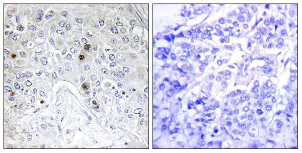
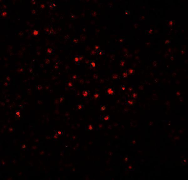
Anti-BATF antibodies have IHC data from paraffin-embedded human breast cancer (A04089-1 IHC caption) and IF data from human spleen tissue (A04089 IF caption).

Real IHC data Immunohistochemical analysis of paraffin-embedded Human breast cancer. Antibody was diluted at 1:100 (4° overnight). High-pressure and temperature Tris-EDTA, pH8.0 was used for antigen retrieval. Negetive contrl (right) obtaned from antibody was pre-absorbed by immunogen peptide.
Anti-B-ATF Antibody
Cat # A04089-1
Real IF data Immunofluorescence of BATF in human spleen tissue with BATF antibody at 20 μg/mL.
Anti-BATF Antibody
Cat # A04089

A04089-1 will render with IHC data from paraffin-embedded human breast cancer; IHC and IF are listed applications, with human and mouse reactivity (A04089-1 catalog and IHC caption). A04089 will render with IF data from human spleen tissue; IF is a listed application, with human, mouse and rat reactivity (A04089 catalog and IF caption).

Which to pick: For tissue IHC, choose A04089-1: its paraffin-section image used Tris-EDTA retrieval at pH 8.0 and antibody at 1:100 overnight at 4 °C; the fixative is unreported (A04089-1 IHC caption). For IF/ICC planning, A04089 has human spleen IF data at 20 μg/mL, but ICC validation is unreported (A04089 IF caption and applications). For cross-species selection, A04089 lists human, mouse and rat reactivity, while A04089-1 lists human and mouse; both are rabbit antibodies with no clone specified, and the pictured results are from human samples (catalog reactivity, host and clone; image captions).

Each figure is that product's own IHC / IF validation image from its datasheet.

References

  1. UniProt Consortium. UniProt entry Q16520 (BATF_HUMAN, Basic leucine zipper transcriptional factor ATF-like).
  2. Human Protein Atlas. BATF tissue IHC expression (reliability: Approved).
  3. Human Protein Atlas. BATF subcellular location (ICC-IF): Localized to the nucleoplasm..
  4. Human Protein Atlas. BATF antibody validation summary (2 antibodies).
  5. Increased expression of the Th17-IL-6R/pSTAT3/BATF/RorγT-axis in the tumoural region of adenocarcinoma as compared to squamous cell carcinoma of the lung. Scientific reports 2014 — PMC4261178.
  6. BATF promotes tumor progression and association with FDG PET-derived parameters in colorectal cancer. Journal of translational medicine 2024 — PMC11165778.
  7. Integration of single-cell and bulk RNA sequencing identifies and validates T cell-related prognostic model in hepatocellular carcinoma. PloS one 2025 — PMC12047759.
  8. Development and validation of a prognostic prediction model for endometrial cancer based on CD8+ T cell infiltration-related genes. Medicine 2024 — PMC11630932.
  9. PubMed PMID:8570175 — UniProt-cited evidence.
  10. PubMed PMID:8630063 — UniProt-cited evidence.
  11. PubMed PMID:9745044 — UniProt-cited evidence.