This website uses cookies to ensure you get the best experience on our website.


- Table of Contents
Plan BBC3 chromogenic IHC in paraffin sections using the 2.5–10 μg/mL antibody range validated in human breast tissue (datasheet A04899-1). Assess cytoplasmic staining in the relevant cell populations (HPA tissue IHC), while accounting for 4 isoforms (UniProt).

Expected localisation, validated protocols, controls and antibodies — the at-a-glance facts below, then the full design guide.
| Expected localisation | Cytoplasmic staining in several tissues (HPA tissue IHC) | |
| Staining pattern | Cytoplasmic in breast glandular cells and lung macrophages (HPA tissue IHC) | |
| Antigen retrieval | Citrate pH 6 HIER, heat-mediated (datasheet A04899-1) | |
| Positive control | Breast+4 more · see all | |
| Negative control | Adipose tissue+4 more · see all |
| Fixation | Use formaldehyde-fixed paraffin sections (selected-SKU IHC image A04899-1) | |
| Caveat | Staining and RNA show only medium consistency (HPA tissue IHC) | |
| Regulation | Regulation is not annotated (UniProt) | |
| Isoform / epitope | 4 isoforms; isoform 3 is nonmitochondrial; epitope coverage is unverified (UniProt) |
The catalog antibody’s citrate pH 6 IHC protocol (datasheet: A04899-1) is accompanied by one published rat IHC protocol (PMC2173793).
| Sample | Formaldehyde-fixed, paraffin-embedded human breast carcinoma tissue (datasheet A04899-1) |
| Fixation | Image formalin-fixed; duration unreported (datasheet A04899-1); verify before use. |
| Sectioning | 4–5 µm sections on charged slides (standard) |
| Deparaffinisation | Xylene, graded ethanol series to water (standard) |
| Antigen retrieval | Heat retrieval: Citrate pH 6 (datasheet A04899-1); 20 min, 95–100 °C (standard) |
| Peroxidase block | 3% H2O2, 10 min, room temperature (standard) |
| Blocking | 10% serum (datasheet A04899-1) |
| Primary antibody | Rabbit anti-BBC3, 2.5-10 μg/mL (datasheet A04899-1) |
| Primary incubation | Overnight at 4 °C (standard) |
| Detection | HRP-polymer secondary, DAB chromogen 5–10 min (standard) |
| Counterstain | Hematoxylin, blue, dehydrate and mount (standard) |
| Expected result | BBC3-positive staining in glandular cells of breast (HPA tissue IHC: High). HPA tissue profile: Cytoplasmic expression in several tissues. No signal in the no-primary control. |
Expect BBC3 staining mainly in the cytoplasm of selected cells, including glandular cells in breast, cervix, colon and rectum; lung macrophages; pancreatic endocrine cells; and cerebellar Purkinje cells (HPA: tissue IHC, high). The HPA tissue profile is Approved, with medium consistency between staining and RNA data (HPA: reliability). UniProt annotates no transmembrane segment and does not assign a subcellular location to isoform 3/4 (UniProt Q96PG8: topology, subcellular annotation).
| Cytoplasmic staining stands out in colon glandular cells, with relatively little staining in adjacent cell types (HPA: colon glandular cells, High). | This matches an observed positive tissue and cell type (HPA: colon glandular cells, High). Use the cell type and compartment together when judging a section; tissue name alone is insufficient because HPA reports staining by cell population (HPA: tissue IHC). |
| Strong, predominantly nuclear or sharply membrane-restricted staining replaces the expected cytoplasmic pattern (HPA: cytoplasmic tissue profile). | Treat this as a localisation mismatch requiring control review, rather than as the expected BBC3 result (HPA: tissue IHC; HPA: cytosol, ICC-IF). UniProt reports no transmembrane segment, but that annotation alone cannot identify the cause of an unexpected staining pattern (UniProt Q96PG8: topology). |
| Prominent staining appears in prostate glandular cells while the positive control stains as expected (HPA: prostate glandular cells, Not detected). | This conflicts with the reported HPA cell pattern and warrants investigation for antibody cross-reactivity or endogenous detection activity (HPA: prostate glandular cells, Not detected; general IHC practice). A Not detected HPA result is a comparison point, not proof that every specimen must be negative (HPA: tissue IHC). |
| Weak, even chromogen covers many cells or the section without a clear cytoplasmic cell pattern (general IHC practice). | Diffuse staining is background until controls support a cell-specific result (general IHC practice). Compare a section lacking primary antibody and inspect the counterstain, detection reagents and wash steps before assigning BBC3 positivity (general IHC practice). |
| No signal appears in colon glandular cells or lung macrophages selected as positive reference populations (HPA: both cell types, High). | First check that the expected cells are present and the staining run worked; absence of signal alone does not establish biological absence (general IHC practice). HPA reports high staining in these populations, while its overall tissue IHC reliability is Approved with medium RNA consistency (HPA: tissue IHC). |
| Cell population within a tissue (HPA: tissue IHC) | HPA reports High staining in colon glandular cells and lung macrophages, but Not detected in prostate glandular cells and adipocytes; score the named cells rather than the whole tissue uniformly (HPA: tissue IHC). |
| Strength and reliability of the reference pattern (HPA: reliability) | The tissue profile is Approved with medium staining-to-RNA consistency and low RNA tissue specificity. Treat HPA intensity categories as observed reference patterns, not absolute cutoffs for an individual specimen (HPA: tissue IHC). |
| Isoform and localisation scope (UniProt Q96PG8; HPA: subcellular) | UniProt lists four BBC3 isoforms and says isoform 3 does not localise to mitochondria, unlike isoforms 1 and 2; it leaves isoform 3/4 location unannotated. HPA's approved cytosol call comes from ICC-IF and does not assign a compartment separately to each isoform (UniProt Q96PG8: isoforms, subcellular annotation; HPA: ICC-IF). |
| Assay-specific antibody evidence (HPA: antibodies) | CAB007752 has Approved IHC status, while HPA070403 has Uncertain ICC status and no listed IHC status. Apply each validation label only to its stated assay; the HPA tissue profile does not establish that any substitute antibody reproduces it (HPA: antibodies, tissue IHC). |
| Situation | Likely cause | Next action |
|---|---|---|
| The positive reference section has no visible cytoplasmic signal (HPA: colon glandular cells, High). | The expected cells may be absent from the section, or a general staining step may have failed (general IHC practice). | Confirm the cells in the counterstained section, then check the primary antibody, detection reagents and run controls before interpreting test sections (general IHC practice). |
| The whole section develops pale, uniform chromogen (general IHC practice). | Nonspecific reagent binding, insufficient washing or endogenous detection activity can produce background (general IHC practice). | Compare a section without primary antibody; review blocking, wash steps and any endogenous enzyme block appropriate to the detection system (general IHC practice). |
| Prostate glandular cells stain strongly even though the reference pattern is Not detected (HPA: prostate glandular cells). | Possible cross-reactivity or detection background; specimen biology can also differ from the HPA reference (HPA: tissue IHC; general IHC practice). | Check compartment and cell identity, compare a positive reference and a section without primary antibody, and avoid calling the signal BBC3 solely from colour intensity (general IHC practice). |
| Signal is concentrated in nuclei or along cell borders rather than in the cytoplasm (HPA: tissue IHC). | The pattern disagrees with HPA's cytoplasmic tissue profile; its cause cannot be assigned from location alone (HPA: tissue IHC; general IHC practice). | Review controls, counterstain and morphology, then compare another section or an independently supported antibody result if available (general IHC practice). |
| A tissue has patchy staining that is difficult to score (general IHC practice). | BBC3 staining varies by cell population: HPA reports High in lung macrophages and Medium in skin fibroblasts, while other listed populations are Low or Not detected (HPA: tissue IHC). | Identify the cell type first and record its compartment and intensity separately; compare only with the matching HPA cell population (HPA: tissue IHC; general IHC practice). |
| Can an IF/ICC image confirm the expected IHC pattern? (HPA: ICC-IF, tissue IHC) | HPA approves cytosolic localisation in ICC-IF, but the supplied antibody statuses differ by assay (HPA: subcellular, antibodies). | Use cytosolic IF/ICC as a localisation comparison; judge paraffin-section chromogenic staining against the tissue IHC cell patterns and the IHC antibody's own validation (HPA: subcellular, tissue IHC, antibodies). |
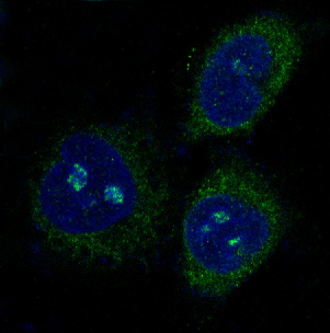
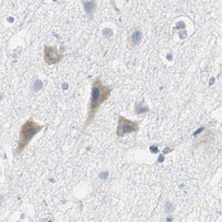
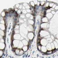
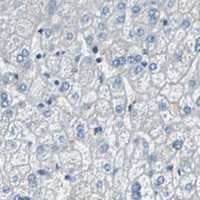
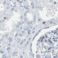


Comprehensive Human Protein Atlas IHC scoring per tissue (reliability: Approved — Medium consistency between antibody staining and RNA expression data.). Rows are taken directly from the HPA tissue chart — click any row's HPA link to view the source.
| Tissue | Cell type | Level | Evidence | Source |
|---|---|---|---|---|
| Breast | Glandular cells | High | Protein (IHC) | HPA → |
| Cerebellum | Purkinje cells | High | Protein (IHC) | HPA → |
| Cervix | Glandular cells | High | Protein (IHC) | HPA → |
| Colon | Glandular cells | High | Protein (IHC) | HPA → |
| Lung | Macrophages | High | Protein (IHC) | HPA → |
| Tissue | Cell type | Level | Evidence | Source |
|---|---|---|---|---|
| Adipose tissue | Adipocytes | Not detected | Protein (IHC) | HPA → |
| Caudate | Glial cells | Not detected | Protein (IHC) | HPA → |
| Kidney | Cells in glomeruli | Not detected | Protein (IHC) | HPA → |
| Liver | Cholangiocytes | Not detected | Protein (IHC) | HPA → |
| Parathyroid gland | Glandular cells | Not detected | Protein (IHC) | HPA → |
Troubleshoot chromogenic IHC for BBC3 isoforms 3/4 in paraffin sections, using the selected antibody’s tissue image and the stated localisation evidence.
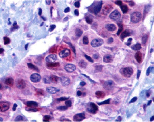
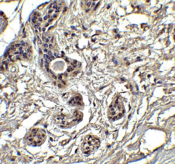
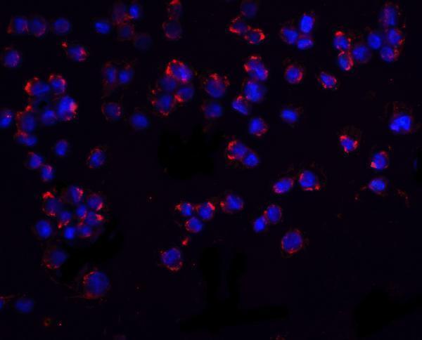
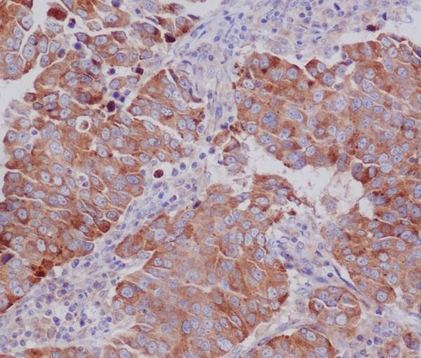
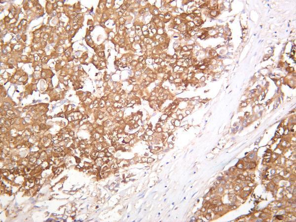
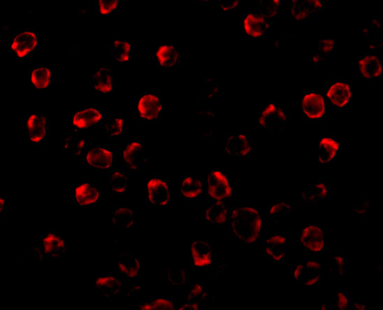
Anti-BBC3 antibodies have IHC images from human breast carcinoma and hepatocellular carcinoma (catalog IHC captions), plus IF images from K562 cells and rat retina (catalog IF captions).
A04899-1 shows formaldehyde-fixed, paraffin-embedded human breast carcinoma IHC (A04899-1 IHC caption); M04899 shows paraffin-embedded human breast cancer IHC, with fixative unreported (M04899 IHC caption). M04899-2 shows human hepatocellular carcinoma staining, with processing and fixative unreported (M04899-2 IHC caption); A04899 shows IF in paraformaldehyde-fixed K562 cells (A04899 IF caption).
Which to pick: For tissue IHC, choose A04899-1 when its documented formaldehyde-fixed, paraffin-section workflow fits your sample (A04899-1 IHC caption); M04899 also has a paraffin-section image, but its fixative is unreported (M04899 IHC caption). For IF/ICC, A04899 lists both applications and shows IF in K562 cells (A04899 applications; A04899 IF caption). For planned human, mouse or rat work, M04899 and M04899-2 are monoclonal options that list all three species and IHC/IF applications (M04899 and M04899-2 catalog entries); their pictured IHC samples are human, and M04899-2’s tissue processing and fixative are unreported (M04899 and M04899-2 IHC captions).